Features of Connective Tissues
Objective 7.5
7.5.1 Describe the basic features and cell types of connective tissue.
Connective tissue is the most abundant tissue in the body. Guess what? This tissue connects things like tissues and organs. It protects and supports the body and body organs, binds organs together, stores energy as fat, and provides immunity.
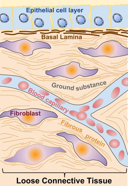
Connective tissue is not densely packed like epithelial tissue. Cells are typically spread out, separated by an extracellular matrix.
Connective tissue is highly vascular with the exception of cartilage, which is avascular, and tendons, which have a limited blood supply.
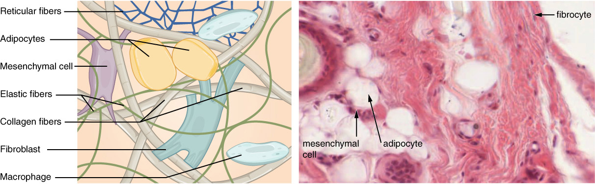
Connective Tissue Cells
Let’s break down these two elements. First, we’ll look at the cellular portion of connective tissue. These cells include:
- fibroblasts, the most numerous connective tissue cells;
- adipocytes, or fat cells;
- mast cells, which are important in inflammation;
- white blood cells, including the immune cells;
- macrophages, which eat debris or invaders;
- plasma cells, which develop from a type of white blood cell and secrete a protein called an antibody that helps attack invaders.
We‘ll look at each of the cell types in turn and briefly discuss the function of each.
Fibroblasts, as the name suggests, lay down the protein fibers: collagen, elastin, and reticular.
That pesky fat. You‘ve tried sucking it out, shaking it out, and dieting it out. And yet, you still have it covering inconvenient and unattractive places. As the French say, tant pis (oh, well).
Body fat is produced by adipocytes and is used for insulation, energy storage, and to enrich diet companies when people resolve to lose weight every January.
The last four types of cells found in connective tissue fit into the category of defensive cells, cells that defend the body against invaders. They patrol the connective tissues and attack any enemies they find there. Cells in this group include mast cells, white blood cells, macrophages, and plasma cells.
Some types of connective tissue contain all of these types of cells, others only one type or even a specialized cell. We’ll look at these differentiations more when we study the different types of connective tissue.
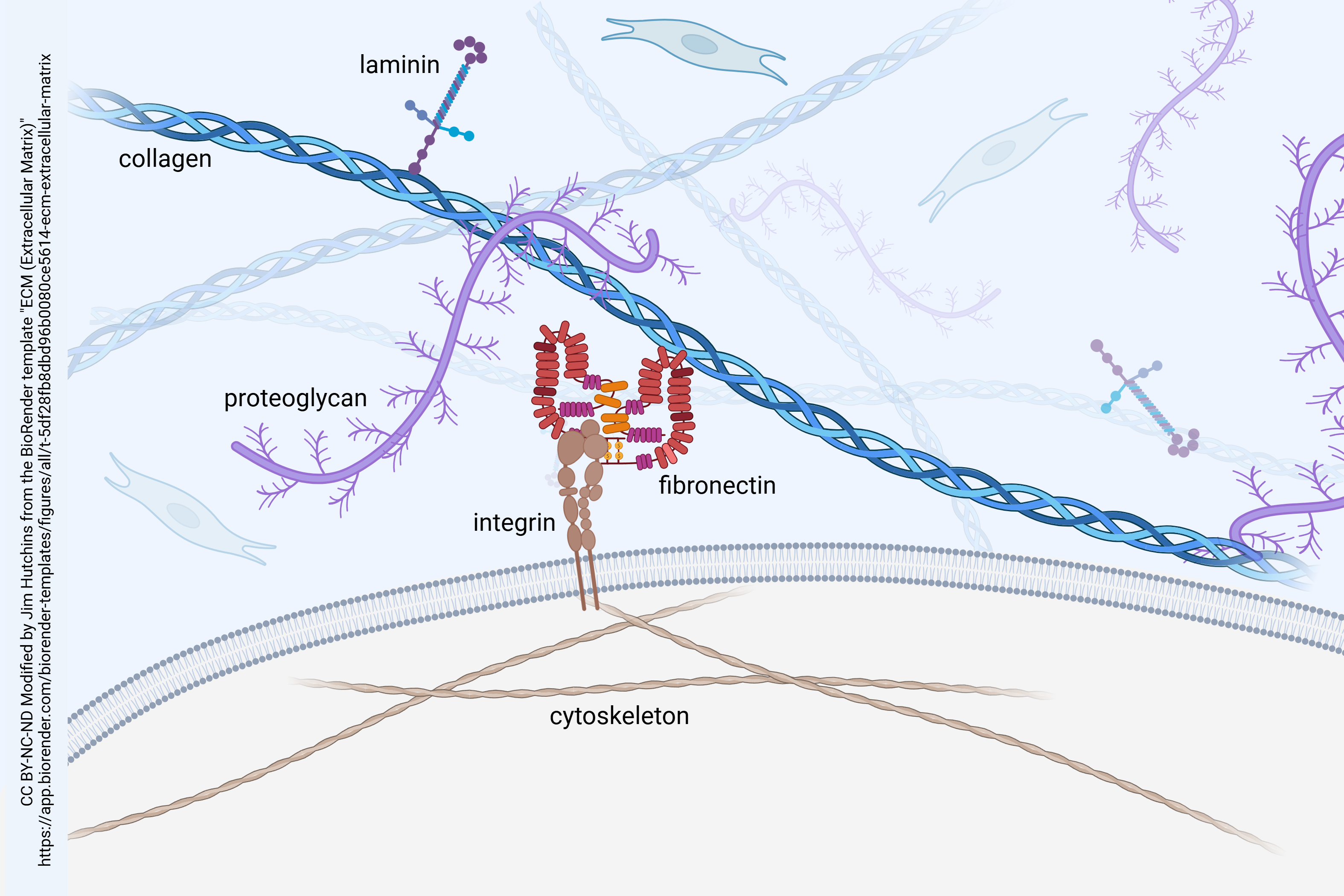
Connective Tissue Extracellular Matrix
Now we’ll turn our attention to the extracellular matrix, generally produced by the cells of the specific connective tissue. The extracellular matrix has two basic components, a ground substance and protein fibers.
Connective Tissue Ground Substance
The ground substance can be liquid, semifluid, gelatinous, fibrous, or calcified. The ground substance provides a medium for the exchange of materials between the blood and connective tissue cells. Substances found in the ground substance can include: hyaluronic acid, chondroitin sulfate, dermatan sulfate, and keratin sulfate.
The ground substance is composed of several proteins:
- fibronectin
- laminin
- proteoglycans
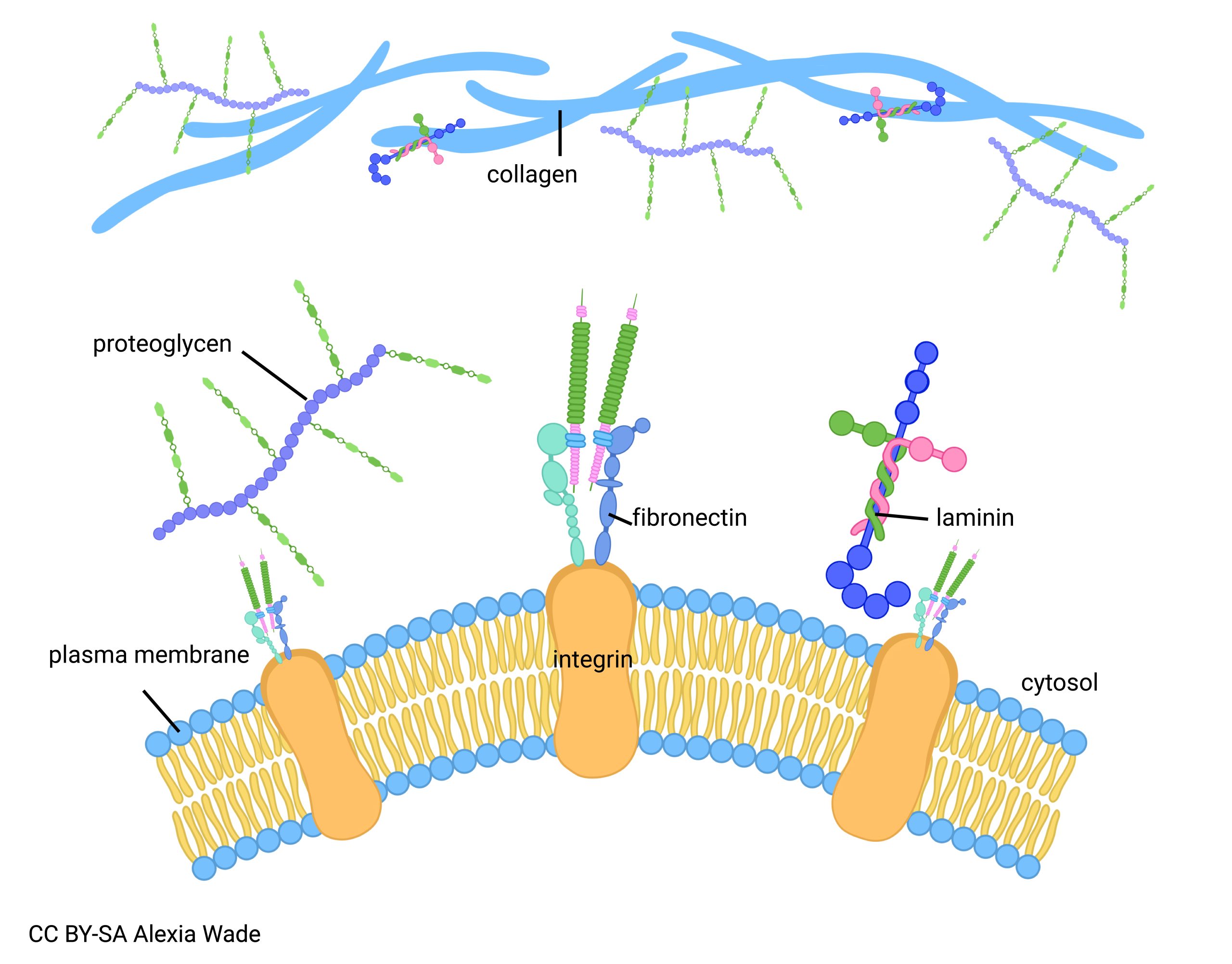
Fibronectin helps cells adhere to each other. It also plays a role in cell growth and migration. So while cells in connective tissue are spread out, this protein helps them stay connected. Fibronectin is often altered in cancerous cells that like to break away from the clan.
Laminin proteins interact with receptors on the surface of cells. They provide both a structural connection between connective tissue and cells, as well as a signaling system to let the cells know what the connective tissue is doing, and vice versa.
The last of these, proteoglycans, are not a specific protein but rather a class of proteins which contain proportionately more sugar (glycans) than protein.
All of these ground substance proteins, acting together, provide a scaffolding for cells and tissues. This scaffolding is not merely structural, like the scaffolding of a building under construction. Rather, the scaffolding is a responsive, signaling structure which both receives information from cells, and sends information to cells.
Connective Tissue Fibers
The common fiber types of connective tissue are: collagen fibers, elastic fibers, and reticular fibers. The proteins that make up these fibers are secreted by fibroblasts. Each of the fibers is made up of a different protein with different properties.
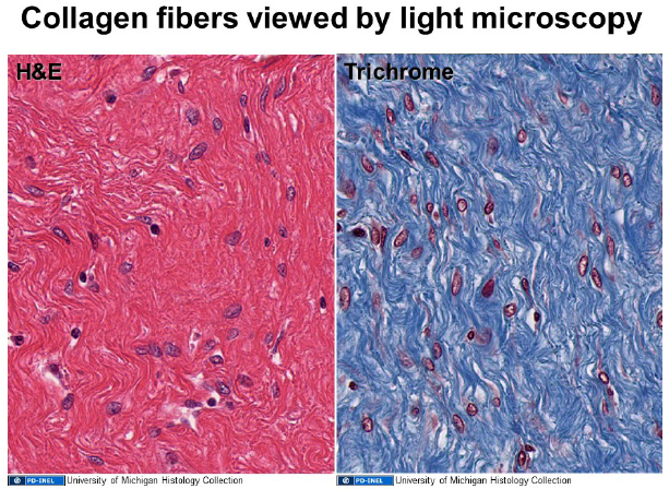
Collagen fibers are made up of collagen (naturally). Collagen comes in several different types (called type I collagen, type II collagen, etc. up to type XXIX collagen) that vary between tissues. Recall that collagen is a triple helix. This structure gives it strength along the fiber (i.e. tensile strength in engineering terms). 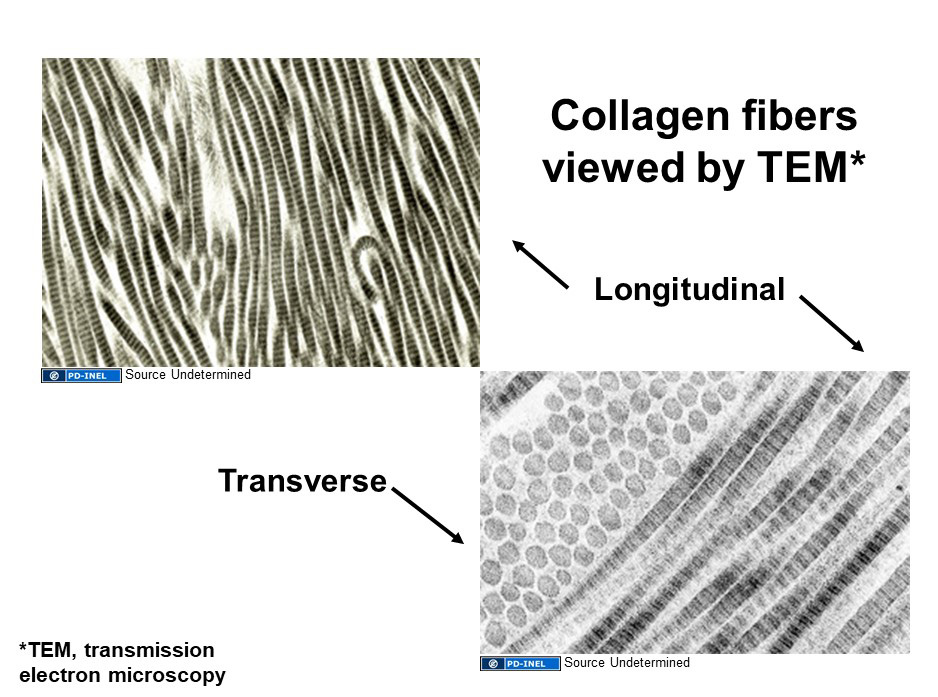
Elastic fibers are made of elastin and are abundant in tissues that need to stretch and then snap back into their previous shape, like blood vessels. We will learn about a type of connective tissue that is rich in elastic fibers.
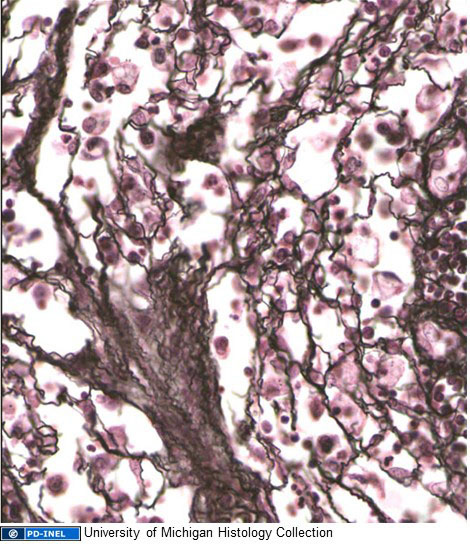
Reticular fibers are made of type III collagen. They were originally named separately because of their silver staining properties, but molecular biologists proved later that reticular fibers are a type of collagen fiber. Structures that stain with silver are called argyrophilic. (Silver, symbol Ag, is argyrum in Latin.)
Next we’ll turn our attention to different types of connective tissue. The following chart may help you keep the different types organized as we proceed through the unit.
Connective Tissue Classifications
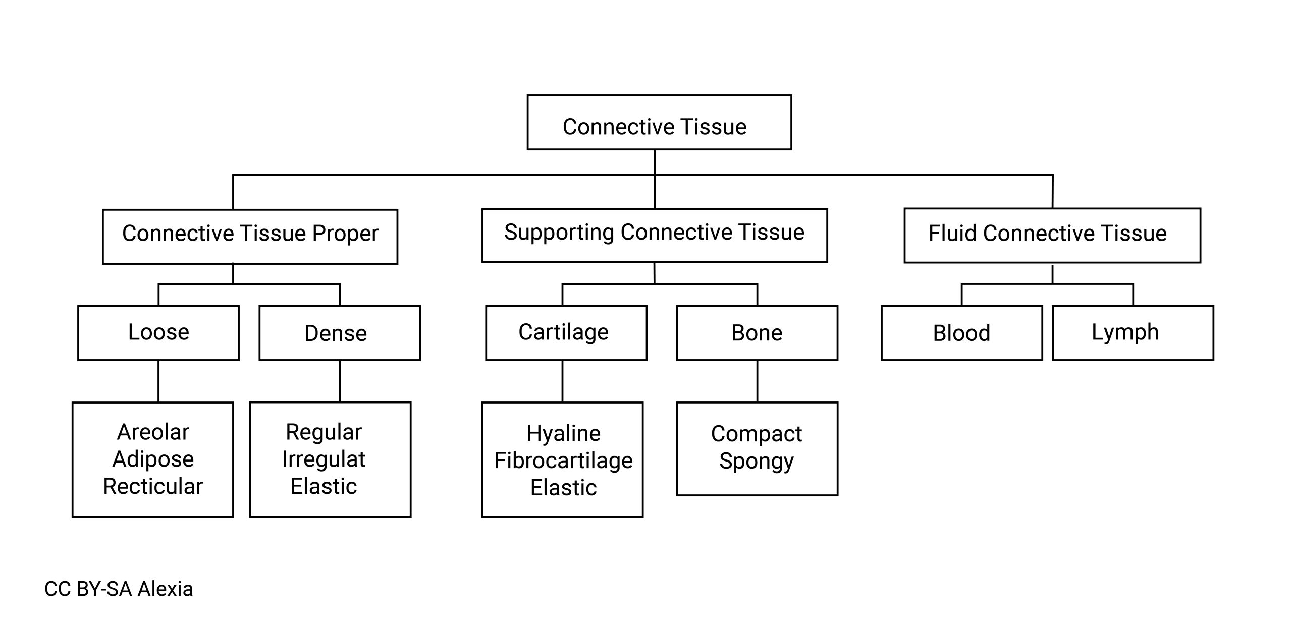
This chart is a classification of connective tissues, that we’ll use as we describe the different types of tissue.
Media Attributions
- Extracellular_Matrix_v1.001-labeled © Sagearbor is licensed under a CC BY-SA (Attribution ShareAlike) license
- Print © Betts, J. Gordon; Young, Kelly A.; Wise, James A.; Johnson, Eddie; Poe, Brandon; Kruse, Dean H. Korol, Oksana; Johnson, Jody E.; Womble, Mark & DeSaix, Peter is licensed under a CC BY-SA (Attribution ShareAlike) license
- U07-040a ECM (Extracellular Matrix) © Hutchins, Jim is licensed under a CC BY-NC-ND (Attribution NonCommercial NoDerivatives) license
- U07-040 linkers © Wade, Alexia is licensed under a CC BY-SA (Attribution ShareAlike) license
- U07-041 collagen TEM © Velkey, Matthew is licensed under a CC0 (Creative Commons Zero) license
- U07-042 collagen LM © Velkey, Matthew is licensed under a CC0 (Creative Commons Zero) license
- U07-043 elastin © Wade, Alexia is licensed under a CC BY-SA (Attribution ShareAlike) license
- U07-044 elastin LM © University of Michigan Histology Collection is licensed under a CC0 (Creative Commons Zero) license
- U07-045 connective tissue org chart © Wade, Alexia is licensed under a CC BY-SA (Attribution ShareAlike) license
