Epidermis
Objective 8.2
8.2.1 Identify the layers of the epidermis.
8.2.2 Compare thin and thick skin. Explain how each of the layers and their cell types (stem cells, keratinocytes, melanocytes, Langerhans cells, Merkel cells, and discs) and substances (keratin, extracellular lipids) contribute to the function of the epidermis.
The Epidermis
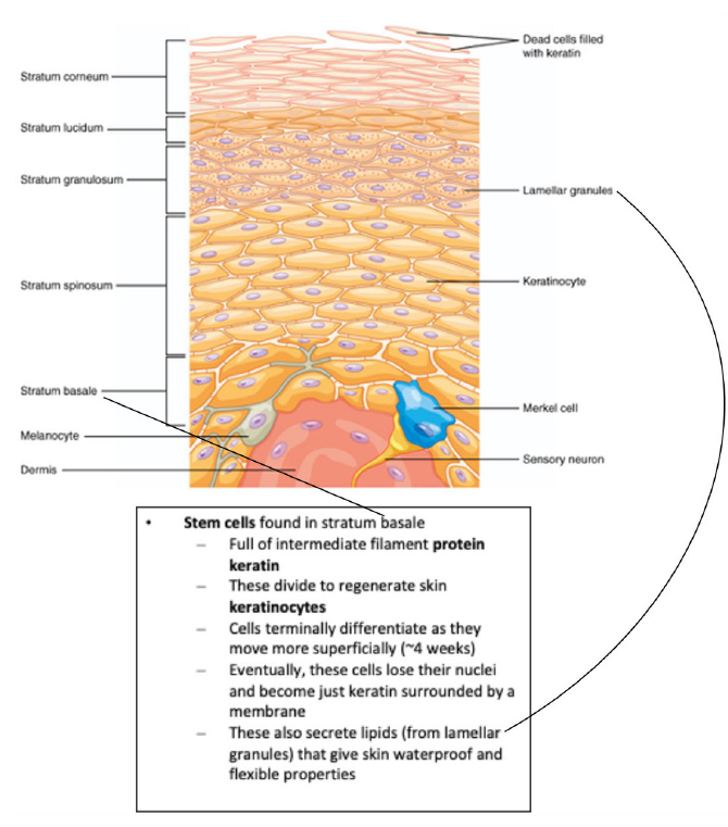
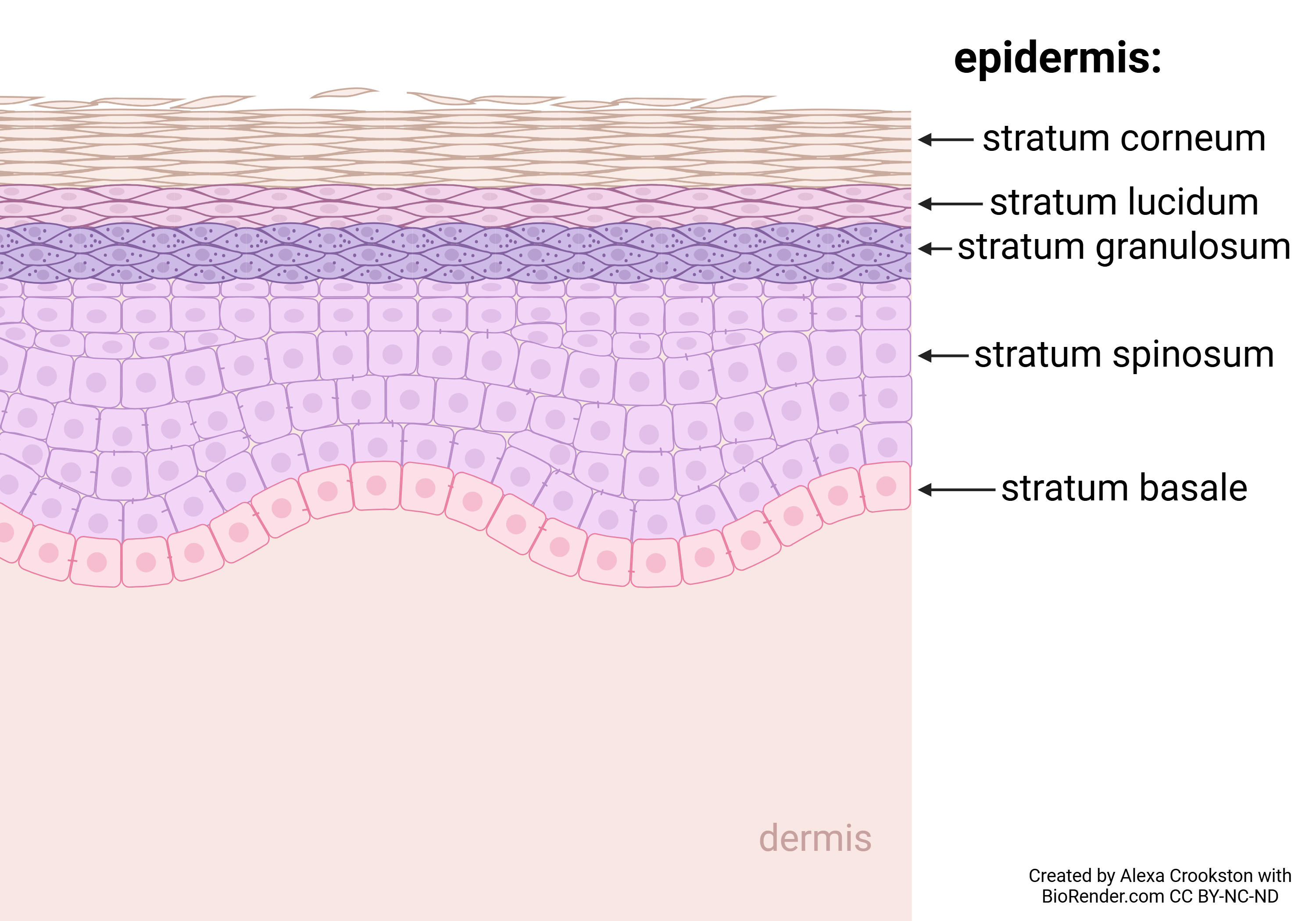
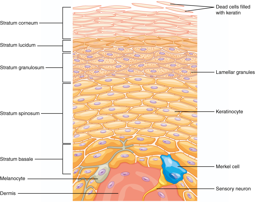
The epidermis contains keratinized stratified squamous epithelium. From deep to superficial, the layers are:
- Stratum Basale:
- Layer from which all others generate (mitosis of stem cells).
- Is the deepest epidermal layer and attaches the epidermis to the basal lamina, below which lie the layers of the dermis.
- Stratum Spinosum: “spiny”
- As the name suggests, the stratum spinosum is spiny in appearance due to the protruding cell processes that join the cells via the intercellular junctions called desmosomes that we learned about in Unit 7. The desmosomes interlock with each other and strengthen the bond between the cells.
- Stratum Granulosum: “grainy”
- Has a grainy appearance due to further changes to the keratinocytes as they are pushed upward from the stratum spinosum.
- Stratum Lucidum: “clear” (only in thick skin)
- Is a smooth, seemingly translucent layer of the epidermis located just above the stratum granulosum and below the stratum corneum. This thin layer of cells is found only in the thick skin of the palms, soles, and digits.
- Stratum Corneum: “horn-like”
- Is the most superficial layer of the epidermis and is the layer exposed to the outside environment. The increased keratinization (also called cornification) of the cells in this layer gives it its name. There are usually 15 to 30 layers of cells in the stratum corneum. This dry, dead layer helps prevent the penetration of microbes and the dehydration of underlying tissues, and provides a mechanical protection against abrasion for the more delicate, underlying layers. Cells in this layer are shed periodically and are replaced by cells pushed up from the stratum granulosum (or stratum lucidum in the case of the palms and soles of feet). The entire layer is replaced during a period of about 4 weeks. Cosmetic procedures, such as microdermabrasion, help remove some of the dry, upper layer and aim to keep the skin looking “fresh” and healthy.
A mnemonic for remembering the names of the epidermal layers, from superficial to deep, is:
“Cher Likes Getting Skin Botoxed” (Corneum, Lucidum, Granulosum, Spinosum, Basale).
All layers of the epidermis contain keratinocytes. In general, living keratinocytes are found in the deeper layers (starting with the stratum basale, the layer that generates keratinocytes through mitosis). As one moves more superficially, dead keratinocytes predominate. The stratum lucidum is found only in thick (hairless) skin. The difference between thick and thin skin will be discussed shortly.
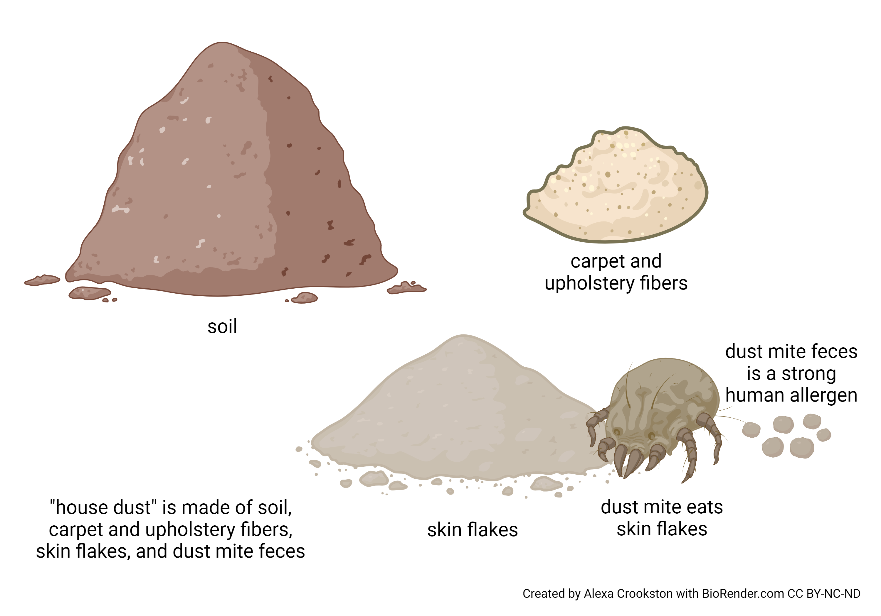
Keratinocytes are constantly made in the stratum basale. As cells are pushed superficially, they lose their nucleus and organelles and become dead bags of keratin. These cells are also called corneocytes. This means these corneocytes are constantly shed from the surface of the stratum corneum. The shed flakes are called squames. Humans shed about 600,000 dead keratinocytes per hour1. These dead skin cells amount to about 2 grams per day or 0.7 kg per year.
An estimated 1/3 or more of “house dust” consists of shed skin flakes 2, 3. Only about half is soil from the outdoors, the remainder is carpet and upholstery fibers. This is not only a cool fact, but is important for human disease. A tiny mite, Dermatophagoides pteronyssinus, eats these skin flakes as its primary source of food. Its feces are a strong allergen for many humans, so the allergy referred to as “house dust” is actually against an allergen shed in mite dung. More than you really wanted to know?
1 http://health.howstuffworks.com/16-unusual-facts-about-the-human-body.htm
2 Butte W and Heinzow B, Rev Environ Contam Toxicol 175:1-46, 2002.
3 Fergusson JE, Forbes EA, Schroeder RJ and Ryan DE, Sci Total Environ 50:217-221, 1986.8-7
Thin vs Thick Skin
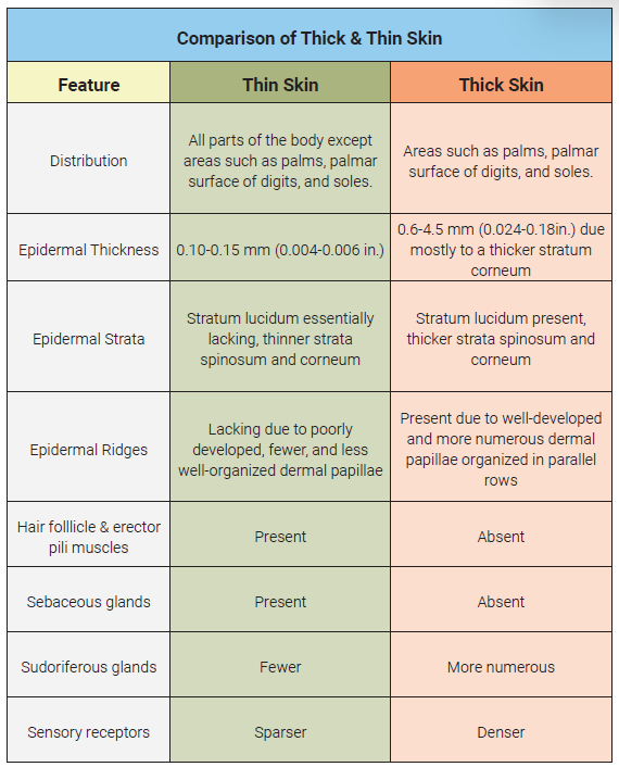
There are two types of skin, each with a different distribution of the four or five skin layers of the epidermis.
Thin skin contains four layers which covers everything except the palms, fingertips, and soles of the feet, and is about 0.1 mm thick. It lacks a stratum lucidum, as mentioned earlier, and the spinosum and corneum are very thin.
Thick skin, also called glabrous (Latin glaber, “bald”) skin, is found on the ventral (palmar) surface of the hands (i.e., palms and fingertips) and on the soles (plantar surface) of the feet. It is from six to 45 times as thick as thin skin; most of the increased thickness is because of the spinosum, lucidum, and corneum.
The Four Cell Types of the Epidermis
- Dead keratinocytes (a cell membrane surrounding the protein keratin) make up about 90% of the cells of the epidermis.
- About 8% of the cells are melanocytes, cells that produce the pigment melanin and carry pigment granules that give the hair and skin its color and helps protect living epidermal cells from ultraviolet (UV) radiation.
- More rarely, we find Langerhans cells, which is the skin’s version of a type of immune cell called a dendritic cell. These cells function as phagocytes to engulf bacteria, foreign particles and damaged cells. Do not confuse these with the beta cells of the islets of Langerhans in the pancreas. (same name, completely different function.)
- The rarest epidermal cell is the Merkel cell (disc), a type of nervous system receptor cell and is responsible for stimulating sensory nerves that the brain perceives as touch. These cells are especially abundant on the surfaces of the hands and feet.
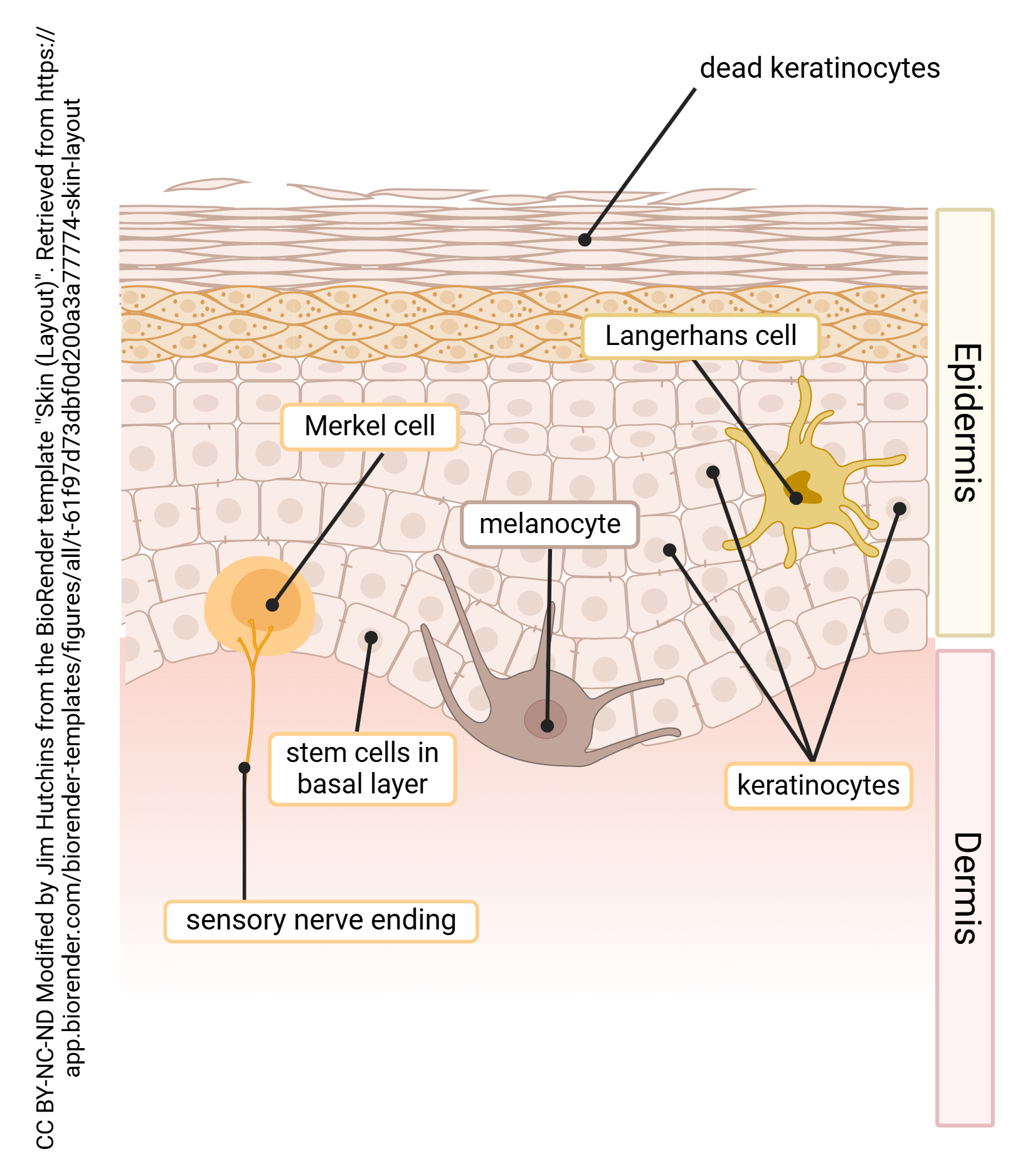
Review the functions of skin listed in Objective 1 and match these four cell types to their functional role.
Media Attributions
- U08-003 skin cells and layers © Betts, J. Gordon; Young, Kelly A.; Wise, James A.; Johnson, Eddie; Poe, Brandon; Kruse, Dean H. Korol, Oksana; Johnson, Jody E.; Womble, Mark & DeSaix, Peter is licensed under a CC BY (Attribution) license
- U08-004 House Dust © Crookston, Alexa is licensed under a CC BY-NC-ND (Attribution NonCommercial NoDerivatives) license
- U08-005 Epidermis Layers © Crookston, Alexa is licensed under a CC BY-NC-ND (Attribution NonCommercial NoDerivatives) license
- U08-006 comparison of thick and thin skin table © Orrock, Marv is licensed under a CC BY-SA (Attribution ShareAlike) license
- U08-007 Cells of the Skin © Hutchins, Jim is licensed under a CC BY-NC-ND (Attribution NonCommercial NoDerivatives) license
