Dermis
Objective 8.4
8.4.1 Identify, describe and list the functions of the layers of the dermis.
8.4.2 Know the tissue types that make up each layer.
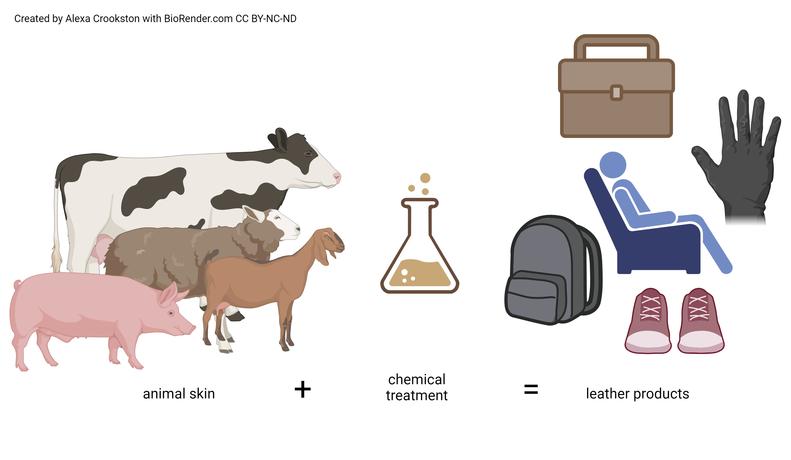
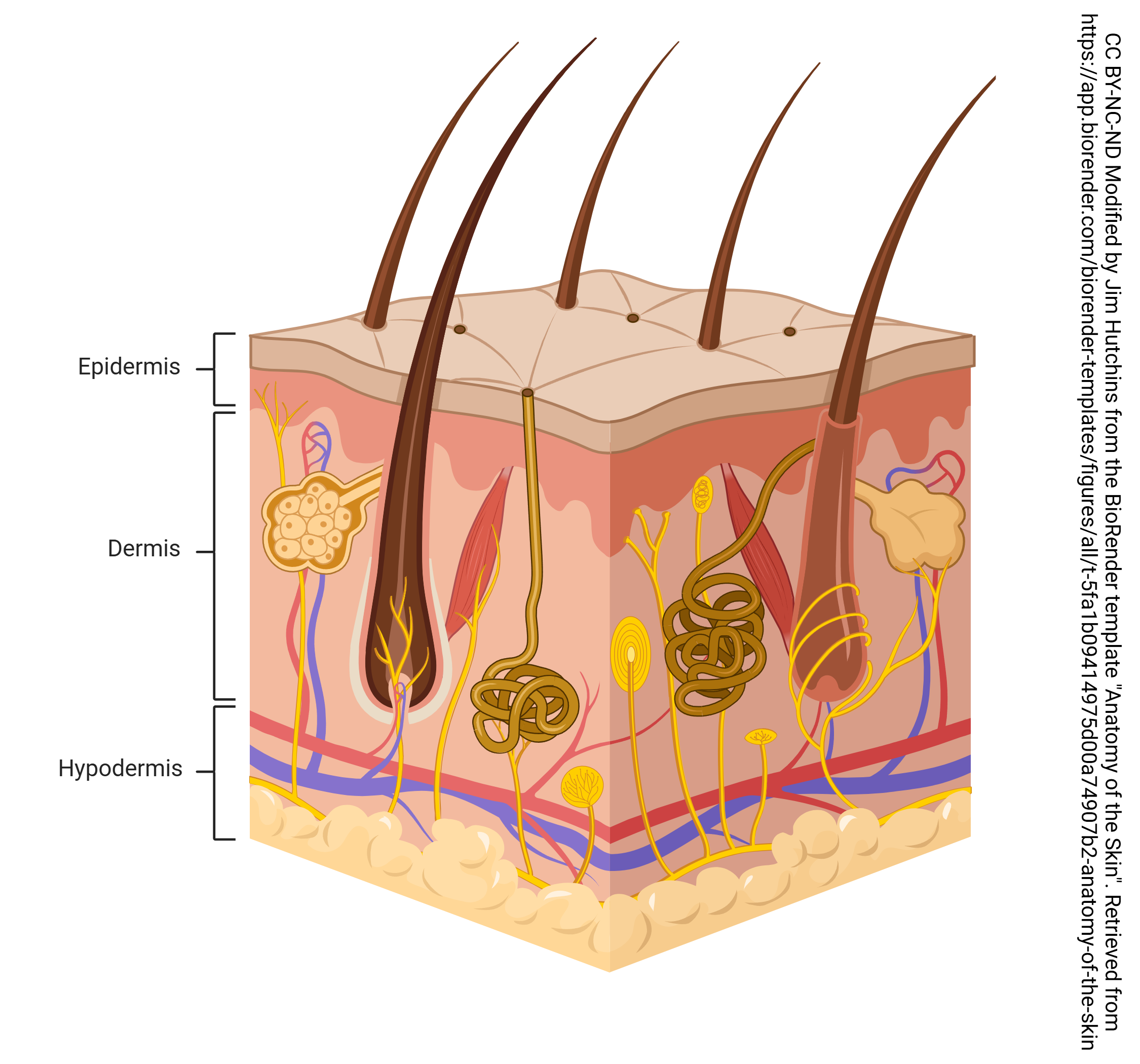
The dermis might be considered the “core” of the integumentary system (derma- = “skin”), as distinct from the epidermis (epi- = “upon” or “over”) and hypodermis (hypo- = “below”). It contains blood and lymphatic vessels, nerves, and other structures, such as hair follicles and sweat glands. The dermis is made of two regions of connective tissue that compose an interconnected mesh of elastin and collagenous fibers, produced by fibroblasts—more on these layers later. Remember from Unit 7 that connective tissue has few cells. It is mostly collagen and elastin fibers. These fibers give the dermis its strength. When the dermis of farm animals is chemically treated, it is called leather.
Dermal Layers
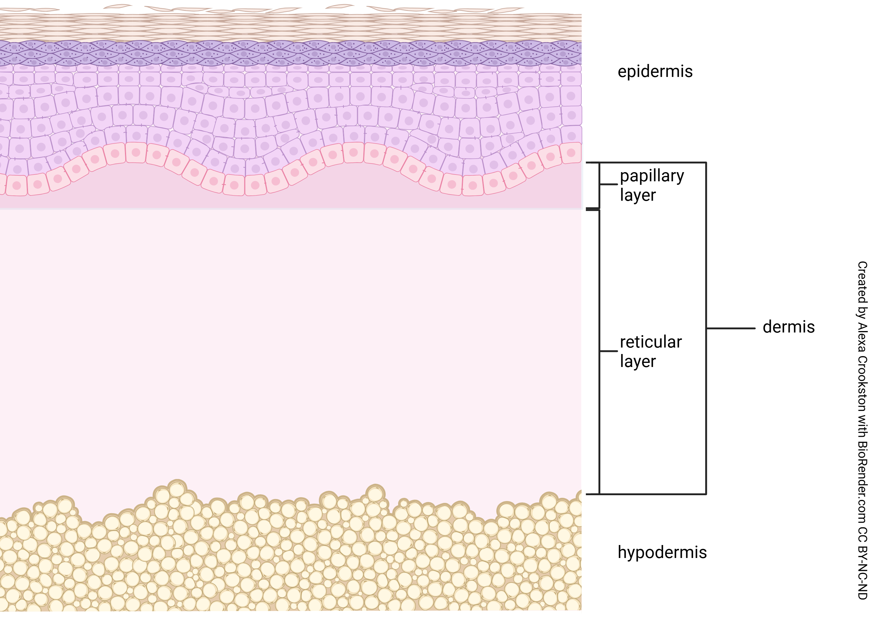
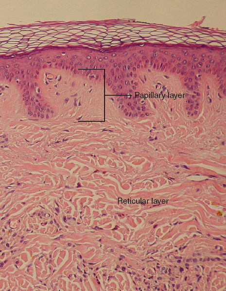
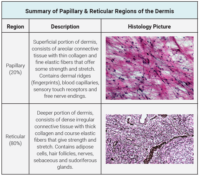
There are two regions of the dermis. The papillary region is made of areolar connective tissue (a type of loose connective tissue) and is about a fifth of the thickness of the dermis. It is thrown up into ridges that penetrate up into the epidermis and each of these tends to contain blood vessels and the sensory structures known as Meissner corpuscles and free nerve endings. We will see later that Meissner corpuscles are for sensing light touch and free nerve endings are used for sensing pain, temperature and itch.
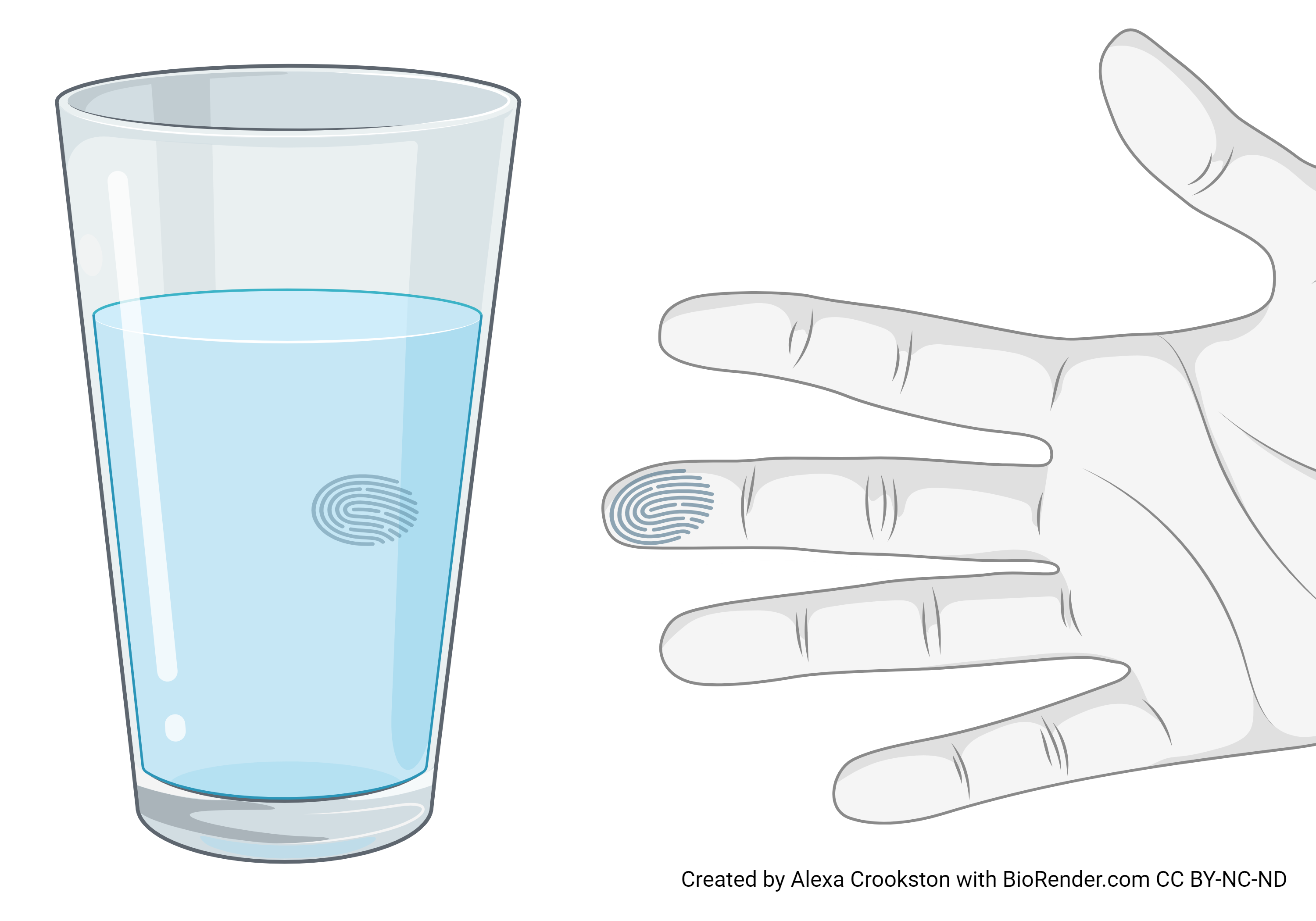
The papillary region is important for its tight attachment to the epidermis. Fingerprints form where the cells of the stratum basale meet the papillae of the underlying dermal layer (papillary layer), resulting in the formation of the ridges on your fingers that you recognize as fingerprints. Fingerprints are unique to each individual and are used for forensic analyses because the patterns do not change with the growth and aging processes. These ridges are found on the palms, fingertips, and soles of the feet.
The remaining 4/5 of the dermis is the reticular region, a layer of dense irregular connective tissue making up about 4/5 of the dermis. The nerves and blood vessels run through the dermis here. Also, hair roots (if present) and glands are found here as well. Where hair roots are present, the stratum basale of the epidermis surrounds the hair root, diving deep into connective tissue layers of the dermis.
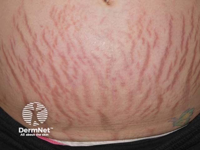
The dense irregular connective tissue of the dermis is strong and resilient; it can be stretched, and it snaps back to its original form. Stretching, such as from obesity or pregnancy, can cause tears in the dermis. These are seen on the skin surface as striae (or, more commonly, “stretch marks”).
As you review the structures that make up the dermis, think about the functions of each.
Media Attributions
- U08-012 Leather © Crookston, Alexa is licensed under a CC BY-NC-ND (Attribution NonCommercial NoDerivatives) license
- U08-013 Dermis Layers © Crookston, Alexa is licensed under a CC BY-NC-ND (Attribution NonCommercial NoDerivatives) license
- 465px-506_Layers_of_the_Dermis 16 © CFCF is licensed under a CC BY (Attribution) license
- U08-014 Fingerprint © Crookston, Alexa is licensed under a CC BY-NC-ND (Attribution NonCommercial NoDerivatives) license
- pregnancy 03 © DermNet NZ is licensed under a CC BY-NC-ND (Attribution NonCommercial NoDerivatives) license
- U08-001 Anatomy of the Skin (1) © Hutchins, Jim is licensed under a CC BY-NC-ND (Attribution NonCommercial NoDerivatives) license
- U08-016 papillary reticular table © Berkshire Community College Bioscience Image Library adapted by Bizzell, Lizz is licensed under a CC BY-SA (Attribution ShareAlike) license

.jpg)