Compact Bone and Spongy Bone
Objective 9.3
9.3.1 Compare and contrast compact bone and spongy bone.
There are two main types of bone, compact bone and spongy bone. Their names give you a pretty good idea of their microscopic structure.
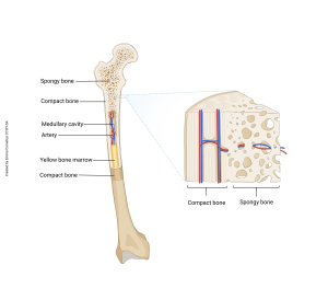
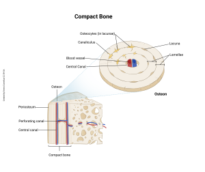
Compact bone is, as the name suggests, compact. This hard, dense tissue makes up the outer part of bones and provides structural support. It accounts for 80% of our total bone mass. The basic structural unit of compact bone is the osteon. These densely packed cylinders of bone tissue each consist of a series of concentric rings called lamellae (singular lamella, Latin for thin plate or layer) and run lengthwise with the long axis of the bone. In the center of each osteon is the central canal, which carries blood and lymphatic vessels. Perforating canals run radially to connect adjacent central canals. Small spaces called lacunae (singular lacuna, Latin for “lake”) lie between the lamellae and each contains an osteocyte. Small channels called canaliculi (singular canaliculus, Latin for “small canals”) connect adjacent lacunae.
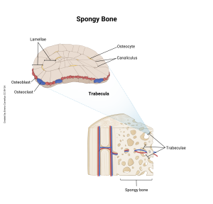
Spongy bone is found on the inside of bones. Its structure is different from compact bone, because it has a different function. The basic structural unit of spongy bone is a trabeculum (plural trabeculae, Latin, little roof beam), “spikey” projections. Rather than the neatly organized osteons of compact bone, the lamellae of spongy bone trabeculae are arranged in several loosely-organized layers and lack a central canal. Like compact bone, spongy bone has lacunae containing osteocytes and canaliculi that connect adjacent lacunae to one another. Osteoclasts and osteoblasts are found around the outside surface of the trabeculae.
Spongy bone lines the medullary cavity of long bones like the femur and humerus. Some bones (skull, pelvis, sternum) and the ends of long bones don’t actually have a marrow “cavity,” but are made up entirely of spongy bone with just a thin shell of compact bone on the outside.
Regardless of where the spongy bone is located, in the holes and spaces between the trabeculae of spongy bone, blood cells are produced in a process called hematopoiesis (red bone marrow). We will study hematopoesis, the formation of blood cells, when we study blood. Within the medullary cavity of long bones, we store fat in the form of triglycerides (yellow bone marrow).
Media Attributions
- U09-009 bone cross section with callout © Cornelius, Emma is licensed under a CC BY-SA (Attribution ShareAlike) license
- U09-009a Compact bone and detail of osteon © Cornelius, Emma is licensed under a CC BY-SA (Attribution ShareAlike) license
- U09-010 Spongy bone and trabecula detail © Cornelius, Emma is licensed under a CC BY-SA (Attribution ShareAlike) license
