Cellular Junctions
Objective 7.10
7.10.1 Describe the five main types of junctions between cells.
7.10.2 Name the most common locations or tissue types where each type of junction is found.
In Unit 5, we looked at the cytoskeleton, the microfilaments, intermediate filaments, and microtubules that give each cell its structure. In this objective, we will look at how cells cooperate together to form tissues. In order for this to happen, the elements of each cell‘s cytoskeleton must be linked to another cell or to the connective tissue supporting the cell.
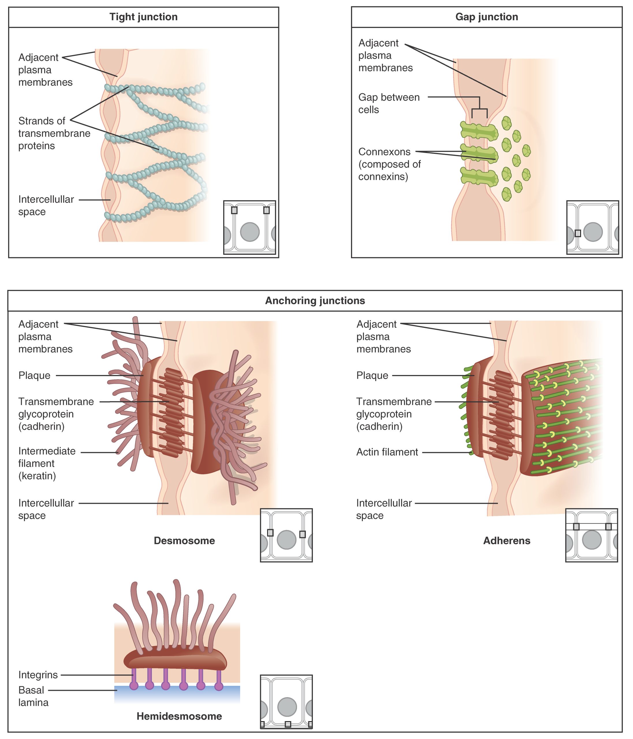
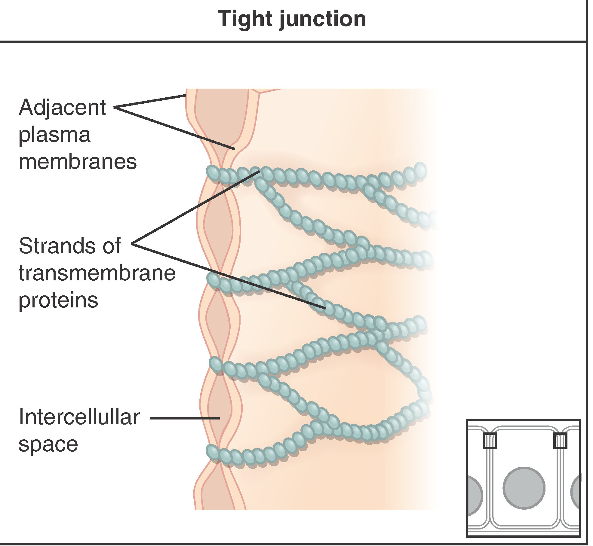
Tight junctions are the Ziploc™ bags of the tissue world. For example, in the intestines, it is important to keep the contents of the intestinal lumen from leaking into the bloodstream. In the skin, tight junctions keep the outside world from gaining access to your connective tissues.
Tight junctions form a leakproof seal between cells where one is needed. Perhaps they also keep the sandwiches inside cells from getting stale and moldy.
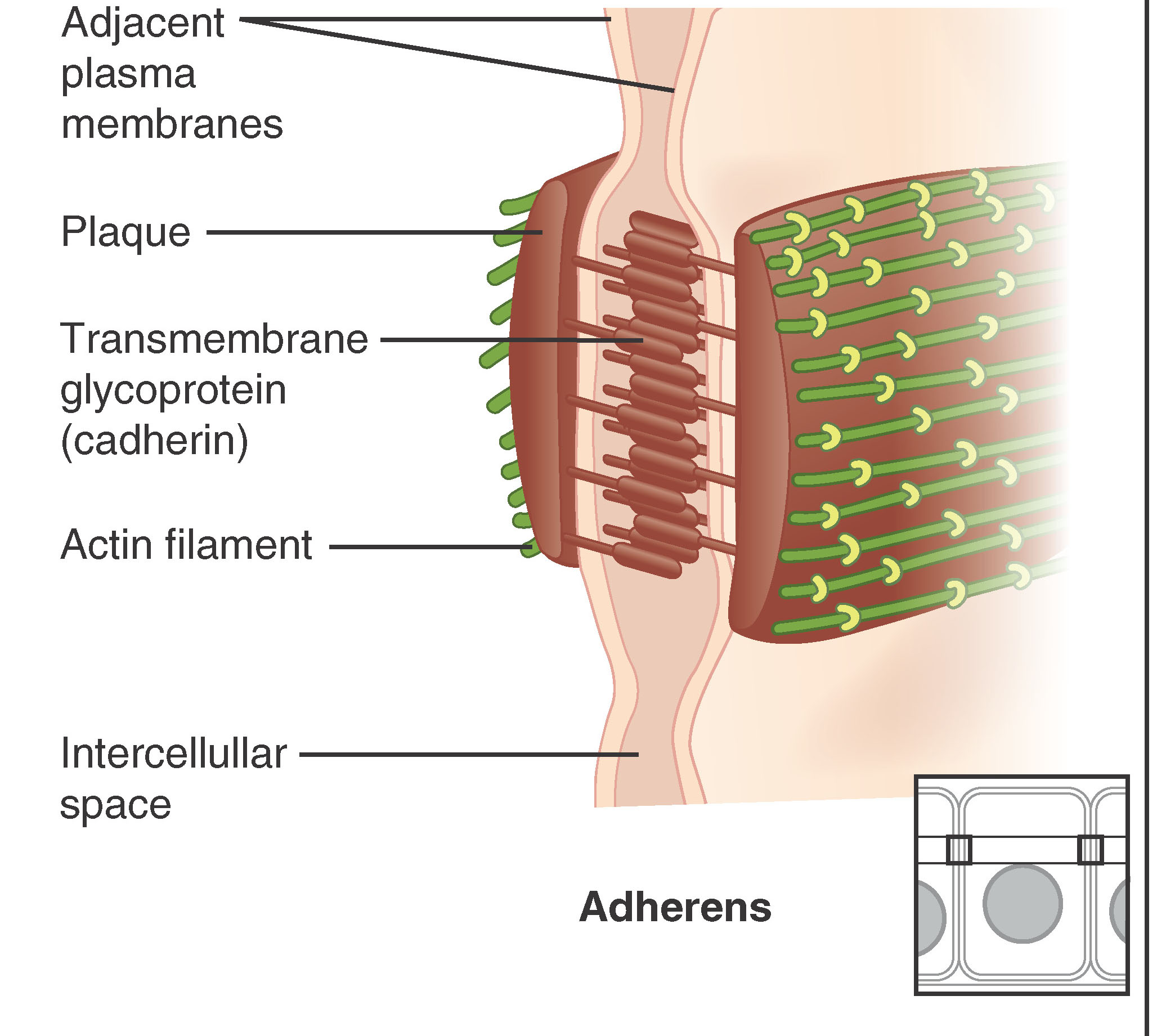
The second type of junction is the adherens junction. These junctions work like a belt to keep the tissues‘ pants from falling down as they expand and contract.
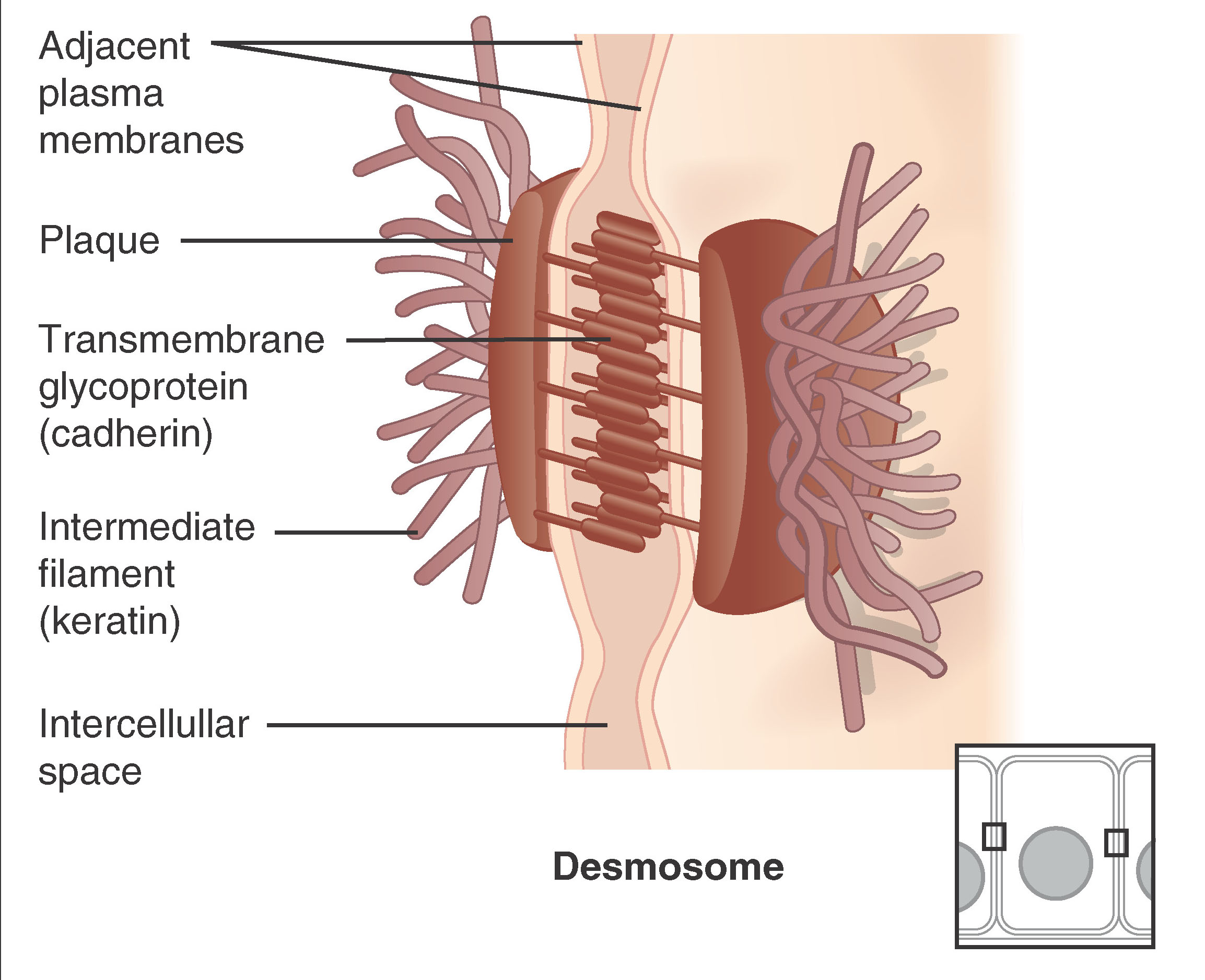
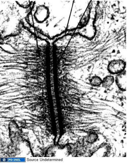
Desmosomes, a third type of cell junction, are used as “spot welds” to hold tissue together against mechanical disruption. Between the cells, the protein cadherin forms a strong linkage. Within the cells, the cadherin is attached to an intermediate filament protein of the cytoskeleton keratin.
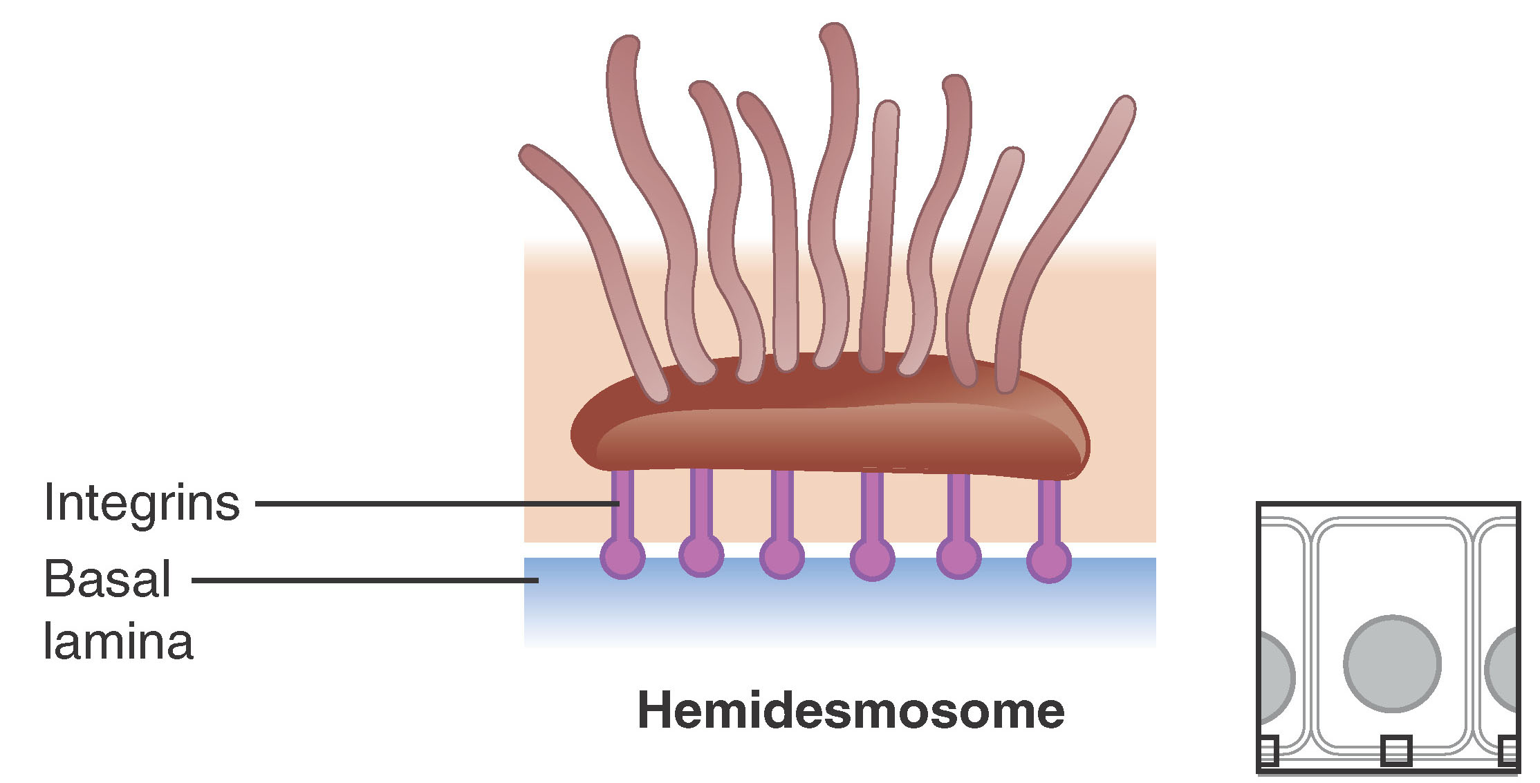
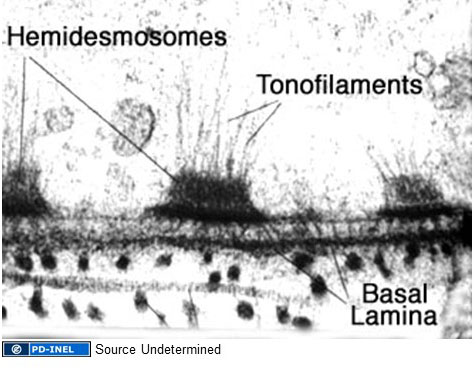
It probably comes as no surprise, then, that a hemidesmosome is half a desmosome. These are found not between two cells, but rather between a cell and its basement membrane and the basal lamina sublayer of the basement membrane. Since there is no cytoskeleton in the basal lamina, the protein integrin replaces cadherin in hemidesmosomes but the appearance in the electron microscope is exactly like half a desmosome.
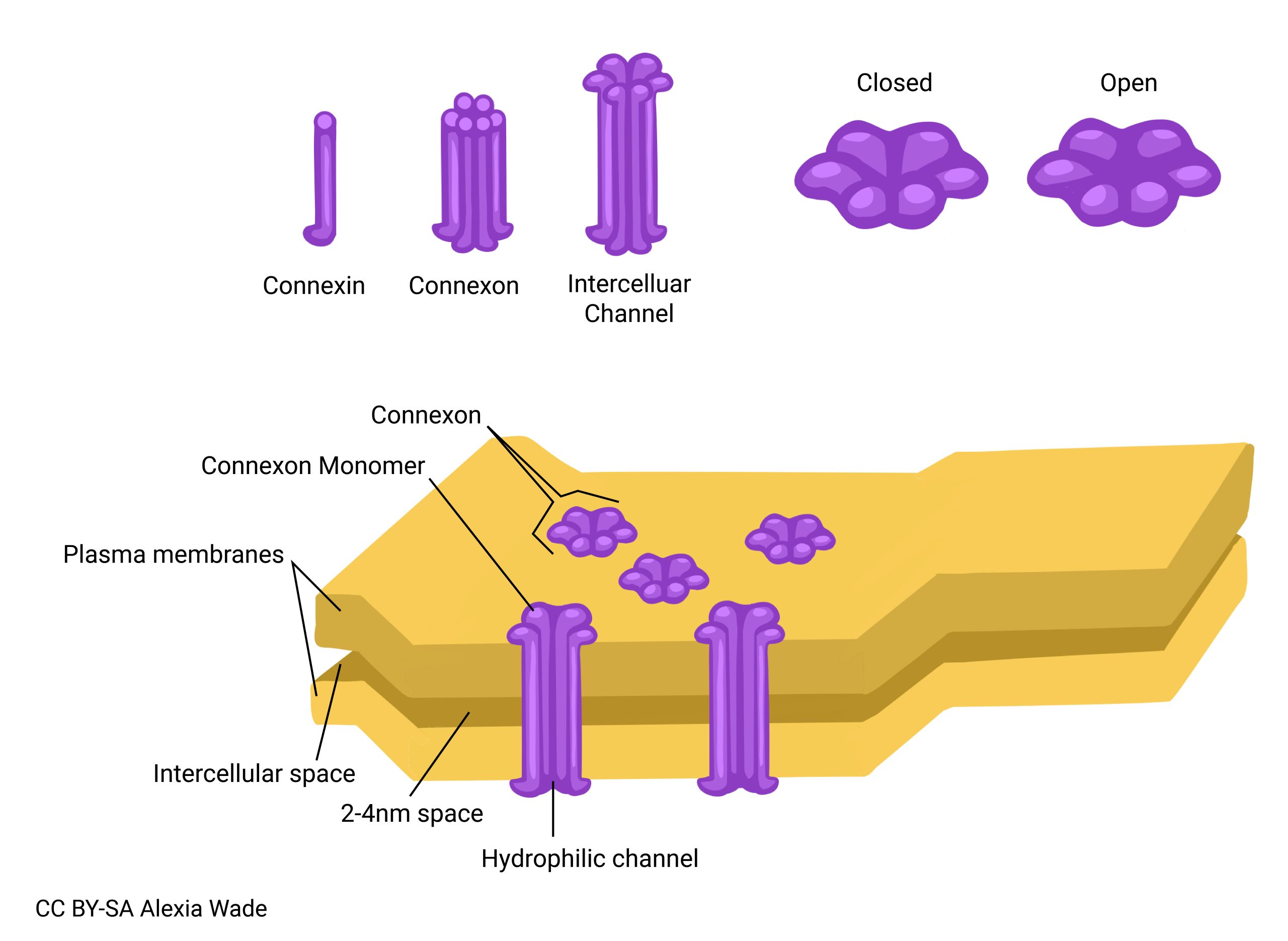
The final type of junction we‘ll consider is the gap junction.
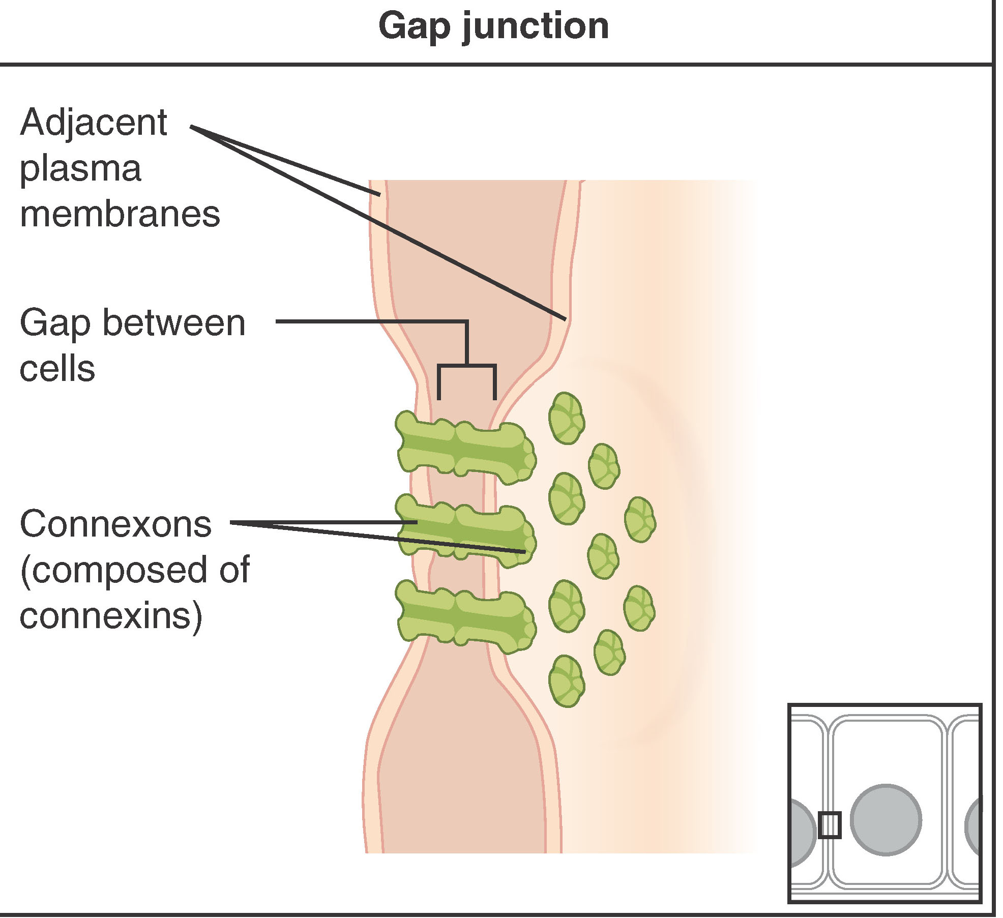
The other junctions serve as mainly mechanical links. In contrast, the gap junction serves as an electrical and biochemical link between two cells. A protein, connexin, forms pore-like structures called connexons. The connexon allows small ions to pass from cell to cell. Monomers and small polymers (for example, calcium ions; the intracellular signaling molecules we studied in Unit 3 such as ATP or cAMP) can pass between cells as well. Heart muscle uses gap junctions to communicate electrical signals so that a group of heart muscle contracts together. Because of this communication, the upper chambers of the heart, the atria, contract all at once. This is followed by the signals traveling to the lower chambers of the heart, the ventricles, causing contraction. This built-in connection between cells could be a problem if a neighboring cell was sick. If this happens, the connexons can seal off like a ship‘s hatch, isolating the damaged cell so that it can die alone without making others sick.
Media Attributions
- U07-064 anchoring junctions © Betts, J. Gordon; Young, Kelly A.; Wise, James A.; Johnson, Eddie; Poe, Brandon; Kruse, Dean H. Korol, Oksana; Johnson, Jody E.; Womble, Mark & DeSaix, Peter is licensed under a CC BY (Attribution) license
- U07-065 tight junction © Betts, J. Gordon; Young, Kelly A.; Wise, James A.; Johnson, Eddie; Poe, Brandon; Kruse, Dean H. Korol, Oksana; Johnson, Jody E.; Womble, Mark & DeSaix, Peter is licensed under a CC BY (Attribution) license
- U07-066 adherens © Betts, J. Gordon; Young, Kelly A.; Wise, James A.; Johnson, Eddie; Poe, Brandon; Kruse, Dean H. Korol, Oksana; Johnson, Jody E.; Womble, Mark & DeSaix, Peter is licensed under a CC BY (Attribution) license
- U07-067 desmosome © Betts, J. Gordon; Young, Kelly A.; Wise, James A.; Johnson, Eddie; Poe, Brandon; Kruse, Dean H. Korol, Oksana; Johnson, Jody E.; Womble, Mark & DeSaix, Peter is licensed under a CC BY (Attribution) license
- U07-068 desmosome_uom © University of Michigan is licensed under a Public Domain license
- U07-069 hemidesmosome © Betts, J. Gordon; Young, Kelly A.; Wise, James A.; Johnson, Eddie; Poe, Brandon; Kruse, Dean H. Korol, Oksana; Johnson, Jody E.; Womble, Mark & DeSaix, Peter is licensed under a CC BY (Attribution) license
- U07-070 hemidesmosome_uom © University of Michigan is licensed under a Public Domain license
- U07-071 gap_junctions © Betts, J. Gordon; Young, Kelly A.; Wise, James A.; Johnson, Eddie; Poe, Brandon; Kruse, Dean H. Korol, Oksana; Johnson, Jody E.; Womble, Mark & DeSaix, Peter is licensed under a CC BY (Attribution) license
- U07-072 connexons © Wade, Alexia is licensed under a CC BY-SA (Attribution ShareAlike) license
