Cellular and Extracellular Components of Blood
Objective 9.2
9.2.1 List the cellular components of bone tissue.
9.2.2 List the extracellular components of bone tissue.
Previously, we learned that connective tissue has cells and an extracellular matrix. The extracellular matrix has fibers and ground substance. Now, let’s apply those concepts to bone tissue.
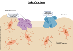
Cells of Bone
The osteogenic cell is a bone stem cell and is the precursor to osteoblasts.
Osteoblasts are dividing cells (Greek οστεου, osteon, bone + -blast, dividing cell) and are responsible for laying down the components of bone. We distinguish them from other cells that start with osteo- by remembering that (osteo)blasts build bone.
As osteoblasts build bone, some of them get “stuck” in the bone they are building and become osteocytes (“bone cells”), which remain stationary in the extracellular matrix and maintain bone integrity. Osteocytes that are “freed” during bone remodeling revert to being osteoblasts.
Osteoclasts constantly chew through bone, releasing enzymes that dissolve the extracellular matrix. Remember: (osteo)clasts chew bone. We mentioned macrophages (“big eaters”) as defensive cells in Unit 7 Objective 5. Osteoclasts are literally siblings of the macrophages, and come from the same stem cell population (Unit 15). These osteoclasts are big eaters too, but they only eat one thing: mineralized bone matrix. They do this by secreting acid which dissolves the ground substance, in this case a mineral instead of an organic compound.
In the bone remodeling process, osteoclasts (chewing bone) are followed by osteoblasts (building bone). In this way, bone is constantly being remodeled and can strengthen when needed and heal when it is broken (albeit slowly…). Both osteoblasts and osteoclasts also play important roles in keeping blood calcium and phosphorus levels within normal limits. To do so, their activity is controlled by hormones, which will be discussed in a later objective.
Extracellular Matrix of Bone
So, now that we’ve talked about the cells of bone, we need to address the extracellular matrix, which has both fibers and ground substance. In bone, we often refer to the fibers as the organic component of bone and the ground substance as the inorganic component.
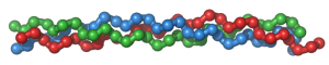
The organic (fiber) portion of bone is mostly type I collagen (remember the Jell-O discussion from Unit 3?). It also contains several minor proteins, but we won’t worry about them.

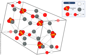
The inorganic (ground substance) portion of bone is primarily a mineral called hydroxyapatite, a compound of calcium (Ca2+), phosphate (PO43–), and hydroxide (OH–) ions. The chemical formula for hydroxyapatite is Ca10(PO4)6(OH)2. In this model (see image) of hydroxyapatite, you can see ten calcium atoms [gray], six phosphate ions [purple phosphorus atoms each connected to four red oxygen atoms], and two hydroxide ions [red and white pairs]. They are held together by ionic bonds and form the rigid, brittle, inorganic ground substance of bone.
With this mix of organic (collagen) and inorganic (hydroxyapatite) components in the ground substance, bone is nature’s composite material. Just like the skin of a modern jet fighter is a composite of metal (inorganic) and non-metal (organic, carbon fibers), bone depends on the proper mixture of organic and inorganic components. If bone has too much mineral and/or not enough collagen, it becomes brittle and fractures easily, as occurs in the disease osteogenesis imperfecta (“brittle bone disease”). If bone has too much collagen and/or not enough mineral, bones are soft and pliable, as in children with rickets or adults with osteomalacia (osteo- + Greek μαλακος, malacos, softness).
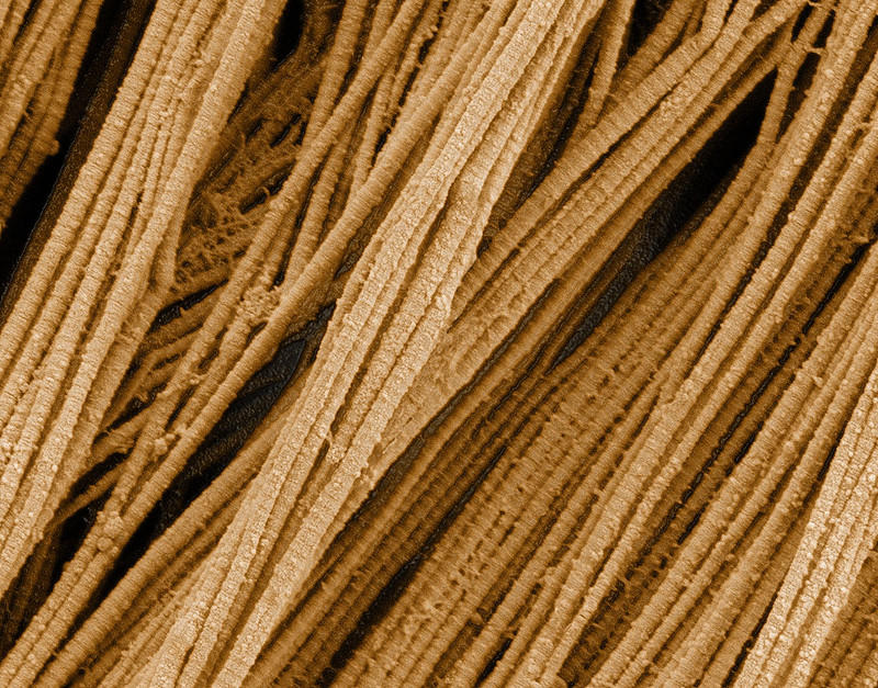
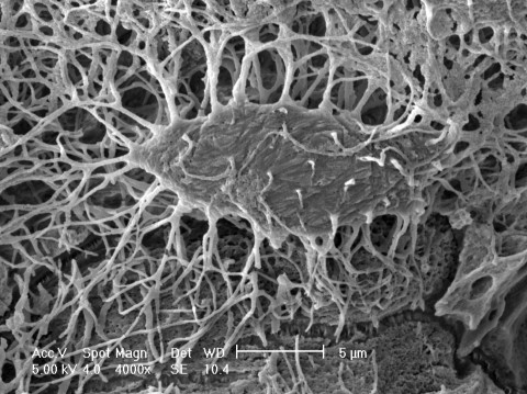
 This image shows a mouse osteocyte with numerous associated canaliculi. The specimen was treated so as to dissolve and remove all of the extracellular matrix (both the collagen and the hydroxyapatite), leaving only the osteocyte and the canaliculi. Where you see open space is where you would find collagen fibers and hydroxyapatite ground substance.
Media Attributions
- U09-002 Cells of the Bone © Barnett, Cierra Memphis is licensed under a CC BY-NC-ND (Attribution NonCommercial NoDerivatives) license
- U09-004 Collagentriplehelix © SteinsplitterBot is licensed under a CC BY-SA (Attribution ShareAlike) license
- U09-005 collagen © National Cancer Institute. Deerinck, Tom & Ellisman, Mark is licensed under a Public Domain license
- U09-006 Hydroxyapatite © Barnett, Cierra Memphis is licensed under a CC BY-NC-ND (Attribution NonCommercial NoDerivatives) license
- U09-007 mouse osteocyte NIAMS Lynda Bonewald © Bonewald, Lynda Ph.D. is licensed under a Public Domain license
