Cells of the Nervous System | Neurons
Objective 3
11.3.1 Compare and contrast the shape of neurons to their function.
 Most artists who draw a “typical” neuron draw a shape we call multipolar. We will look at neurons at a molecular level of detail in Unit 13. For now, we’ll name some microscopic parts of the neuron that are important for its function:
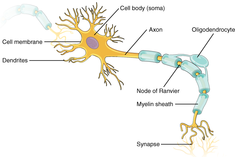
- dendrites, tree-like extensions of the cell body;
- cell body, or soma, the area where the nucleus and most organelles reside;
- axon, the “cable” of the neuron
It’s important to remember that very few actual neurons operate exactly the way we’re describing here. Rather, we’re taking the properties of 80 billion neurons with thousands of different shapes and making some general “rules” about what the different parts do. Along the way, we’ll find plenty of exceptions to these general rules.
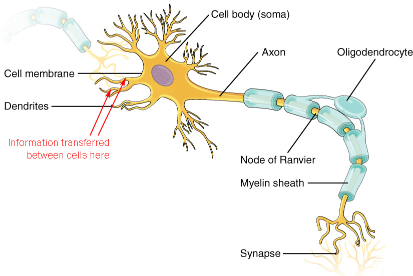
A multipolar neuron has an extensively branched tree surrounding its cell body. Fittingly, these branches are called dendrites (Greek δενδρον, dendron, “tree”). The dendrites are, in general, where a neuron receives information. Even neurons without an obvious tree receive information in a modified, unrecognizable dendrite (such as the lamellated corpuscle we saw in the skin).
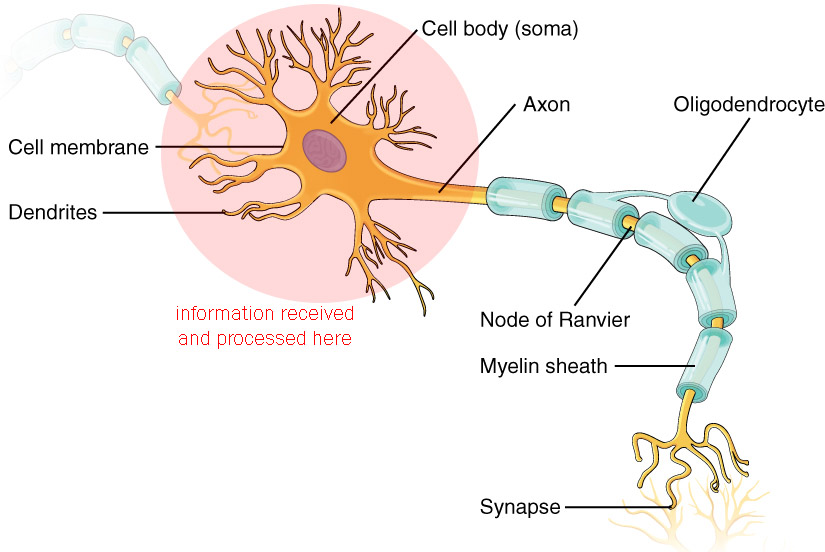
The dendrites are also used in the initial stages of information processing. The dendrites and soma together determine what information is going to be transmitted to another cell or to a target organ. We’ll discuss the details of this process in Unit 13 Objective 10.
Information coming into a fine, faraway dendritic branch has less of an effect on the output of a neuron than information coming into a thicker, nearer branch.
The metabolic center of a neuron, which includes the cell nucleus, is the cell body or soma. The cell body aids in the processing of information.
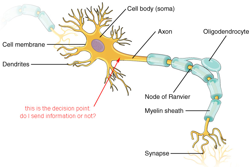
After information is gathered at the dendrites, and processed via the dendrites and cell body, a decision has to be made: do we give that information to another cell? This decision is made at a point that naturally enough has the nickname of the decision point.
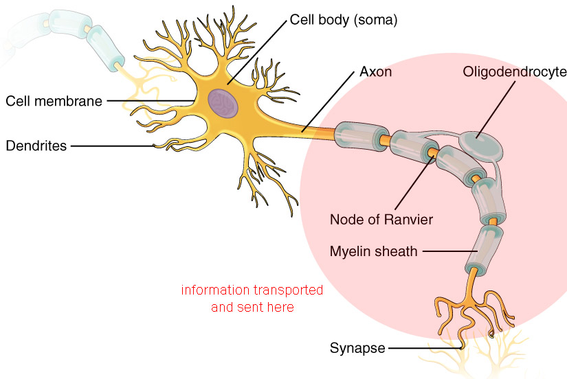
The output of a neuron travels through its axon. If the information must travel a long distance, the axon is myelinated: it is covered by an insulated sheath. In some neurons, the myelin sheath is interrupted periodically at points called the nodes of Ranvier.
The axon ends in one or more axon terminals (or terminal knobs or terminal boutons). These are the sites where information is sent to the next neuron in line, or to muscles or glands, a specialized structure called the synapse.
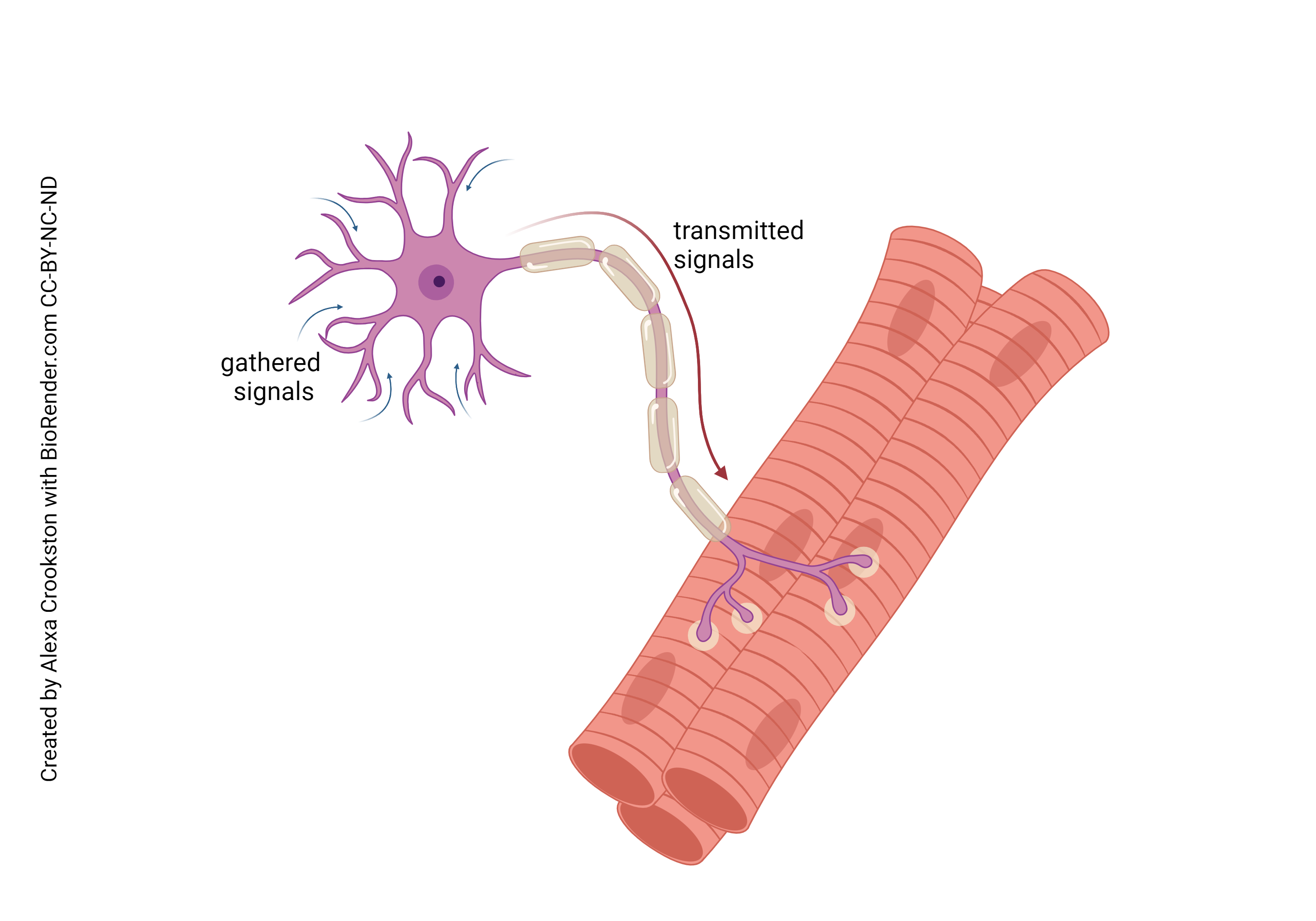
This is an example of a neuron we’ll learn about later that incorporates all the features listed. This is the motor neuron which we were introduced to in Unit 10 Objective 5. Information is received on the dendrites; processed through the dendrites and cell body; transmitted via the axon; and at the connection between the neuron and its target (in this case, muscle), the information is sent to the target using a chemical neurotransmitter.
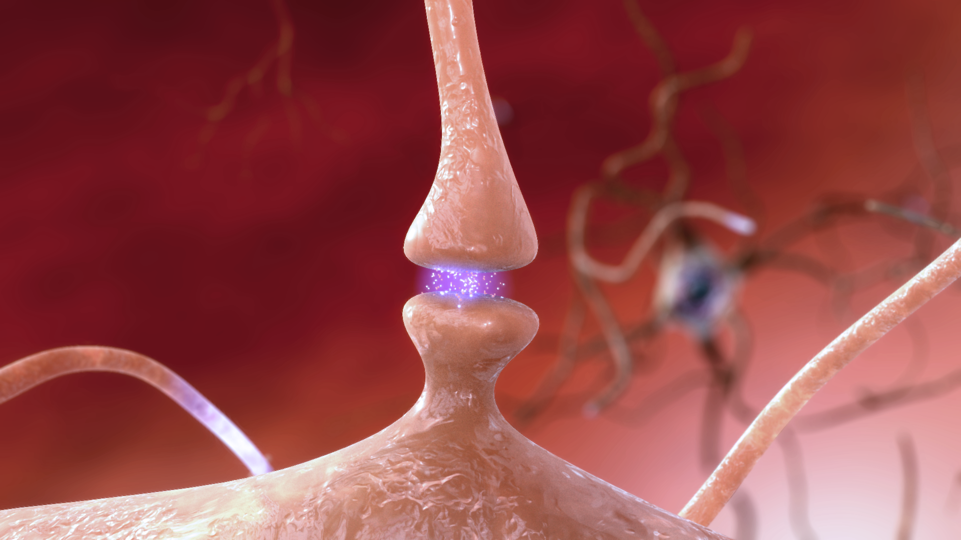 We will study the molecular machinery of the synapse in Unit 13. For now, all we need to know is that an individual neuron makes a lot of synapses; 10,000 synapses would be a rough average number for a typical neuron. Each of these synapses transfers information to a target cell using chemical signals called neurotransmitters.
The side of the synapse which sends information is the presynaptic side. The axon terminals of an α motor neuron would be an example. The side which receives information is the postsynaptic side. The muscle cell of the neuromuscular junction would be an example. The postsynaptic side contains neurotransmitter receptors which can either change the electrical properties of the postsynaptic cell, or the biochemical properties, or both.
All of the neurons we have seen so far are multipolar. They have multiple processes (“sticky-outy-things”) coming out of the cell body.
Along with the multipolar neurons we’ve been looking at, there are two more shapes of neuron that deserve special mention.
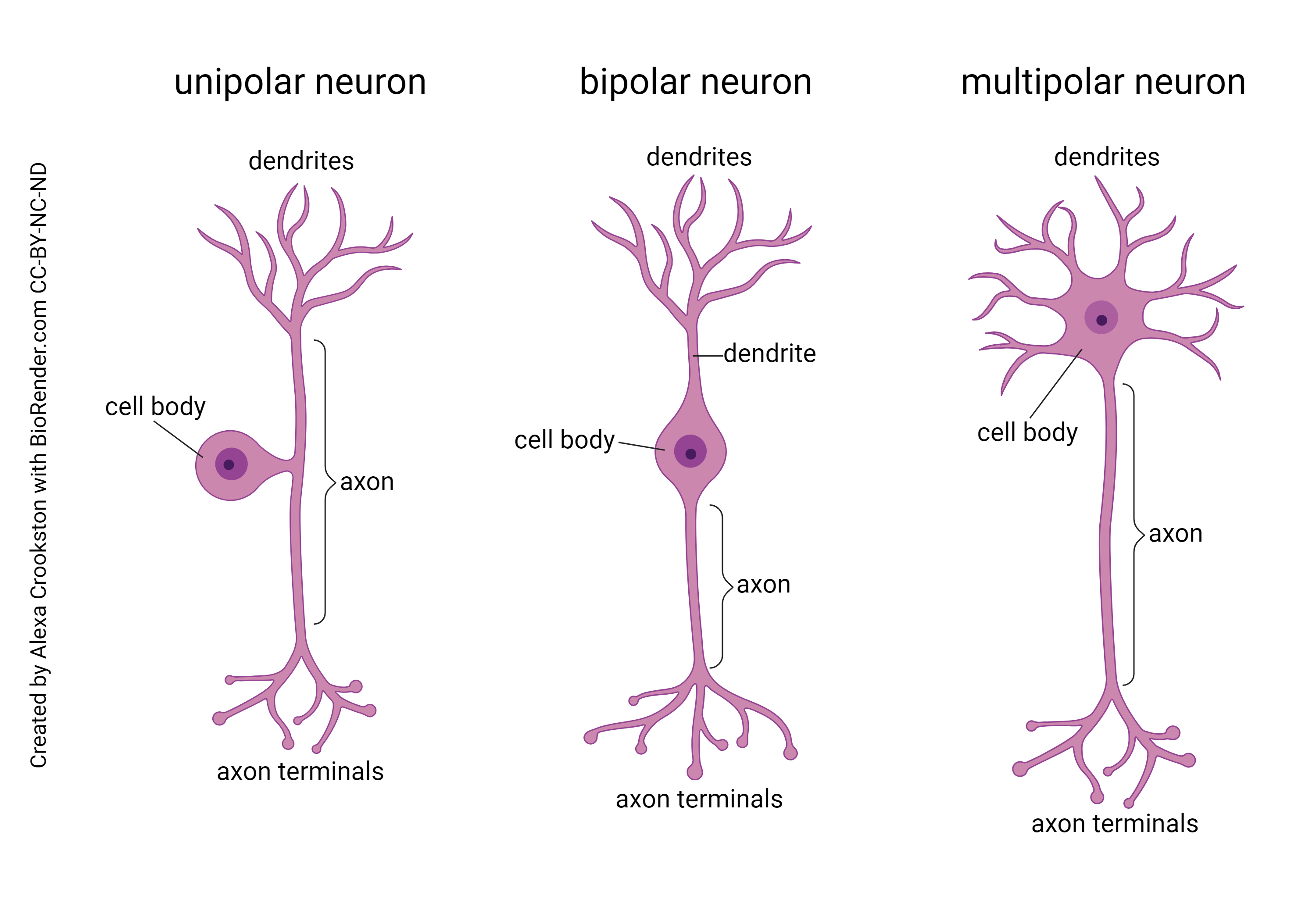
Bipolar neurons have two main processes, one emerging from each side of the cell body. As the name implies, the dendritic tree and the axon branches are close to mirror images of each other. They are found in places where information is relayed without much modification.
Unipolar neurons (also called pseudo-unipolar neurons) move the cell body off to the side, so the information is not processed or transformed as it bypasses the cell body. This type of neuron picks up information from the sensory surfaces of the skin and sends it, unmodified, to be dropped off in the spinal cord or brainstem. They are called unipolar because a single process emerges from the cell body.
In summary, to recognize the difference between multipolar, bipolar, and unipolar neurons, you should ask yourself: how many sticky-outy-things come out of the cell body?
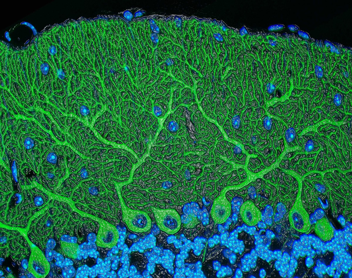
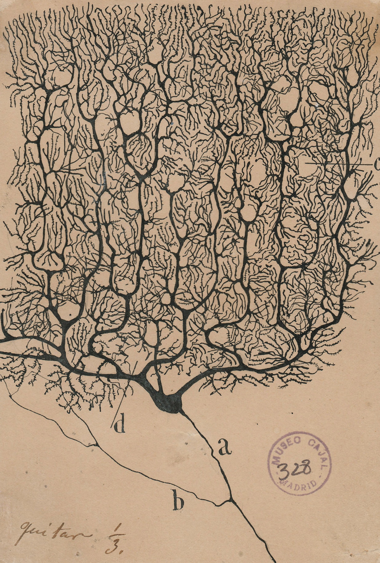
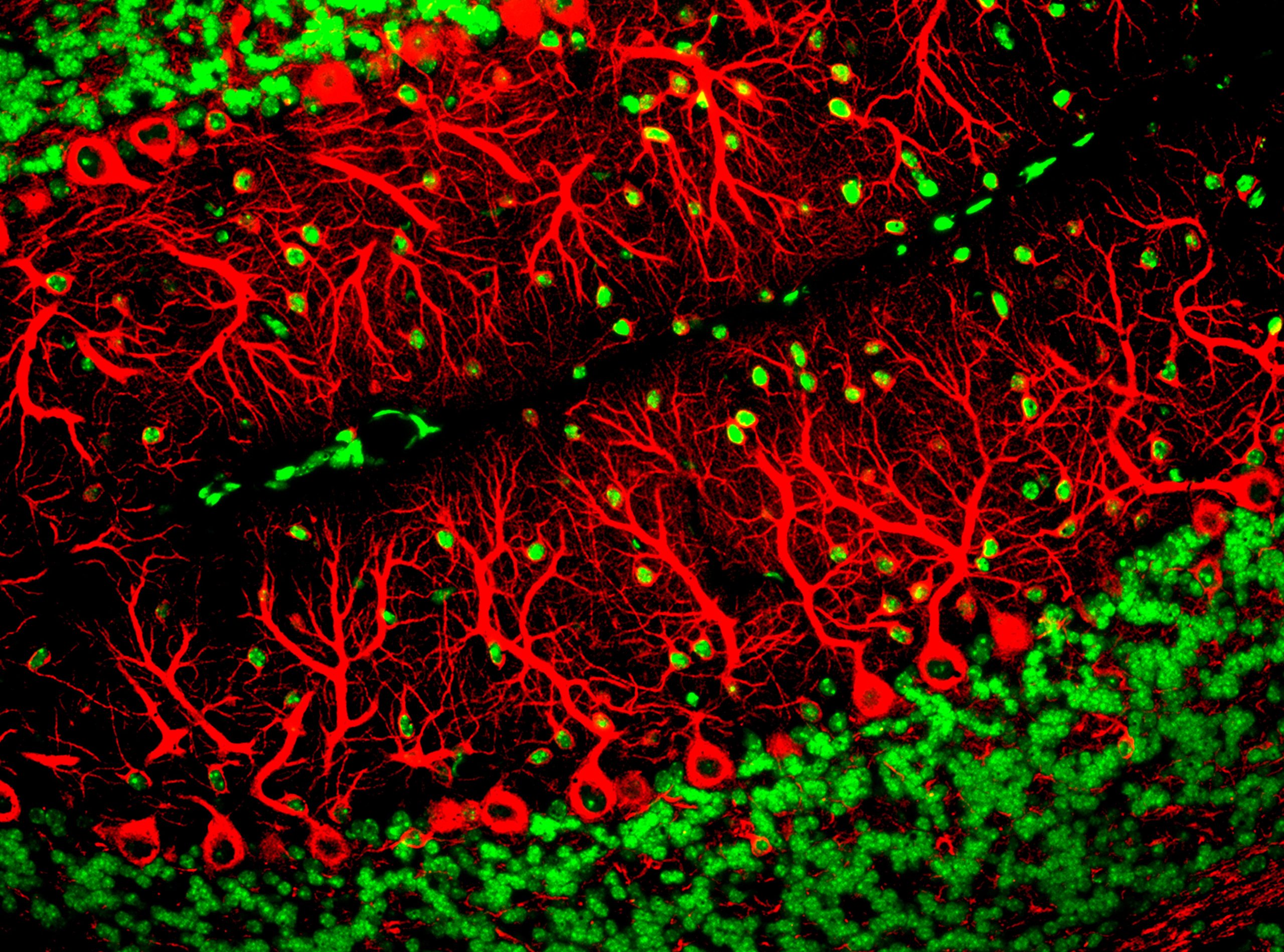
These images are of Purkinje cells
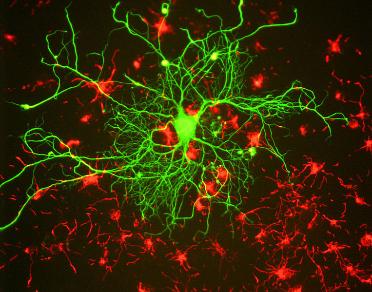
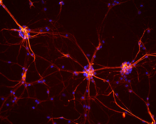
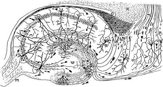
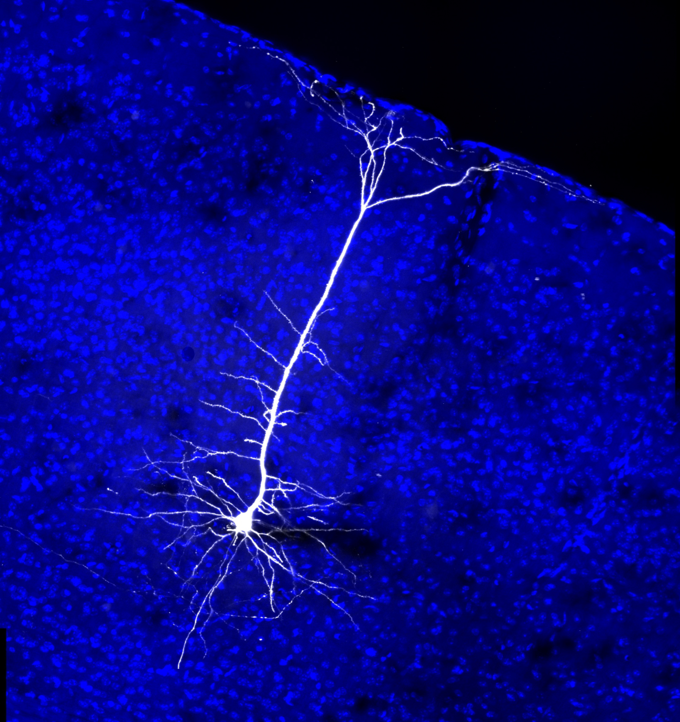
These images show the variety of neuronal shapes. You can admire them, or you could speculate on what each of the shapes is doing.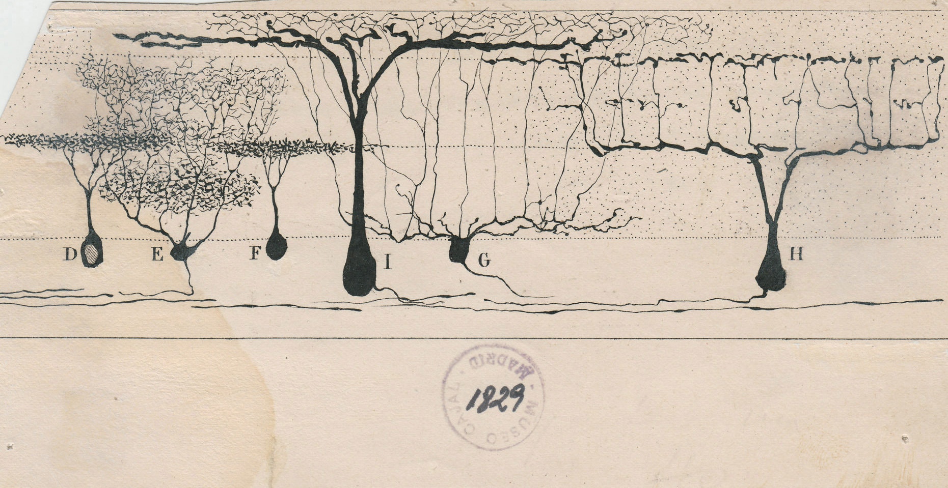
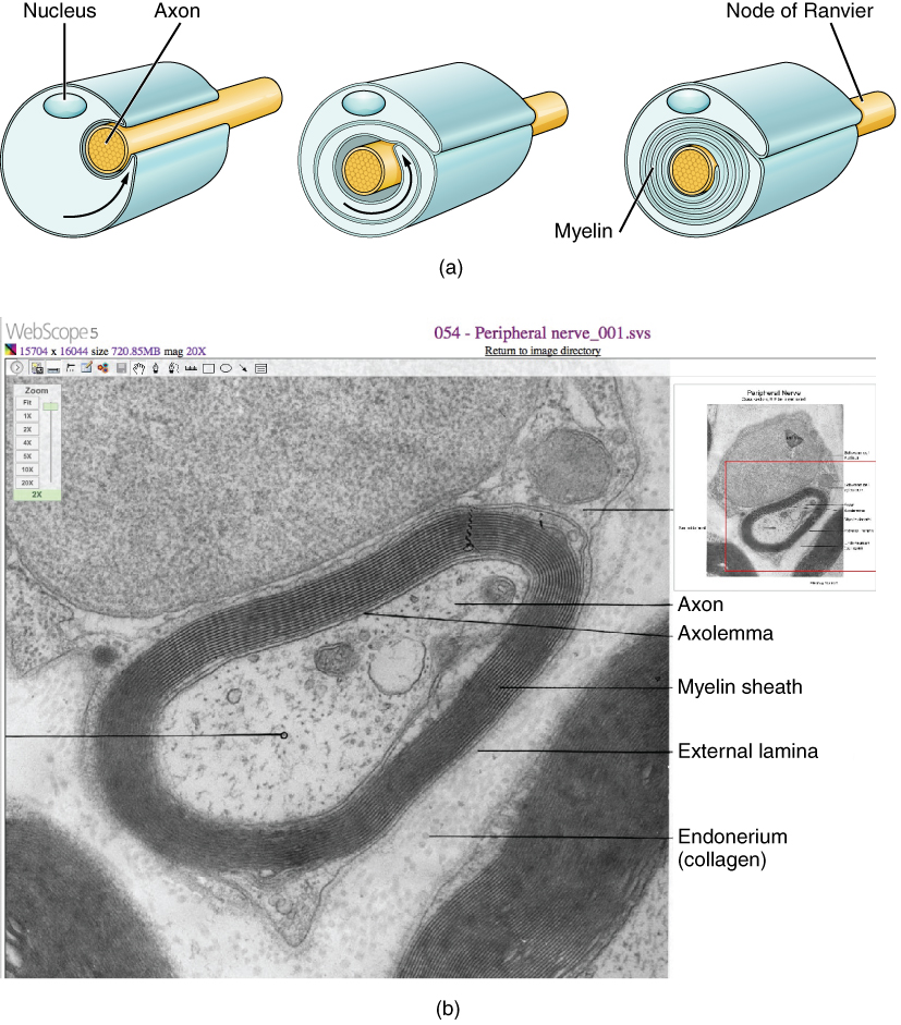 Some axons have an insulating wrapping surrounding them. We will see the function of this wrapping in Unit 13, but for now, all you need to know is that myelinimproves the speed and fidelity of information flow down the axon, from the cell body to the synaptic terminal. There is a difference between the myelin wrapping in the CNS, which does not support axonal regrowth in case of injury, and myelin wrapping in the PNS, which does. Both kinds of myelin are mostly (80%) lipid, but PNS myelin has specialized protein (like the recognition and adhesion proteins we studied in Units 4 and 7) which supports axonal regrowth.
Accordingly, it’s a different cell type that makes myelin in the CNS (oligodendrocytes) and PNS (Schwann cells). We’ll study these cells in the next objective.
Myelin, like most lipids, appears white when you cut into it. This is the basis for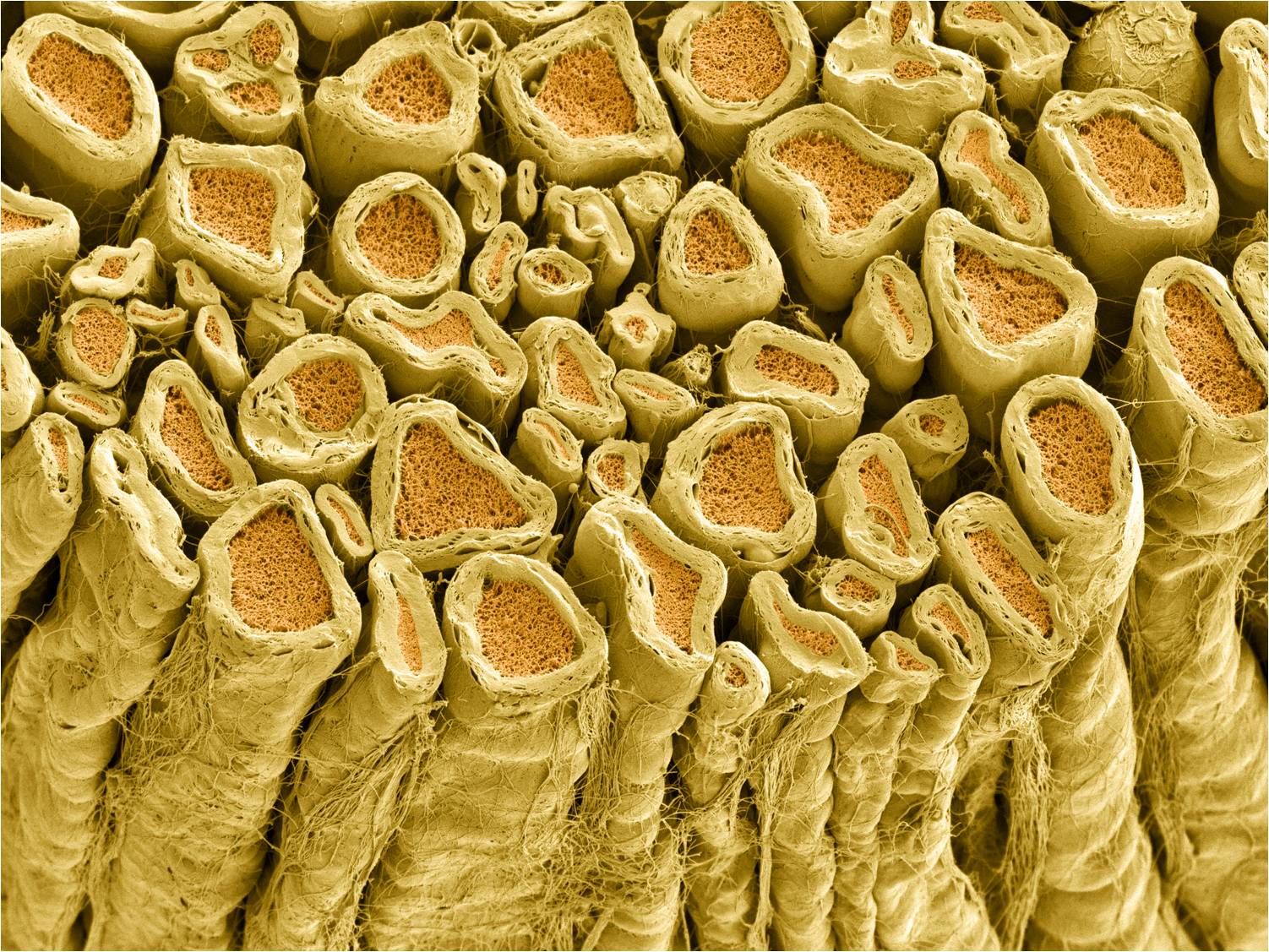 so-called white matter and gray matter in the brain. (It’s really less-pink-matter and darker-pink-matter in a freshly cut or living brain, but those aren’t the terms we are given.)
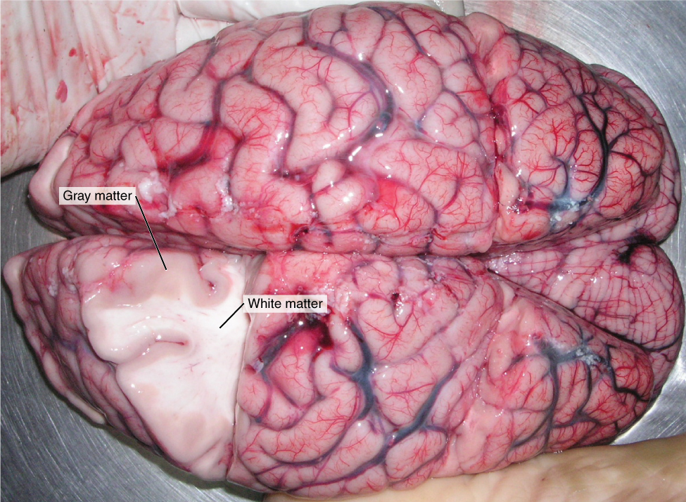 White matter is rich in myelinated axons and generally indicates the presence of one or more fiber tracts. Remember a tract is a bundle of neuronal axons traveling to and from the same place.
Gray matter is rich in cell bodies and dendrites. Because those are (in general) the processing parts of the neuron, most processing is done in the gray matter.
Media Attributions
- U11-018 Parts of a Neuron © Betts, J. Gordon; Young, Kelly A.; Wise, James A.; Johnson, Eddie; Poe, Brandon; Kruse, Dean H. Korol, Oksana; Johnson, Jody E.; Womble, Mark & DeSaix, Peter is licensed under a CC BY (Attribution) license
- U11-019 Parts of a Neuron Information Transfer © Betts, J. Gordon; Young, Kelly A.; Wise, James A.; Johnson, Eddie; Poe, Brandon; Kruse, Dean H. Korol, Oksana; Johnson, Jody E.; Womble, Mark & DeSaix, Peter adapted by Jim Hutchins is licensed under a CC BY (Attribution) license
- U11-020 Parts of a Neuron Information Received © Betts, J. Gordon; Young, Kelly A.; Wise, James A.; Johnson, Eddie; Poe, Brandon; Kruse, Dean H. Korol, Oksana; Johnson, Jody E.; Womble, Mark & DeSaix, Peter adapted by Jim Hutchins is licensed under a CC BY (Attribution) license
- U11-021 Parts of a Neuron Information Decision Point © Betts, J. Gordon; Young, Kelly A.; Wise, James A.; Johnson, Eddie; Poe, Brandon; Kruse, Dean H. Korol, Oksana; Johnson, Jody E.; Womble, Mark & DeSaix, Peter adapted by Jim Hutchins is licensed under a CC BY (Attribution) license
- U11-022 Parts of a Neuron Information Transported © Betts, J. Gordon; Young, Kelly A.; Wise, James A.; Johnson, Eddie; Poe, Brandon; Kruse, Dean H. Korol, Oksana; Johnson, Jody E.; Womble, Mark & DeSaix, Peter adapted by Jim Hutchins is licensed under a CC BY (Attribution) license
- U11-023 Motor Neuron (action) © Crookston, Alexa is licensed under a CC BY-NC-ND (Attribution NonCommercial NoDerivatives) license
- U11-024 New Healthy Neuron © National Institute on Aging, National Institutes of Health is licensed under a CC BY-NC (Attribution NonCommercial) license
- U11-025 Synapse © National Institute on Aging, National Institutes of Health is licensed under a CC BY-NC (Attribution NonCommercial) license
- U11-029 Neuron Shapes © Crookston, Alexa is licensed under a CC BY-NC-ND (Attribution NonCommercial NoDerivatives) license
- U11-026 Purkinje Cells Cerebellum © Deerinck, Tom and Ellisman, Mark, National Center for Microscopy and Imaging Research (NCMIR) is licensed under a CC BY-NC (Attribution NonCommercial) license
- U11-028 Purkinje Cell © Santiago Ramón y Cajal is licensed under a Public Domain license
- U11-027 Purkinje Cells Scaled © Ma, Yinghua and Vartanian, Timothy, Cornell University, Ithaca, N.Y. is licensed under a CC BY-NC-ND (Attribution NonCommercial NoDerivatives) license
- U11-031 Neuron in Tissue Culture © Shaw, Gerry is licensed under a CC BY-SA (Attribution ShareAlike) license
- U11-032 Mouse Neurons © NICHD/Jeong, S. is licensed under a CC BY-SA (Attribution ShareAlike) license
- U11-033 Hippocampus © Santiago Ramón y Cajal is licensed under a Public Domain license
- U11-034 Projection Neuron © Scanziani, Massimo Dr. is licensed under a CC BY-NC (Attribution NonCommercial) license
- U11-035 Retina of the Lizard © Santiago Ramón y Cajal is licensed under a Public Domain license
- U11-036 Myelination © Betts, J. Gordon; Young, Kelly A.; Wise, James A.; Johnson, Eddie; Poe, Brandon; Kruse, Dean H. Korol, Oksana; Johnson, Jody E.; Womble, Mark & DeSaix, Peter is licensed under a CC BY (Attribution) license
- U11-037 Myelinated Axons in Rat Spinal Root © Deerinck, Tom and Ellisman, Mark/ National Center for Microscopy and Imaging Research (NCMIR) is licensed under a CC BY-NC (Attribution NonCommercial) license
- U11-038 White and Gray Matter © Betts, J. Gordon; Young, Kelly A.; Wise, James A.; Johnson, Eddie; Poe, Brandon; Kruse, Dean H. Korol, Oksana; Johnson, Jody E.; Womble, Mark & DeSaix, Peter is licensed under a CC BY (Attribution) license
