Bone Formation
Objective 9.5
9.5.1 Identify the key features of a long bone.
9.5.2 Compare and contrast endochondral and intramembranous ossification.
9.5.3 Identify the region of the bone that is capable of growth in the immature skeleton.
First, we’ll examine long bones, bones that are (by definition) longer than they are wide. Long bones are roughly cylindrical in shape, like the humerus, radius,
ulna, and metacarpals of the upper extremity; the femur, tibia, fibula, and metatarsals of the lower extremity; and the phalanges (finger and toe bones) of both upper and lower extremities.
The Greek word φυστς or physis means “nature,” and there are three natural parts of a long bone.
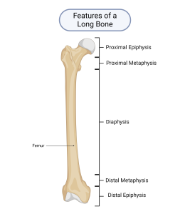
At each end of a long bone is an epiphysis (epi-, on top of). The epiphyses (plural) are the knobby ends of the bone which form part of a joint. Moving from the ends of the bone toward the middle, next to each epiphysis is the short meta- or “transition” region, the metaphysis. Connecting the two metaphyses (plural) in the middle of the bone is the dia-or “through” region, the diaphysis (shaft). We will focus on the epiphyses; but first, let’s consider how bones form during embryonic and fetal development, and how they grow through late adolescence.
Bone Formation
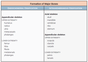
During embryonic and fetal development, bones form in one of two ways — by either endochondral ossification or intramembranous ossification. As you can see in the table, endochondral ossification is used to make long bones longer. Flat, square, and irregular bones are made by intramembranous ossification.
Endochondral Bone Formation
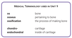
Endochondral (endo-, within + chondral, cartilage) ossification is bone formation using a cartilage model or scaffold which determines the location and general shape of the bone. As the bone tissue develops, it gradually replaces the cartilage with spongy bone, the outer layers of which remodel to form compact bone. Let’s consider this in a bit more detail.
In fetuses and very young children, many of the future bones are completely cartilage. For long bones, at some point, a primary ossification center appears in the center of the cartilaginous scaffold, where the diaphysis will be. The primary ossification center enlarges, gradually replacing the cartilage ground substance with the organic/inorganic composite (collagen/hydroxyapatite) ground substance of bone.
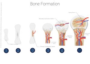
Later, two secondary ossification centers develop, one in each epiphyseal region, near the bone’s articulating surfaces. As the primary ossification center expands toward the epiphyses, and both secondary ossification centers expand (more slowly) toward the primary ossification center, the primary and a secondary eventually meet at the distal border of each metaphyseal region.
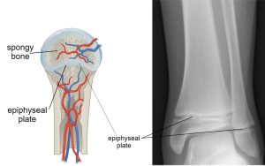
Where the primary ossification center pushes up against a secondary ossification center, the cartilage caught between them becomes the epiphyseal plate. This is where the bone continues to grow (lengthwise growth) throughout childhood and adolescence, hence it is also referred to as the growth plate. The epiphyseal plate is under the control of human growth hormone (hGH), which we’ll study in Unit 14, the Endocrine System. For now, all you need to remember is as long as high concentrations of hGH are present in the blood, the epiphyseal plate continues its growth (we say the plates are “open”). When hGH levels drop (toward the end of adolescence), the plates mineralize or “close.”
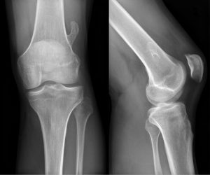
In adults, who are done growing in height, the remnant of the epiphyseal plate can sometimes (not always) be seen as the epiphyseal line, a landmark in conventional X-rays of adult bone. The formation of the primary and secondary ossification centers, as well as the longitudinal growth at the epiphyseal plates, is all part of the endochondral ossification process.
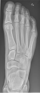
As seen here, it’s fairly easy to distinguish cartilage from bone in X-rays. Cartilage and the organic part of bone are mostly carbon, hydrogen, and oxygen; phosphorus and calcium, found in hydroxyapatite, are better at stopping X-rays from passing through the tissue.
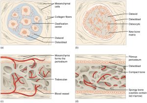
Intramembranous Bone Formation
The other strategy for bone development is intramembranous ossification. As with endochondral ossification, the future bone begins as a cartilaginous model or scaffold. However, with intramembranous ossification, instead of a primary ossification center forming in the middle with a secondary ossification center at each end, multiple ossification centers form throughout the cartilage at roughly the same time. They start as small islands of spongy bone and then spread outward to meet each other. Along the outside of the bone, the spongy bone is remodeled to a thin shell of compact bone, with spongy bone remaining in the center. This is how bones of other shapes develop (short bones, flat bones, irregular bones, and sesamoid bones, i.e. not long bones). Bones that form in this manner do not have a medullary cavity, but they do have lots of red bone marrow in the spaces between the spongy bone trabeculae.
Media Attributions
- U09-017 medical terminology © Bizzel, Lizz is licensed under a CC BY-SA (Attribution ShareAlike) license
- U09-018 Features of a Long Bone © BioRender is licensed under a CC BY-NC-ND (Attribution NonCommercial NoDerivatives) license
- U09-020 bones table © Bizzel, Lizz is licensed under a CC BY-SA (Attribution ShareAlike) license
- U09-019 Bone Formation © Barnett, Cierra Memphis is licensed under a CC BY-NC-ND (Attribution NonCommercial NoDerivatives) license
- U09-021 and U09-022 may need to be split brad winterton composite of radiopaedia images © Gilo1969 adapted by WSU Design Team is licensed under a CC BY-SA (Attribution ShareAlike) license
- U0923 replace knee xray © Hellerhoff is licensed under a CC BY-SA (Attribution ShareAlike) license
- U09-024 Bone and Cartilage Foot © Jones, Jeremy & Hacking, Craig is licensed under a CC BY-SA (Attribution ShareAlike) license
- U09-025 intramembranous formation openstax © Betts, J. Gordon; Young, Kelly A.; Wise, James A.; Johnson, Eddie; Poe, Brandon; Kruse, Dean H. Korol, Oksana; Johnson, Jody E.; Womble, Mark & DeSaix, Peter is licensed under a CC BY (Attribution) license
