Axial Skeleton
Objective 9.8
9.8.1 Identify the axial skeleton and the bones of the skull, vertebral column, and ribs.
Just as with so many things in the human body, we divide the skeleton into two parts or groups – the axial skeleton and the appendicular skeleton. First, we will briefly define each. Then, we’ll look, a bit more in depth, at the various bones that make up each.
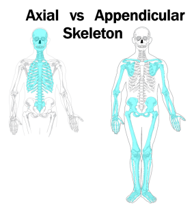
The axial skeleton consists of the bones in the center of the body, from head to coccyx. In other words, the axial skeleton includes the skull, vertebrae, ribs, and sternum.
(The appendicular skeleton, which we will cover in the next objective, consists of the upper and lower extremities. Thus, the bones of the arm, forearm, wrist, and hand are appendicular, and we include the scapula and clavicle because they attach the upper extremity to the axial skeleton. Similarly, the bones of the thigh, leg, ankle, and foot are appendicular, as is the pelvis, which is actually three pairs of bones fused together, because it forms the socket of the hip joint and attaches the lower extremity to the axial skeleton.)
Skull
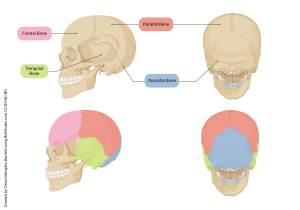
The skull is a relatively complicated fusion of several different bones. Each of the bones of the skull form separately during fetal development, then fuse together, as we will see later. In these lateral and posterior views of the skull, identify the frontal bone, the parietal bones, the temporal bones, and the occipital bone.
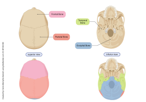
Now identify those same bones (frontal, parietal, temporal, and occipital). These views of the skull are superior (looking down from above) and inferior (looking up from below). Notice the following: 1) on the superior view, we can’t see the temporal bones and we can barely see the occipital bone on the posterior-inferior part of the skull; 2) on the inferior view we can see neither the frontal bone nor the parietal bones. Look back at the lateral view in the image to understand why this is so.
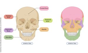
This frontal view shows the frontal, parietal, and temporal bones, as well as four additional bones we now add to the list. The lacrimal bone is located in the medial corner of each orbital (eye socket). Lacrimal refers to tears (crying), so the lacrimal bone is just deep to the tear ducts you see in the medial corner of each eye when you look in the mirror. The maxilla is the upper jawbone. The mandible is the lower jawbone. The zygomatic bone, with a prominent zygomatic arch, forms the high cheekbones for which some people get paid lots of money.
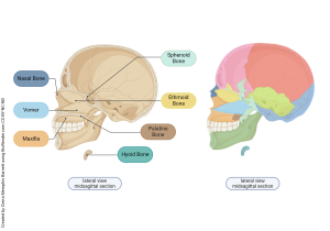
Now, imagine we split the skull exactly in half along the midline, a midsagittal section, and look at the inside surface of the skull. Notice the four large flat bones we’ve already identified in other views: frontal bone, parietal bone, temporal bone, and occipital bone.
Additionally, we see the:
- ethmoid bone, part of the nasal sinuses; the perpendicular plate of the ethmoid is easiest to see here;
- nasal bone, a small bone that forms the bridge of the nose; the bulk of the visible nose is formed by cartilage; the bony part can be felt right where glasses rest on your nose;
- sphenoid bone, the bone with complex shape in the center of the diagram;
- vomer, a bone between the ethmoid, maxilla, and palatine;
- palatine bone, forming the roof of the mouth (hard palate); and
- hyoid bone, a horseshoe-shaped, “floating” bone that makes up part of the larynx in the neck.
That completes the list of skull bones. We need to look at some specific features of three of them (ethmoid bone, sphenoid bone, and occipital bone) from one more view and also name a couple of holes in these bones. To see what we need to in this image, we make a horizontal cut and look down from above (superior view, transverse section).
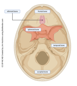
In order for nerves and blood vessels to pass in and out of the skull, it’s necessary to have both large and small (and in-between) holes in the skull. These holes are called foramina (singular: foramen, Latin for — wait for it — hole… or window). There are also shallow depressions called fossae (singular: fossa, Latin for ditch).
The ethmoid bone looks a bit different here (rather small) than in the previous image (much larger in lateral view). We are looking down at the upper-most part of the ethmoid bone, where it joins other bones to form the floor of the cranial vault; directly below are the nasal sinuses. This part of the ethmoid bone is called the cribiform plate and looks a bit like a small rectangle of sponge, with numerous small holes called olfactory foramina. The neurons that give us our sense of smell pass from the nasal mucosa just below the bone, through the olfactory foramina of the cribiform plate, and become part of the olfactory nerve, carrying nerve impulses to the smell center of the brain.
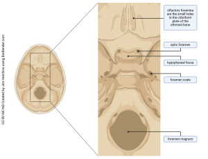
In the sphenoid bone (Greek σφην, sphen-, wedge + -oid, like), the optic foramen forms a hole for the optic nerve to pass from the eye to the brain (can’t see the two optic foramina in this view as they are sort of hidden below a small “shelf” of bone); the hypophyseal fossa forms a shallow depression that holds and protects the pituitary gland (hypophysis); and an oval-shaped hole, the foramen ovale, which allows the passage of several nerves and blood vessels.
The foramen magnum (Latin: huge hole) of the occipital bone allows the spinal cord to exit the skull and enter the vertebral canal.
Vertebral Column
Continuing with the axial skeleton, we now turn to the vertebral column.
Vertebrae (singular: vertebra, Latin, jointed) are stacked to form the vertebral column which runs the length of the back. The vertebrae are named by region, then number, from superior to inferior.
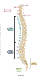
The most superior seven vertebrae are called cervical (“neck”) vertebrae. The region is designated by the letter C (for cervical) and each vertebra in the cervical region has a number, so these vertebrae are designated C1 through C7.
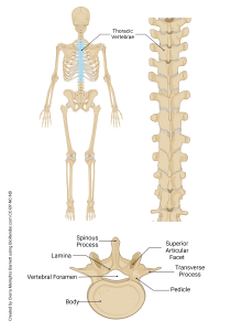
Next are the 12 thoracic vertebrae. The thoracic vertebrae are different from other vertebrae in that each is the attachment point for a pair of ribs (12 thoracic vertebrae →12 pairs of ribs). The thoracic vertebrae are designated T1–T12.
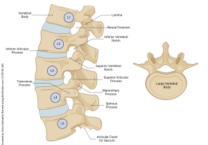
The next five vertebrae are the lumbar vertebrae, L1-L5. These are the last vertebrae that move (have joints between them) as the sacral vertebrae, which come after the lumbar region, are fused into a single piece of bone. In other words, S1-S5 are joined in an immobile, locked state. Also, the number of sacral vertebrae is variable, with most people having five but some having only four. Finally, several small coccygeal vertebrae are also fused and form the coccyx (tailbone), which is the end of the vertebral column.
Now let’s look at the structure of individual vertebrae and the joints between them.
The largest part of a vertebra is the vertebral body.
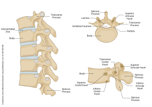
Two transverse processes extend laterally from each vertebra and a pedicle connects each transverse process to the vertebral body.
Along the midline of each vertebra is a spinous process, which protrudes posteriorly. This is connected to the two traverse processes by the laminae (singular lamina).
Taken together, the medial (deep, inside) surfaces of the body, processes, pedicles, and laminae form a large hole called the vertebral foramen that allows passage of the spinal cord.
A joint between each two adjacent vertebral bodies is filled with a disc of fibrocartilage that has a soft center, like a jelly donut. This is called the intervertebral disc.
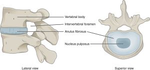
There is also a passageway, called the intervertebral foramen, bounded by parts of the more superior vertebra, the more inferior vertebra, and the intervertebral disc. The intervertebral foramen allows the passage of information carried by spinal nerves from the body into the central nervous system, as well as out of the spinal cord and to the muscles.
The intervertebral foramen is the location where local anesthesia is applied (for example, in an epidural nerve block used during childbirth) and is also the location where inflammation or a rupture of the intervertebral disc can press on nerves and cause extreme pain and even paralysis.
Vertebrae vary considerably in shape; some even seem to be “missing” parts. In the next few images, we’ll look at unique features of a few vertebrae, starting with the first two cervical vertebrae.
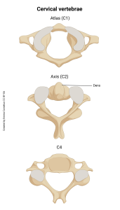
In Greek mythology, Atlas was responsible for holding the entire Earth on his shoulders. According to the ancient Greeks, Atlas lived (and held up the earth) somewhere in the Atlantic Ocean, so the Atlantic is named after Atlas. We also refer to books of maps and books of anatomical structures as atlases.
Since your whole world is in your skull, and the first cervical vertebra (C1) holds up that whole world, C1 is called the atlas. The joint between the occipital bone of skull and the atlas is called the atlanto-occipital joint. This is the joint allows us to nod. Go ahead and nod if you understand what I’m saying. The C2 vertebra also has a special name – axis. Just as the Earth rotates around its axis, the joint between the atlas and the axis (C1 and C2) is the joint that allows us to rotate our head from side to side. A transverse ligament attached to the atlas (C1) wraps around a tooth (dens) on C2 that protrudes up into the vertebral foramen of C1.
The remaining cervical vertebrae, C3-C7, have a unique feature in common. The posteriorly-projecting spinous process of each is bifid, meaning it is forked. Also, the last of the cervical vertebrae, C7, is called vertebra prominens because it’s prominent. If you feel down along the back of your neck, just higher than your shoulder blades, you’ll feel a distinct bony bump that seems to protrude farther than the bumps just above and below it. That is the spinous process of C7 and is often used as a landmark.
The 12 pairs of ribs make joints with (articulate with) the 12 thoracic vertebrae. There are two points at which each rib (left and right) touches a vertebra: the transverse process of a vertebra touches the tubercle of a rib, and the body of a vertebra touches the head of a rib. The vertebrae also make joints with each other at inferior articular and superior articular facets.
Lumbar vertebrae support a lot of weight (everything above the waist) and so they have to be strong. For this reason, they have a thick, blocky body.
To finish our study of the axial skeleton, we now examine the thorax (chest). We already mentioned the 12 pairs of ribs: the T1 vertebra articulates with the first pair of ribs, T2 with the second pair, and so forth. Ribs are, therefore, numbered superior to inferior.
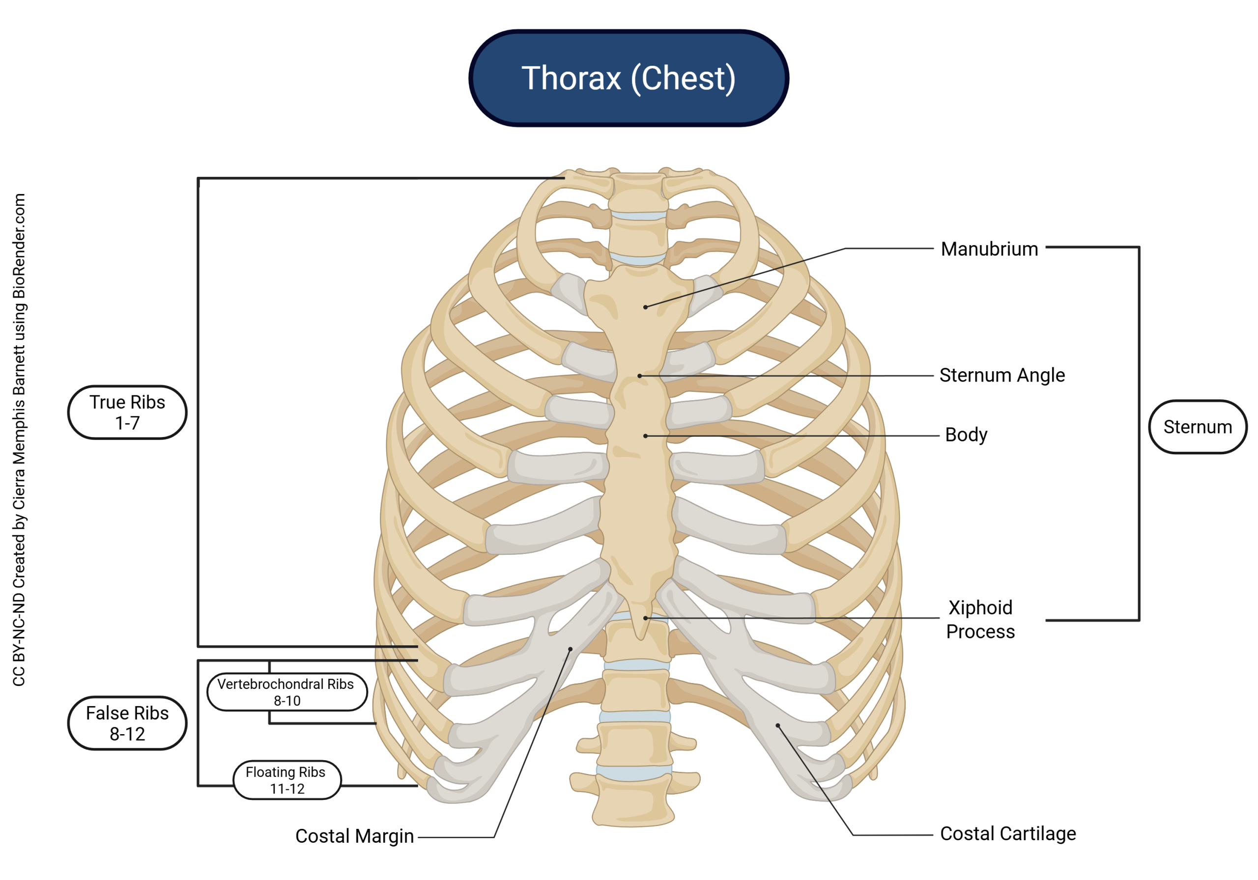
Ribs 1-7 are called true ribs because their anterior ends connect directly to the sternum. The connection between true ribs and the sternum is not bone to bone, but rather rib to hyaline cartilage to sternum. Because it is part of the rib, this hyaline cartilage is usually referred to as costal cartilage (Latin, costa, rib).
Ribs 8-10 are called false ribs because their costal cartilage does not attach directly to the sternum. Rather, their costal cartilage connects to the costal cartilage of rib 7 which, in turn, attaches directly to the sternum.
Ribs 11 and 12 are also false ribs, as well as being referred to as floating ribs because they float; ie, their anterior ends are not attached to anything.
Clinical Connection
Comparison of Fetal/Infant & Adult Skull
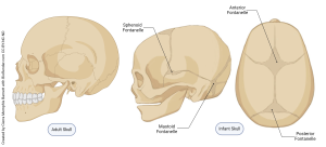
As mentioned before, the skull is a fusion of many different bones that form separately. In the fetus and newborn, these bones have not yet fused together but are joined by bands of fibrocartilage between the developing bones. These are called fontanels. This arrangement allows the head to fit through the birth canal and also allows for continued growth of the brain after birth.
The easiest fontanel to find on a newborn is the anterior fontanel, the place where the frontal bones join the parietal bones. The anterolateral fontanel (one on each side of the head) is where the temporal, parietal, and frontal bones are joined. The posterior fontanel is along the midline between the parietal and occipital bones; and the posterolateral fontanel is where the parietal, occipital, and temporal bones meet.
During the first few months of life, the fontanels fuse and become bone. The remnants of these joints remain as sutures of the skull, zigzag lines that hold the bones of the skull together tightly.
Media Attributions
- U09-047 U09-048 needs to be split © LadyofHats is licensed under a Public Domain license
- U09-049 Skull Bones Lateral and Posterior Views © Barnett, Cierra Memphis is licensed under a CC BY-NC-ND (Attribution NonCommercial NoDerivatives) license
- U09-050 Skull Bones Superior and Inferior View © Barnett, Cierra Memphis is licensed under a CC BY-NC-ND (Attribution NonCommercial NoDerivatives) license
- U09-051 Skull Bones Anterior View © Barnett, Cierra Memphis is licensed under a CC BY-NC-ND (Attribution NonCommercial NoDerivatives) license
- U09-052 Skull Bones Lateral View Midsagittal Section © Barnett, Cierra Memphis is licensed under a CC BY-NC-ND (Attribution NonCommercial NoDerivatives) license
- U09-053 bones of the skull base © Hutchins, Jim is licensed under a CC BY-NC-ND (Attribution NonCommercial NoDerivatives) license
- U09-053a foramina of the skull base © Hutchins, Jim is licensed under a CC BY-NC-ND (Attribution NonCommercial NoDerivatives) license
- U9-054 Vertebral Column © Hutchins, Jim is licensed under a CC BY-NC-ND (Attribution NonCommercial NoDerivatives) license
- U09-056 Vertebrae Features © Barnett, Cierra Memphis is licensed under a CC BY-NC-ND (Attribution NonCommercial NoDerivatives) license
- U09-055 intervertebral disc openstax © Betts, J. Gordon; Young, Kelly A.; Wise, James A.; Johnson, Eddie; Poe, Brandon; Kruse, Dean H. Korol, Oksana; Johnson, Jody E.; Womble, Mark & DeSaix, Peter is licensed under a CC BY (Attribution) license
- U09-059 U09-060 U09-061 Cervical Vertebrae © Cornelius, Emma is licensed under a CC BY-SA (Attribution ShareAlike) license
- U09-058 flickr image atlas © rubyblossom. is licensed under a CC BY-NC-SA (Attribution NonCommercial ShareAlike) license
- U09-064 Thoracic Vertebrae Features © Barnett, Cierra Memphis is licensed under a CC BY-NC-ND (Attribution NonCommercial NoDerivatives) license
- U09-065 Lumbar Vertebrae Features © Barnett, Cierra Memphis is licensed under a CC BY-NC-ND (Attribution NonCommercial NoDerivatives) license
- U9-066 Thorax © Barnett, Cierra Memphis is licensed under a CC BY-NC-ND (Attribution NonCommercial NoDerivatives) license
- U09-067 Infant Skull and Fontanelles © Barnett, Cierra Memphis is licensed under a CC BY-NC-ND (Attribution NonCommercial NoDerivatives) license
