Appendicular Skeleton
Objective 9.9
9.9.1 Identify the appendicular skeleton.
9.9.2 Identify the bones of the upper extremity, shoulder girdle, lower extremity, and pelvic girdle.
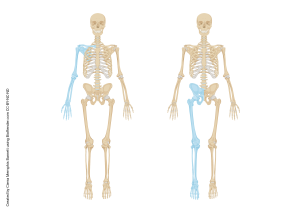
Enough with the axial skeleton; let’s move on to the bones of the appendicular skeleton. Remember, as mentioned before, the appendicular skeleton consists of the upper and lower extremities. Thus, the bones of the arm, forearm, wrist, and hand are appendicular, and we include the scapula and clavicle because they attach the upper extremity to the axial skeleton. Similarly, the bones of the thigh, leg, ankle, and foot are appendicular, as is the pelvis (which is actually three pairs of bones fused together), because it forms the socket of the hip joint and attaches the lower extremity to the axial skeleton.
Even though the names are different between the upper and lower extremities, there are similarities in structure between humerus (upper) and femur (lower); between the radius and ulna (upper), and the tibia and fibula (lower); between the carpals (upper) and the tarsals (lower); between the metacarpals (upper) and metatarsals (lower); and among the phalanges (upper and lower).
Upper Extremity
The shoulder (pectoral) girdle is supported by the clavicle (collarbone) and the scapula (shoulder blade). Together, these form the socket, called the glenoid fossa, into which fits the head of the humerus.
The longest and thickest bone of the upper extremity is the humerus. The head of the humerus fits into the glenoid fossa to form the shoulder joint. Anatomists call the structures between the shoulder joint and the elbow joint (like the humerus) the arm; those between the elbow and the wrist are the forearm (anatomists tend to get a bit testy if you misuse these terms…).
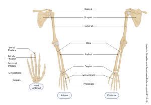
The bones of the forearm, the radius and ulna, articulate with the distal humerus at the elbow joint.
The bones of the wrist are called carpals. You may know the expression carpe diem, Latin for seize the day. Carpals, therefore, seize things; ie, they have a lot to do with the amazing motion capabilities of our hands. There are eight carpal bones arranged in two rows of four (you do not need to learn the names of the individual carpals).
Moving distally from the carpals, there are five metacarpals, one for each digit. These five bones form the structure of the hand.
The phalanges are so named because they line up like Roman soldiers in a phalanx formation. Each of the phalanges, except the thumb (pollex), has three phalangeal bones (proximal, medial, distal); the thumb has two (proximal and distal).
Lower Extremity
The pelvis supports the lower extremity. Just as for the upper extremity, anatomists have specific terms for regions of the lower extremity: between the hip and knee is the thigh; between the knee and the ankle is the leg (if you’re talking to an anatomist, don’t use leg and lower extremity inter-changeably).
The three paired bones of the pelvis are the ilium, which forms a large, flat, curved, flared bone laterally; the ischium (hip); and the pubis. Two notable landmarks we’ll see and use later are the ischial tuberosity (the bones you sit on, sometimes jokingly called the sitz bones) and the pubic symphysis, which joins the two halves of the pelvis at the midline. Remember that the pubic symphysis is fibrocartilage.
All three pelvic bones come together to form the large acetabulum (hip socket), into which the head of the femur fits to form the hip joint.
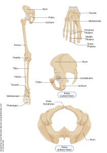
The largest bone of the body is the femur; the large, long bone of the thigh. The knee joint is covered by the sesamoid patella (kneecap).
The two bones of the leg are the tibia and fibula. The tibia is the larger, medial bone of the leg. The fibula is smaller and lateral. Remember you can tell a little fib, and the l in fibula reminds us the fib is a lie and the fibula is lateral.
The ankle bones are called tarsals. The Greek ταρσος, tarsus means broad, flat basket, which in someone’s imagination describes the bones of the ankle. (It’s also the geography of the ancient city of Tarsus, where Paul the Apostle was born.) There are seven tarsals (and, like the carpals, you don’t need to remember the individual names).
Distal to the tarsals are the metatarsals, one for each phalange. Like the upper extremity, there are 14 phalanges: three each for the smaller digits, and two for the great toe (hallux).
Clinical Connection
Comparison of Adult Male & Female Skeletons
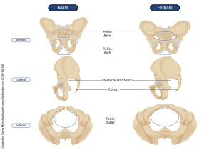
The pelvis is one of the few bony structures that is substantially different between male and female. This makes identification of the differences between the male and female pelvises important for forensic medicine and anthropology.
In females, the angle between the ischial tuberosities, called the pubic arch, is generally greater than 90°, while in males the angle is less than 90°. The pelvic brim (pelvic inlet), the oval region defined by the medial aspect of the ilia, is larger in females as well. The medial-lateral distance and anterior-posterior distance of the pelvic outlet, viewed from below (inferiorly), is also larger in females. Finally, viewed laterally, the female coccyx projects further posteriorly, giving females a larger greater sciatic notch. All of these adaptations are important for childbirth, allowing more room for a baby to pass through the birth canal without encountering the obstacle of bone.
Media Attributions
- U09-070 U09-071 Upper and Lower Extremity Similarities © Barnett, Cierra Memphis is licensed under a CC BY-NC-ND (Attribution NonCommercial NoDerivatives) license
- U09-072 Upper Extremity Bones © Barnett, Cierra Memphis is licensed under a CC BY-NC-ND (Attribution NonCommercial NoDerivatives) license
- U09-073 Lower Extremities © Barnett, Cierra Memphis is licensed under a CC BY-NC-ND (Attribution NonCommercial NoDerivatives) license
- U09-074a Male vs Female Pelvis © Barnett, Cierra Memphis is licensed under a CC BY-NC-ND (Attribution NonCommercial NoDerivatives) license
