Accessory Skin Structures | Nails and Hair
Objective 8.7
8.7.1 Describe the structure and function of the accessory skin structures.
Nails
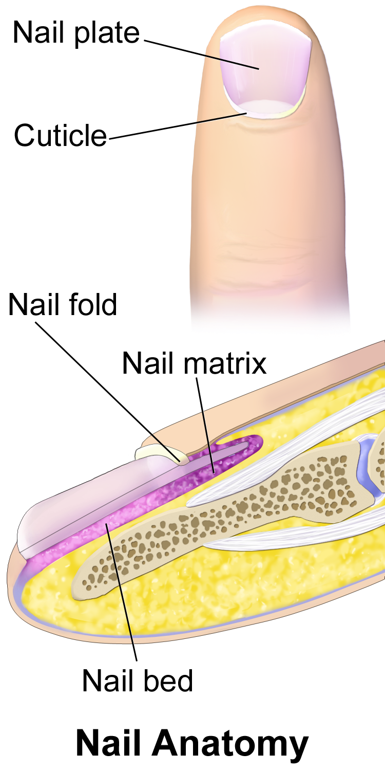
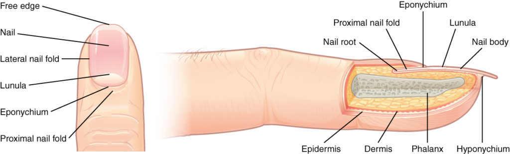
At the distal end of the dorsal surface of fingers and toes, there is a fold of epidermis that gives rise to a hard, dense covering made of keratin that is tightly packed to form a clear, tough nail body.
At the proximal end, a nail matrix pushes out the nail. An eponychium (cuticle) fills the space where the epidermis ends. A lunula (“little moon”) is sometimes seen near the cuticle as a slightly lighter area. At the free edge, another fold of epidermis with blood vessels dives under the surface of the nail. Past that interface (the hyponychium), the free edge of the nail loses contact with living tissues and can be trimmed without blood or pain.
Hair
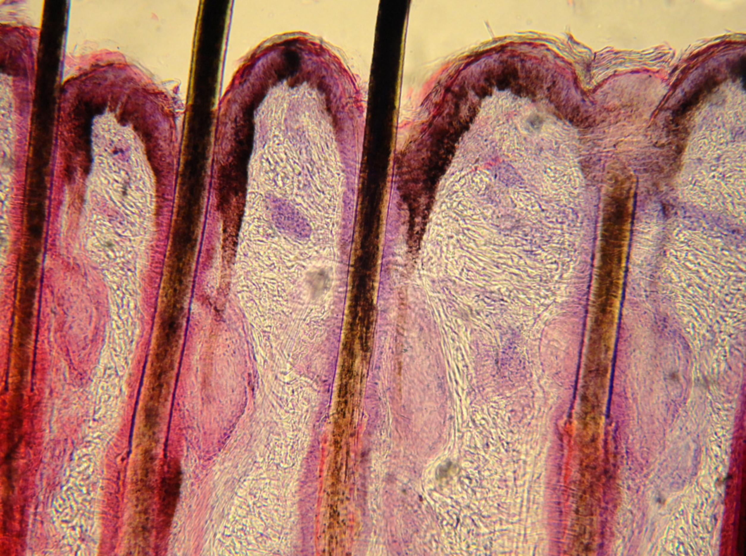
Hair, like skin and nails, is a specialized form of keratin growing out of the epidermis. It is primarily made of dead, keratinized cells.
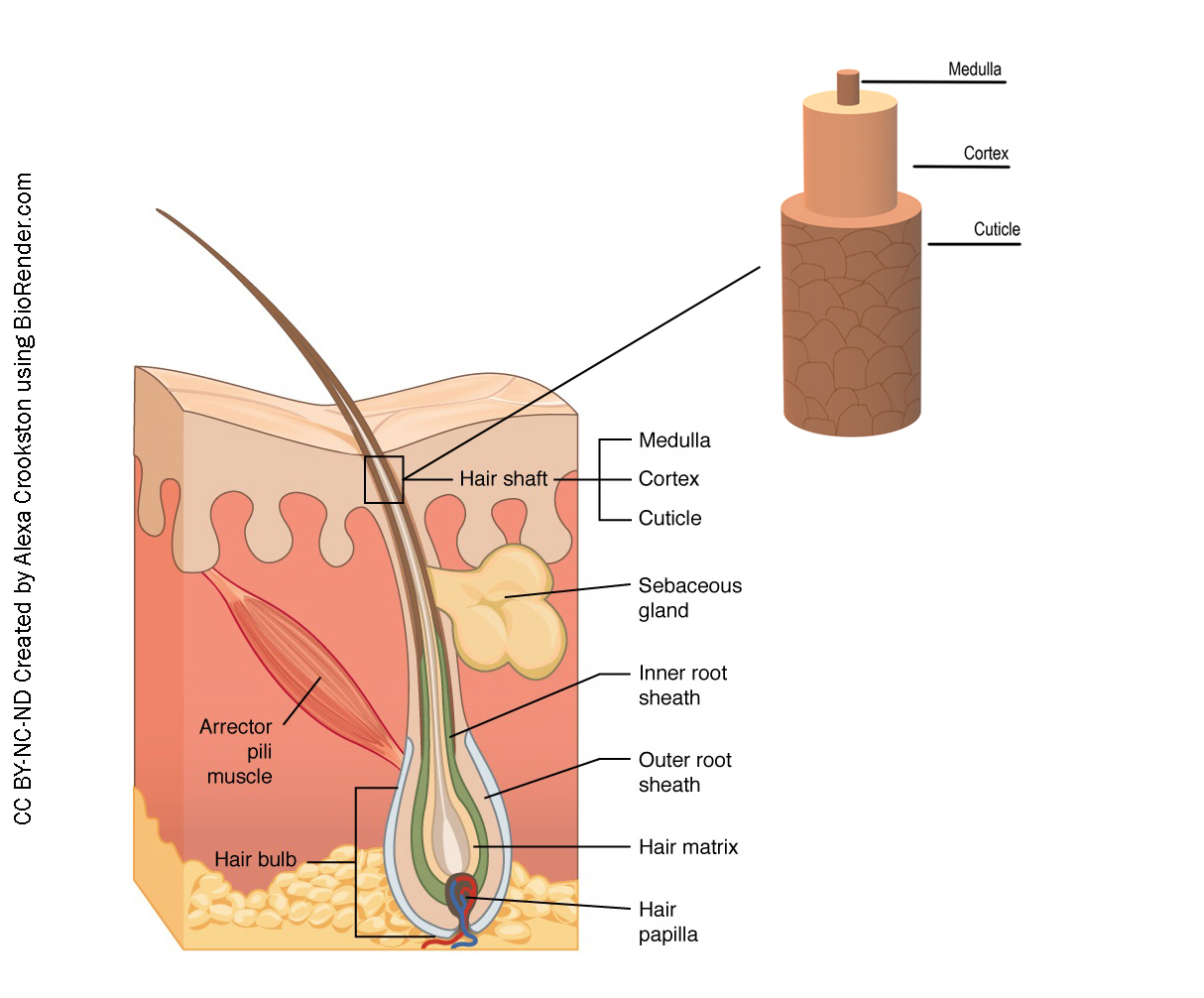
The external (visible) portion of hair is made up of three concentric layers: the medulla in the center, a cortex further out, and finally a covering, or cuticle, on the outside.
Beneath the surface of the skin, the hair follicle and inner and outer dermal root sheath surround the medulla, cortex and cuticle. At the base, there are blood vessels and melanocytes and a sebaceous gland.
Hair serves a variety of functions, including protection, sensory input, thermoregulation, and communication.
For example, hair on the head protects the skull from the sun. The hair in the nose and ears, and around the eyes (eyelashes) defends the body by trapping and excluding dust particles that may contain allergens and microbes. Hair of the eyebrows prevents sweat and other particles from dripping into and bothering the eyes. Hair also has a sensory function due to sensory innervation by a hair root plexus surrounding the base of each hair follicle.
Hair is extremely sensitive to air movement or other disturbances in the environment, much more so than the skin surface. This feature is also useful for the detection of the presence of insects or other potentially damaging substances on the skin surface. Each hair root is connected to a smooth muscle called the arrector pili that contracts in response to nerve signals from the sympathetic nervous system, making the external hair shaft “stand up.” The primary purpose for this is to trap a layer of air to add insulation. This is visible in humans as goose bumps and even more obvious in animals, such as when a frightened cat raises its fur.
Hair Color
Color comes from pigment granules (brown or black eumelanin; blonde or red pheomelanin) trapped along with the keratin protein in the hair shafts. White or gray hair occurs when pigment production is reduced or halted and air bubbles replace pigment.
Hair Growth
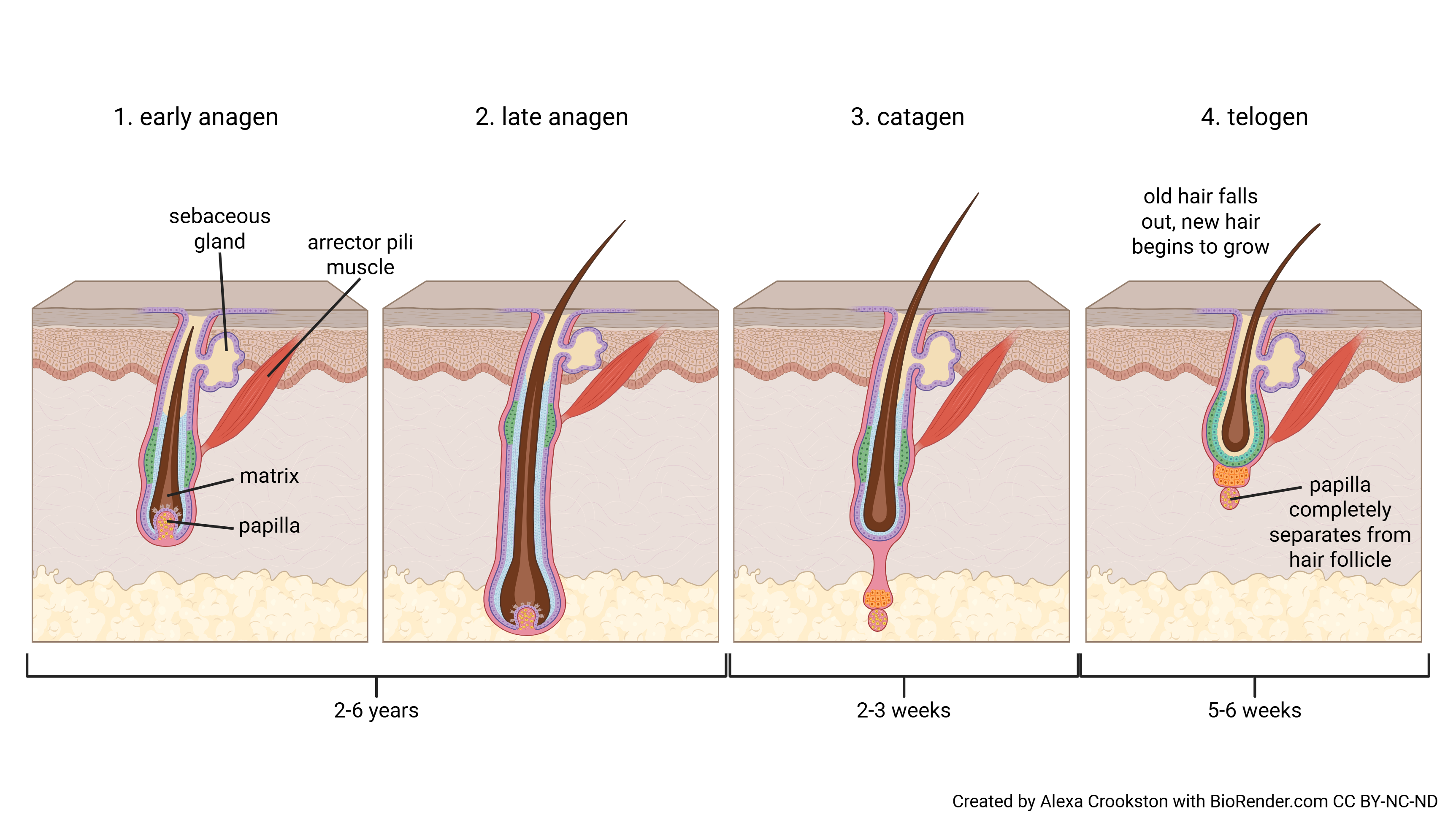
Hair grows and is eventually shed and replaced by new hair. This occurs in three phases. The first is the anagen phase, during which cells divide rapidly at the root of the hair, pushing the hair shaft up and out. The length of this phase is measured in years, typically from 2 to 6 years. The catagen phase lasts only 2 to 3 weeks, and marks a transition from the hair follicle’s active growth. Finally, during the telogen phase, the hair follicle is at rest and no new growth occurs. At the end of this phase, which lasts about 3 months, another anagen phase begins. The basal cells in the hair matrix then produce a new hair follicle, which pushes the old hair out as the growth cycle repeats itself. Hair typically grows at the rate of 0.3 mm per day during the anagen phase. On average, 50 hairs are lost and replaced per day. Hair loss occurs if there is more hair shed than what is replaced and can happen due to hormonal or dietary changes. Hair loss can also result from the aging process, or the influence of hormones.
Media Attributions
- U08-026 nail anatomy Blausen_0406_FingerNailAnatomy © BruceBlaus is licensed under a CC BY (Attribution) license
- 507_Nails-1024×309 © Ernstmeyer K, & Christman E is licensed under a CC BY (Attribution) license
- U08-028-Hair-Follicles. © Crookston, Alexa is licensed under a CC BY-NC-ND (Attribution NonCommercial NoDerivatives) license
- U08-029 Histology_of_Felidae_hair_follicles_-_40X_view © Juan Carlos Fonseca Mata is licensed under a CC BY-SA (Attribution ShareAlike) license
- U08-029 Human Hair Growth © Crookston, Alexa is licensed under a CC BY-NC-ND (Attribution NonCommercial NoDerivatives) license
