Hypodermis (Subcutaneous Layer)
Objective 8.5
8.5.1 Identify and describe the subcutaneous layer.
8.5.2 Know the tissue types that make up the subcutaneous tissue.
The hypodermis is also called subcutaneous tissue. We may use both interchangeably in this unit. This layer is not typically included in the skin proper, but because it is so intimately associated with skin, we will briefly consider its properties here.
Subcutaneous tissue is a loose connective tissue made up of areolar and adipose tissues. Blood vessels pass through this layer on their way to the dermis. Pacinian corpuscles, also called lamellated corpuscles, are found in the lower dermis and upper part of the subcutaneous layer. They are a kind of nerve ending that sense vibration (Objective 6).
The subcutaneous layer is a common place to give injections. It is generally abbreviated subQ.
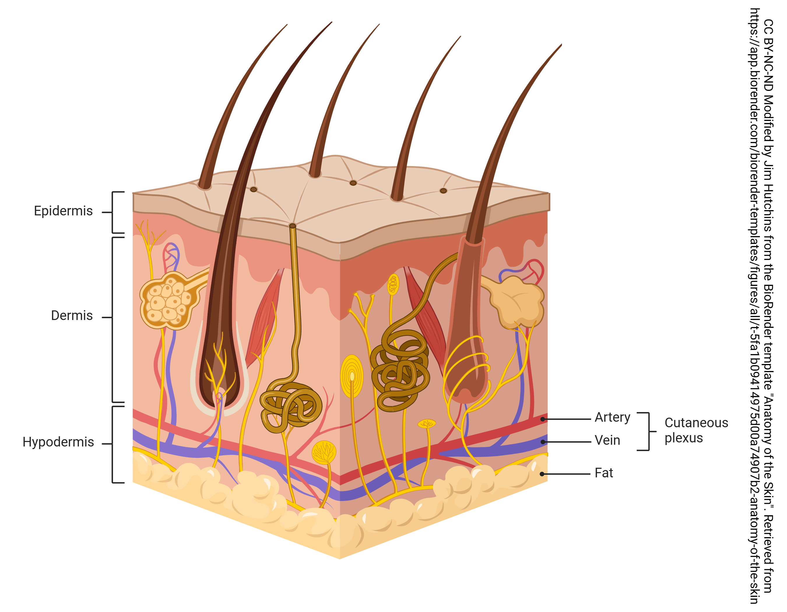
Recall that the subcutaneous layer of the skin is a typical example of an areolar connective tissue, which is a loose connective tissue made up of collagen, reticular and elastic fibers with adipose (fat) cells suspended in the fiber matrix. Blood vessels and nerves traverse the layer.
What is the function of the fat found here? Why is it located in this layer, between integument and deeper tissues (often muscle)? Is this layer always the same thickness, or does it vary in thickness depending on the individual?
Media Attributions
- U08-019 Anatomy of the Skin © Hutchins, Jim is licensed under a CC BY-NC-ND (Attribution NonCommercial NoDerivatives) license
