Active Transport Across Membranes
Objective 4.6
4.6.1 Summarize the essential features of active transport, and compare and contrast primary and secondary active transport.
Active transport, by definition, requires energy. Active transport is used to move an ion against its concentration gradient. We were introduced to active transport earlier when we discussed carriers. Carrier proteins are involved in facilitated diffusion as well as active transport. The difference is that the carriers for active transport require energy from ATP.
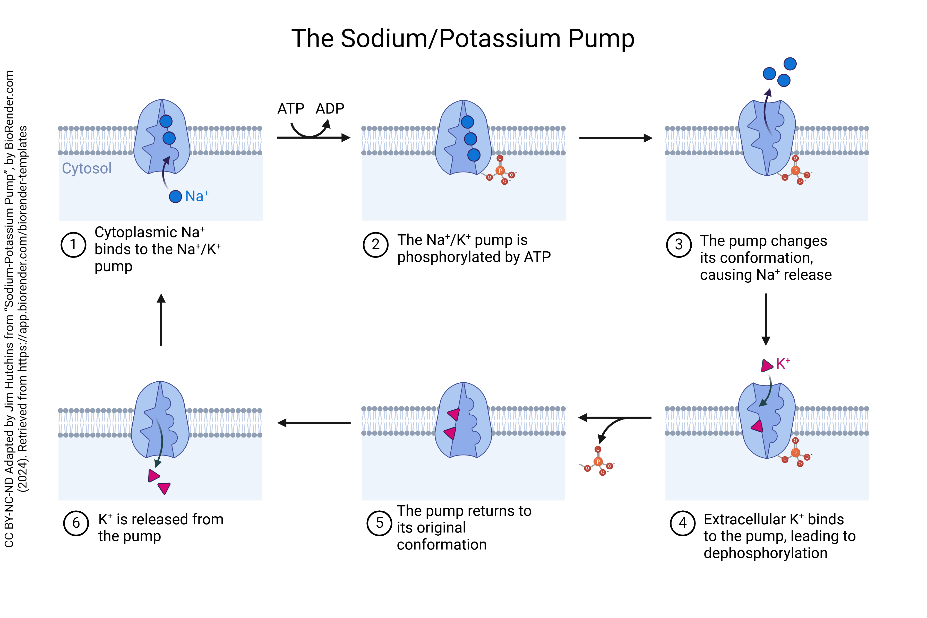
Na+/K+ Pump
The most important of these carriers is a protein called the Na+/K+ pump, or Na+/K+ ATPase. The Na+/K+ ATPase maintains the normal gradient of these ions, with Na+ high outside and K+ high inside the cell. Three Na+ ions rest in a pocket of the pump. These Na+ ions are expelled as ATP is split into ADP + phosphate, releasing the energy needed to drive the pump. Then, two K+ ions bind outside the cell and these are released inside the cell.
Secondary Active Transport: Symport & Antiport
Secondary active transport involves two types of carrier proteins, symporters and antiporters. In secondary active transport we use the energy stored in the Na+ concentration gradient established by primary active transport to move other solutes against their gradients. In an antiport system, two ions move in opposite directions.
In the antiport system shown below, the entry of sodium down its concentration gradient is coupled to the exit of calcium, or hydrogen, against their concentration gradients. Does this require energy? There are two ways to arrive at a “yes, this requires energy” answer.
In the left frame of the antiport image below:
- Calcium is moving against its concentration gradient, from where it is at low concentration, 0.00000001 M inside the cell, to where it is at high concentration, 0.002 M outside the cell. Moving any substance against its concentration gradient requires energy.
- In order to “set up” the concentration gradient for sodium which drives this process, we have to use the sodium/potassium pump to keep the sodium concentration high outside the cell. This uses ATP directly. The antiport system shown here then “runs down” the sodium “battery” which we had to charge using ATP. Each sodium ion that enters in this antiport system has to be pumped back out, and pumping them out requires energy.
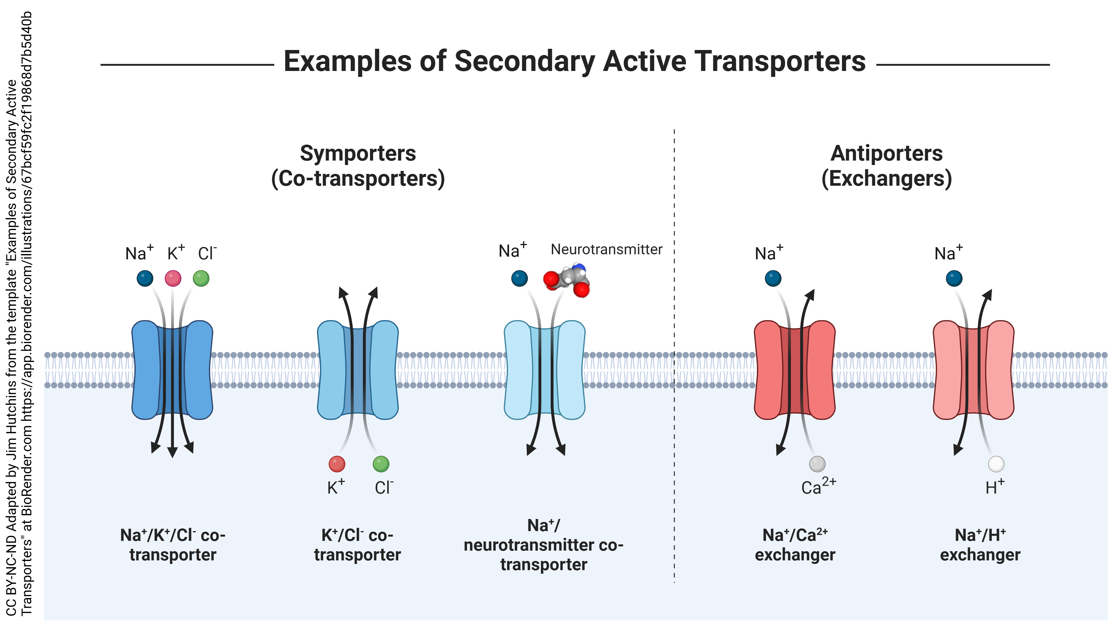
The next example, shown above, is called a symport system. In this type of secondary active transport, ions or a molecule and an ion move in the same direction. A pre-existing Na+ gradient created by primary active transport is the “motor” that drives the pump. Since it took energy to set up the Na+ gradient, this is still an energy-requiring process (which is why we classify it as active transport.) In the image above, as sodium is allowed to diffuse into the cell it again moves with a lot of energy. This energy is used to carry other ions or molecules into the cell.
In the above image, the pre-existing sodium “battery” is being used to move potassium (left frame of the symport image) or a neurotransmitter (right frame of the symport image) from where they are at low concentration (outside the cell) to where they are already at high concentration (inside the cell). Remember, chloride is in highest concentration outside the cell, so it is also flowing down its gradient. The middle frame of the symport image shows a pre-existing potassium “battery” being used to move chloride from where is is at low concentration (inside the cell) to where it is already at high concentration (outside the cell).
Once again, each sodium that enters has to be pumped back out again, which requires energy. This is called a symport system because the ions, or the ion and molecule, are moving in the same direction. In this case, Na+, K+, and Cl- (left frame of symport image), K+ and Cl- (middle frame of symport image), and Na+ and a neurotransmitter are moving in the same direction.
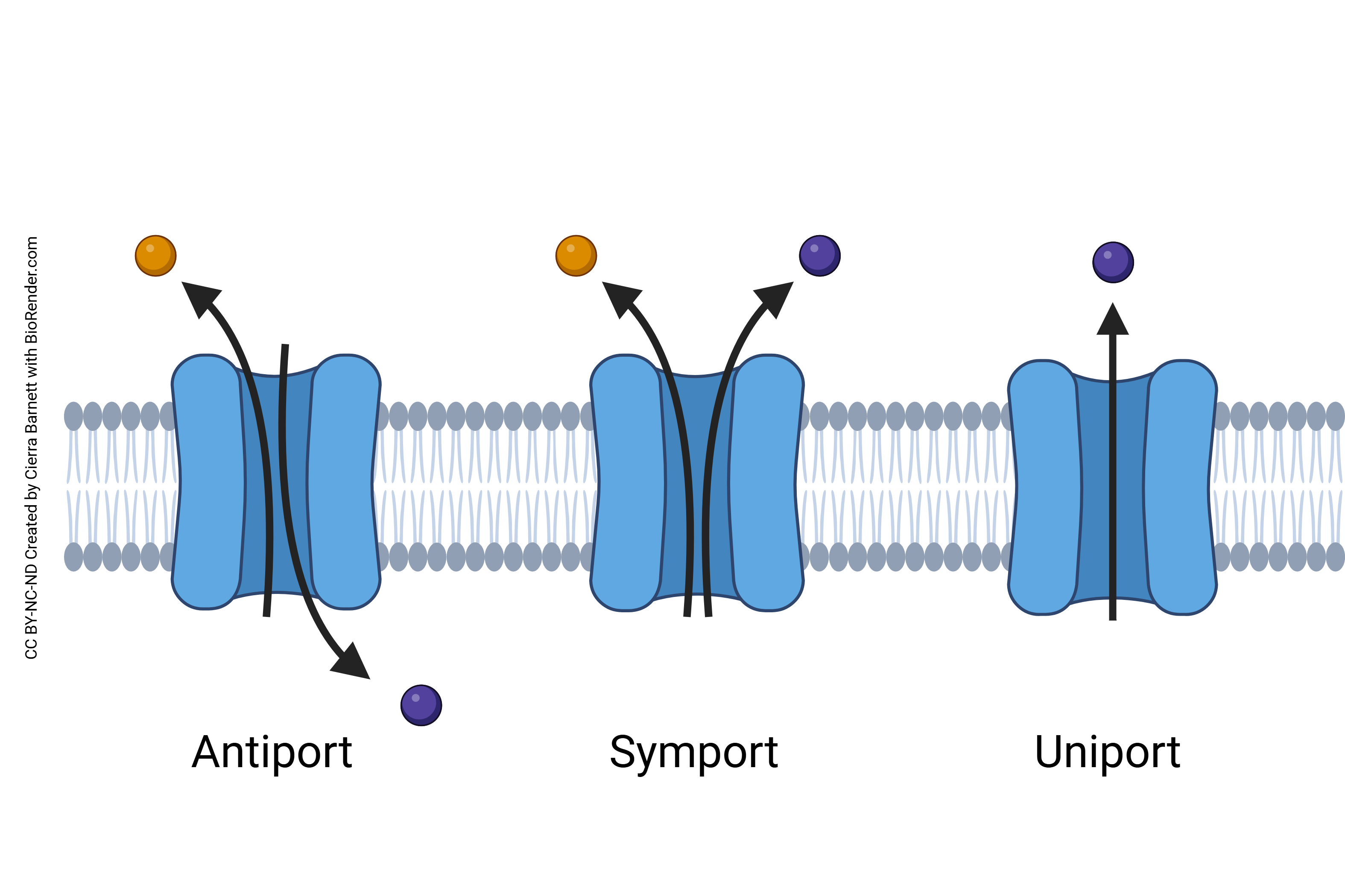
The picture below summarizes the direction of movement in symport, antiport, and uniport systems.
Endocytosis & Exocytosis
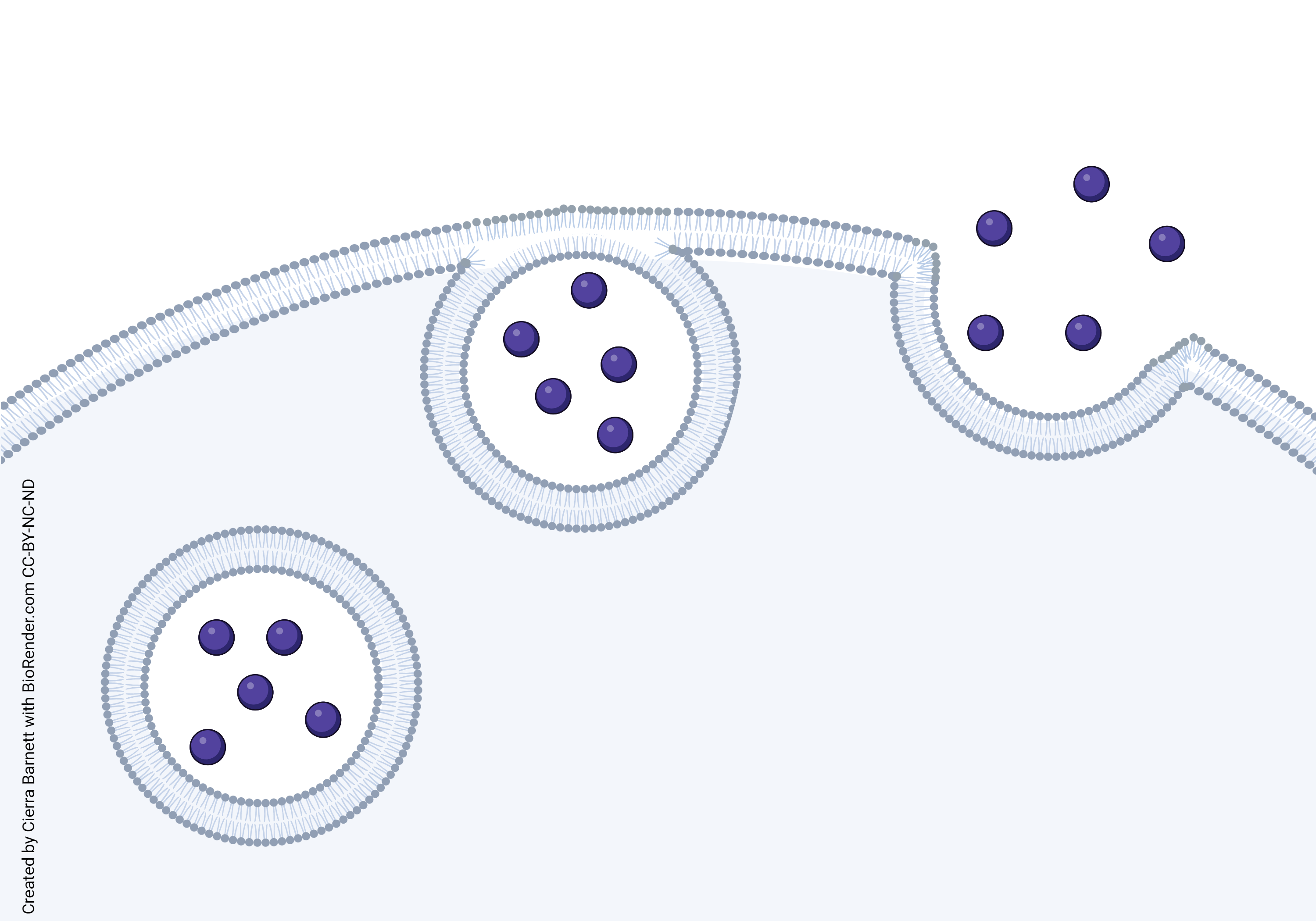
The active transport processes shown previously used protein pumps. The ones we’ll consider next use larger structures called vesicles (Latin: “little bladders” or “blisters”). If material is to be taken into a cell, it is called endocytosis. If material is expelled from a cell, it is called exocytosis. All forms of endocytosis and exocytosis require energy from ATP.
Vesicular transport is used to move large quantities of small things, or to move things too large to fit inside of a protein (for example, materials much larger than the protein itself).
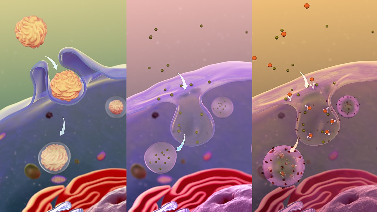
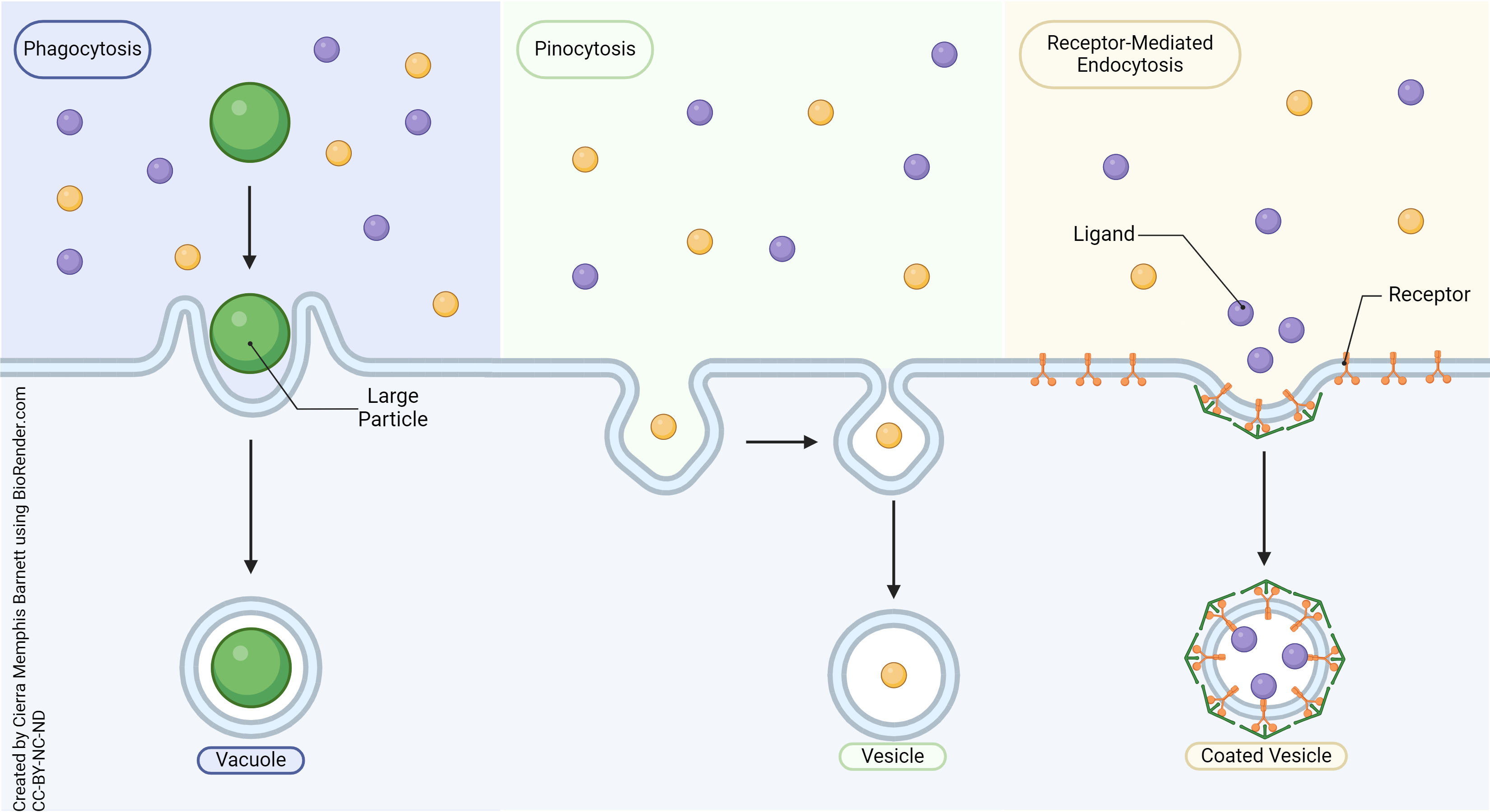
Phagocytosis and pinocytosis are types of endocytosis. Phagocytosis (Greek φαγειν or phage- in: “eating”) is a important defense of body against invaders. When invaders are detected specialized white blood cells surround and kill the invading cells.
Pinocytosis (Greek πινω or pino-: “drinking”) is akin to phagocytosis, but is used to bring liquids into the cell. It is also called bulk-phase endocytosis.
In pinocytosis, the cell makes a pit, then seals it. In the process, a vesicle full of liquid is incorporated into the cell.
In receptor-mediated endocytosis, cells surface proteins (receptor proteins) bind a substance of interest. Underneath the receptors is a protein called clathrin. When the ligand binds to the receptor, clathrin changes shape to pinch off the vesicle which envelopes the ligand in a coated vesicle. For example, low-density lipoprotein (LDL) particles are taken into the cell by this process so that the lipid may be used for cell metabolism.
This picture depicts (from L to R) phagocytosis, pinocytosis, and receptor-mediated endocytosis.
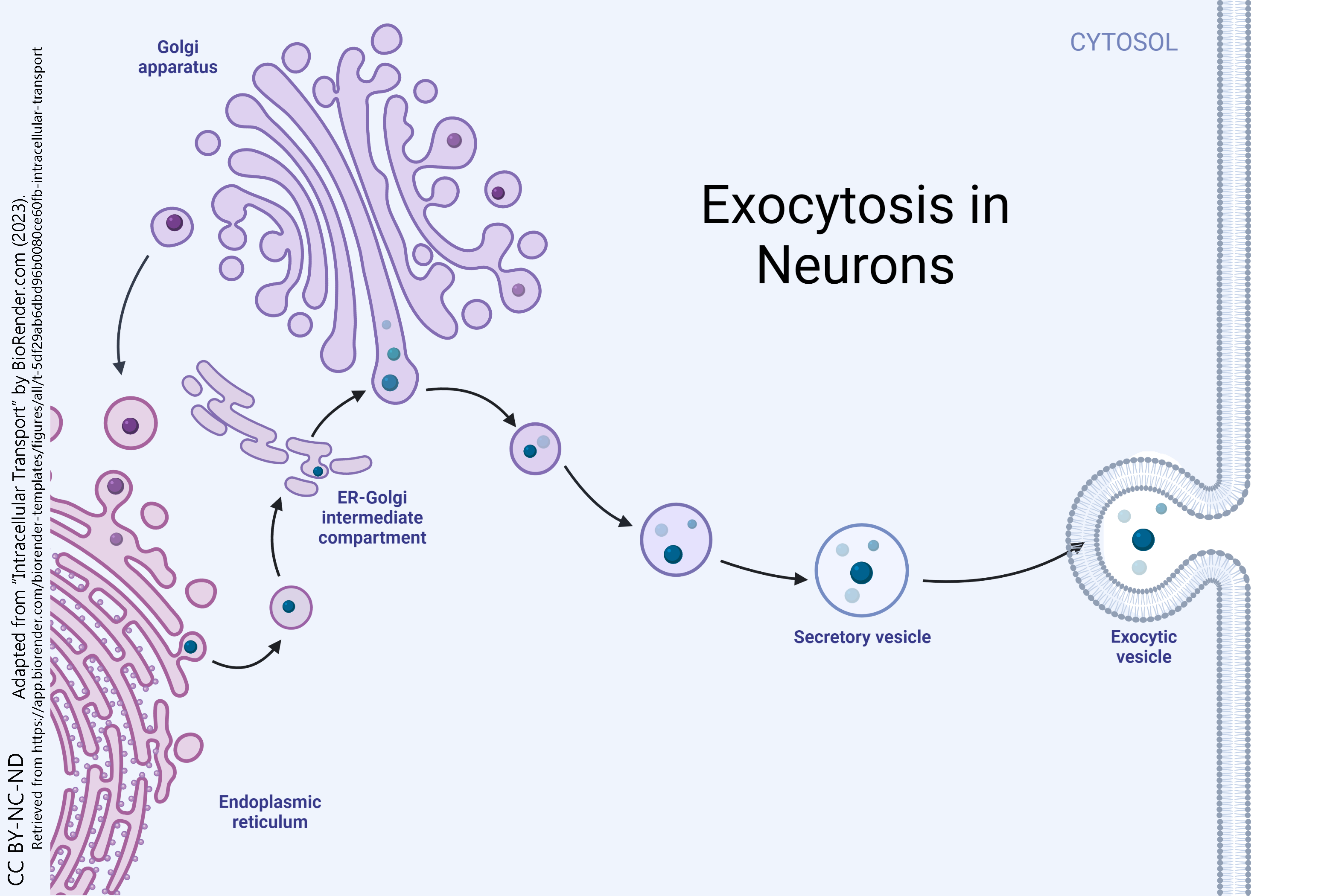
Exocytosis is used to send materials out of the cell in bulk. Both endocytosis and exocytosis require cellular energy, so they are both classified as active transport processes.
The material to be expelled from the cell is packaged into a vesicle. The phospholipid bilayer of the vesicle then fuses with the phospholipid bilayer on the surface of the cell, and this allows the exported material to flow out of the cell.
Two common examples which we will study in detail are exocytosis of digestive enzymes from cells of the pancreas and exocytosis of neurotransmitter signaling molecules from nerve cells.
The pancreatic acinar cells produce and secrete many enzymes that digest food. The tiny black granules in this electron micrograph are secretory vesicles filled with enzymes that will be exported from the cells via exocytosis.

Media Attributions
- U04-040 copy of U13-017 Sodium-Potassium Pump © Hutchins, Jim is licensed under a CC BY-NC-ND (Attribution NonCommercial NoDerivatives) license
- U04-996 Examples of Secondary Active Transporters © Hutchins, Jim is licensed under a CC BY-NC-ND (Attribution NonCommercial NoDerivatives) license
- U04-041 Symport and Antiport Systems © Barnett, Cierra Memphis is licensed under a CC BY-NC-ND (Attribution NonCommercial NoDerivatives) license
- U04-044 1280px-A_depiction_of_various_types_of_Endocytosis © Scientific Animations is licensed under a CC BY-SA (Attribution ShareAlike) license
- U04-042 Phagocytosis, Pinocytosis, Receptor-Mediated Endocytosis © Barnett, Cierra Memphis is licensed under a CC BY-NC-ND (Attribution NonCommercial NoDerivatives) license
- U04-046 copy of U13-061 Cell Membrane in Exocytosis © Barnett, Cierra Memphis is licensed under a CC BY-NC-ND (Attribution NonCommercial NoDerivatives) license
- U04-047 Exocytosis in Neurons © BioRender is licensed under a CC BY-NC-ND (Attribution NonCommercial NoDerivatives) license
- Screenshot 2025-02-13 at 4.43.55 PM (2) © Betts, J. Gordon; Young, Kelly A.; Wise, James A.; Johnson, Eddie; Poe, Brandon; Kruse, Dean H. Korol, Oksana; Johnson, Jody E.; Womble, Mark & DeSaix, Peter adapted by Maddison Johnston is licensed under a CC BY (Attribution) license
