Prokaryotic and Eukaryotic Cells
Objective 4.1
4.1.1 Compare and contrast eukaryotic and prokaryotic cells.
4.1.2 Describe the structure of prokaryotic cells.
4.1.3 Describe cell wall characteristics and staining techniques that differentiate prokaryotic cells.
All living things are made up of cells. Your body has about 100 trillion cells and about 10 trillion of these are human cells. (What are the other 90 trillion?) Cells are organized into tissues and organs. Just as we learned in Unit 1 that the function of organs depends on their structure, we’ll learn in this unit how the function of cells depends on the subcellular structures they’re composed of and how membranes regulate what goes into and comes out of the cell.
(Remember from unit 3 that a cell membrane is made up of a phospholipid bilayer that makes the membrane semi-permeable. More on this in objective 3.)
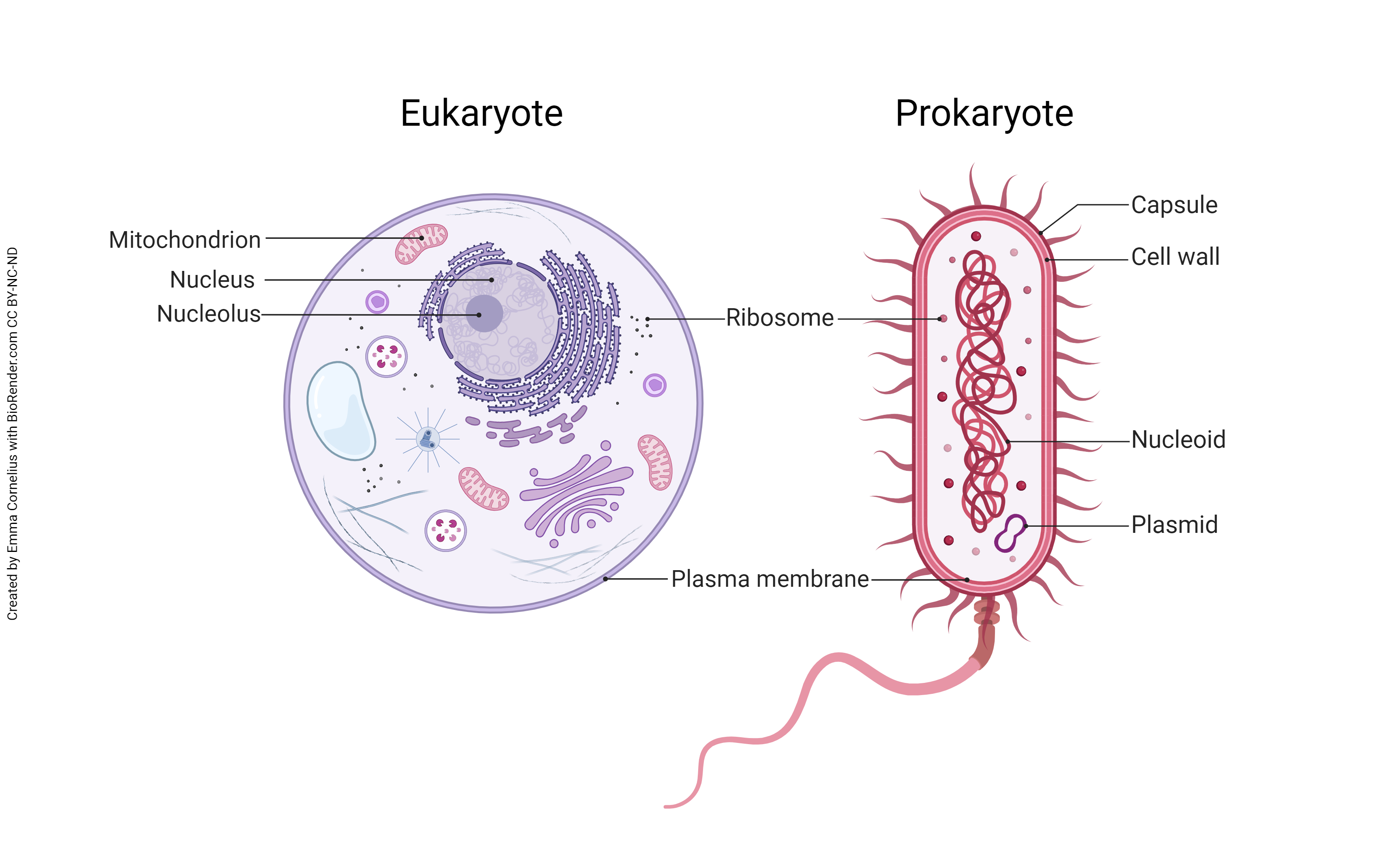
There are two broad divisions of living cells. Humans are eukaryotes; the name means they have a “true” nucleus. Eukaryotic cells also have membrane-bound organelles, subcellular structures we’ll study in detail. Fungi (for example, molds, mushrooms and yeast) are eukaryotes; plants are eukaryotes; and animals are eukaryotes.
Prokaryotes include bacteria and are mostly unicellular (one-celled) organisms. They don’t have a true nucleus. Their DNA is spread throughout the cell and is not collected into packed structures. They differ in some metabolic pathways from eukaryotes. This distinction is especially important in human disease and its treatment. It’s relatively easy to design drugs that kill or disable prokaryotes, because their cell physiology is so different. It’s more difficult to design drugs that kill or disable eukaryotes, because those pathogens (disease-causing organisms) are similar to our cells, with similar genes and metabolic pathways.
Comparison of Prokaryotic & Eukaryotic Cells
Prokaryotic Cells
The prokaryotes belong to the Kingdom Monera and consist of bacteria and cyanobacteria (blue-green algae). Prokaryotes are cells without a true nucleus. All bacteria and other prokaryotic organisms are the earth’s most abundant form of life. Scientists estimate there are 5×1030 (5,000,000,000,000,000,000,000,000,000,000) bacteria on Earth. Bacteria inhabit nearly every environmental niche on the planet and are among the smallest organisms. The average size range of bacteria is 0.5 to 2.0 microns in diameter. For comparison, the human red blood cell is about 7.5 microns in diameter. Despite their small size, bacteria have an extensive surface area for the absorption of nutrients.
In this objective, we will gain a basic familiarity with bacteria, only one of several medically important microbes. A more in-depth study of bacteria and an introduction to viruses, parasites, fungi, and helminths will be left to a separate course.
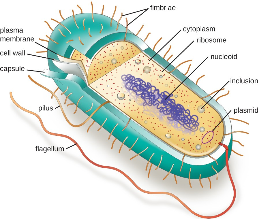
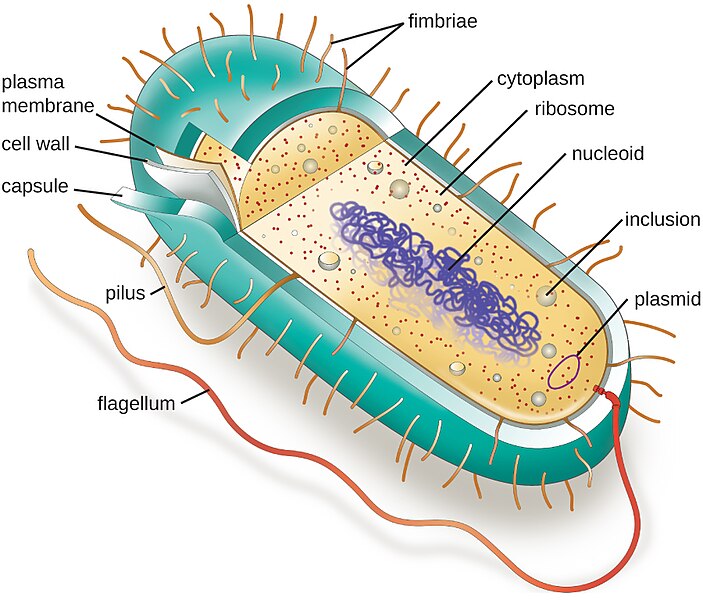 The prokaryotes do not have a membrane-bound nucleus, membrane-bound organelles, or DNA that is organized into chromosomes. Chromosomes carry all of the genetic information in cells. Prokaryotes do not have a nucleus, but instead, have a nuclear region called the nucleoid, which is the cell’s chromosome. This is a circular structure consisting mostly of DNA with small amounts of RNA and protein.
Prokaryotes have inclusion bodies, which store molecules essential to cell functions such as glycogen.
Ribosomes, the site of protein synthesis in the cell, consist of RNA and protein. They are abundant in the cytoplasm of bacteria, often grouped in long chains called polyribosomes. Ribosomes are nearly spherical, stain densely, and contain a large subunit and a small subunit.
Prokaryotic cells, like the eukaryotes, are filled with cytoplasm, which makes up much of the cell inside the cell membrane. The cytoplasm consists primarily of water and substances either suspended or dissolved in the water. These substances include lipoproteins, carbohydrates, lipids, and various enzymes.
Some of the prokaryotes have the ability to secrete a capsule, a protective structure that serves as a defense mechanism. Tightly bound to the cell wall, typical capsules consist of complex polysaccharide molecules that form a gel-like covering that surrounds the bacterial cell. Human phagocytes (cells that eat foreign invaders) have a difficult time attacking an encapsulated organism. In medically important bacteria, therefore, the capsule is considered a major contributor to their ability to cause disease (pathogenicity). For example, Streptococcus pneumoniae is a bacterium that produces a capsule. This bacterium causes pneumococcal pneumonia. It is difficult for the cells of the immune system to attack this microbe because of the capsule.
In addition to these cellular structures, some prokaryotes contain genetic material in structures called plasmids. Plasmids are self-replicating extrachromosomal DNA that carry one or more pieces of genetic information, not required to sustain life. The ability of bacteria to exchange this extrachromosomal genetic material has allowed many strains of bacteria to share their resistance to antibiotics.
The Cell Wall
The majority of bacteria possess semi-rigid cell walls that are unique to the prokaryotic bacterial cell. The main function of the cell wall is to provide shape and stability. Cell walls of all bacteria contain a large polymer called peptidoglycan. Peptidoglycan is an immense, covalently-linked molecule arranged in layers resembling multiple layers of a chain-link fence which gives the cell support. Many antibiotics in use today, such as the penicillins, target the bacterial cell wall as their site of bacteriocidal (bacteria-killing) activity.
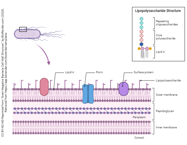
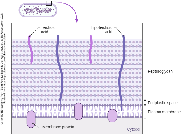
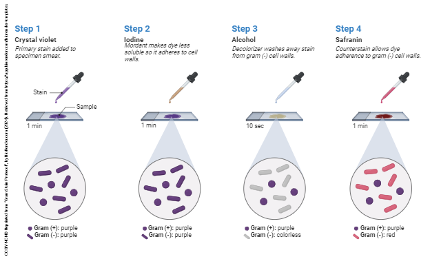
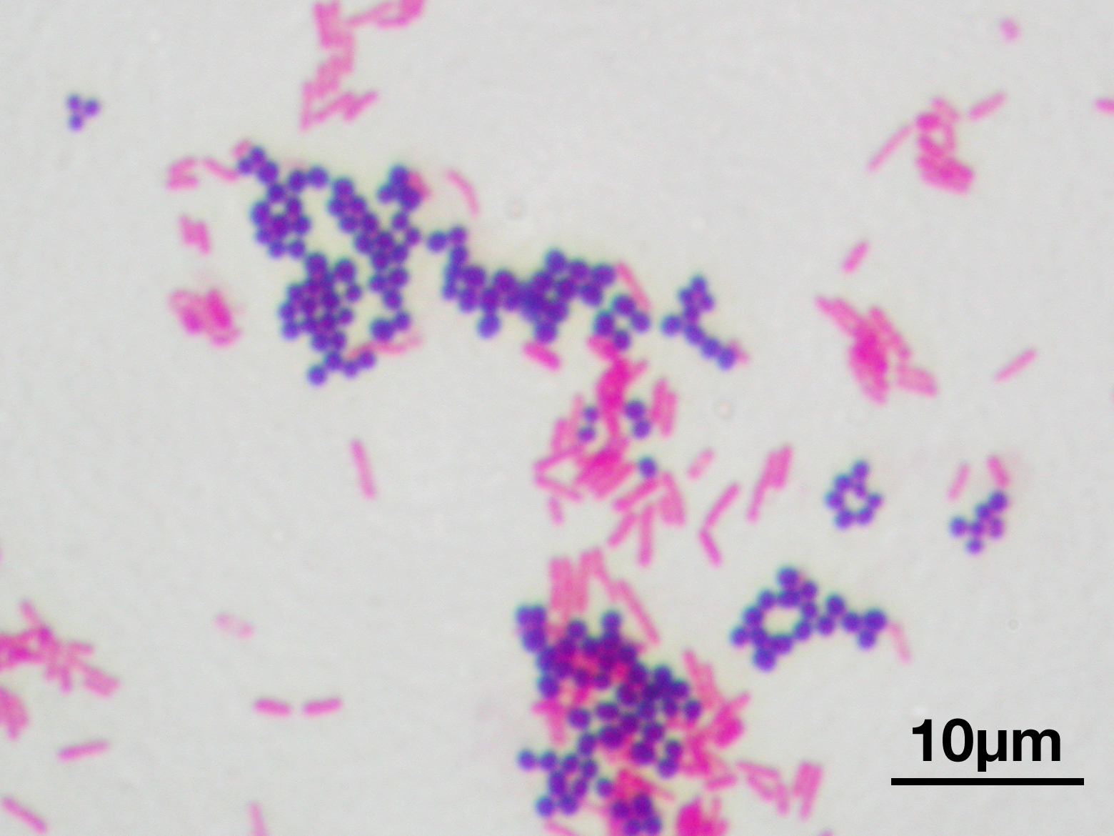
We can differentiate bacteria based on their cell wall characteristics by a staining technique called a Gram stain. In this procedure, the sample is smeared on a slide and fixed with heat. The slide is stained with crystal violet (purple) and fixed with iodine (helps to retain the stain). The slide is washed with alcohol or acetone and then counterstained with safranin, a pink stain. Organisms with a thick peptidoglycan cell wall retain the purple crystal violet stain and remain purple. Organisms with a thin peptidoglycan layer and high lipoprotein content release the crystal violet when washed with alcohol. These organisms retain the pink safranin stain. We call organisms that retain the purple stain, Gram-positive organisms. Those that are pink due to the counterstain are Gram-negative organisms. Important Gram-positive organisms include Streptocococcus spp. (Strep sore throats, Pneumococcal pneumonia), Staphlococcus spp. (wound infections, food poisoning, toxic shock syndrome), and Clostridium spp. (tetanus, botulism, and life-threatening inflammation of the colon). Important Gram-negative organisms include E-coli, the number one cause of outpatient urinary tract infections.
 The importance of the Gram staining technique cannot be overstated. If a physician suspects a bacterial infection, a culture report will take 24 to 48 hours (the little critters need to have time to grow). A Gram stain can give a physician immediate information that can determine the appropriate antibiotic course.
The Cell Membrane (Plasma Membrane)
The cell membrane (also called the plasma membrane) is present in all living cells, both prokaryotic and eukaryotic. The membrane, as with the eukaryotic cell membrane, contains many complex molecules, including phospholipids, long-chain fatty acids, and proteins. As in the eukaryotic cell, the main functional activity of the cell membrane is to regulate materials into and out of the cell primarily by transport mechanisms.
About half of all known bacteria possess organelles of locomotion called flagella (singular flagellum). Organisms displaying flagella are capable of various degrees of movement and are said to be motile. Pili (singular: pilus) are tiny, tube-like projections from the cell’s surface associated with adherence and sometimes the exchange of genetic information.
Media Attributions
- U04-001 Comparison of prokaryote and eukaryote © Cornelius, Emma is licensed under a CC BY-NC-ND (Attribution NonCommercial NoDerivatives) license
- U04-002 choose one prokaryotic cell openstax © OpenStax is licensed under a CC BY-NC-SA (Attribution NonCommercial ShareAlike) license
- U04-005 Gram Negative Bacteria Cell Wall Structure © BioRender is licensed under a CC BY-NC-ND (Attribution NonCommercial NoDerivatives) license
- U04-004 Gram-Positive Bacteria Cell Wall Structure © BioRender is licensed under a CC BY-NC-ND (Attribution NonCommercial NoDerivatives) license
- U04-003 Gram Stain Protocol © BioRender is licensed under a CC BY-NC-ND (Attribution NonCommercial NoDerivatives) license
- U04-006 Gram_stain_01 © Y tambe is licensed under a CC BY-SA (Attribution ShareAlike) license
