1 The Heart
Cardiovascular System – Heart Word Parts
Click on prefixes, combining forms, and suffixes to reveal a list of word parts to memorize for the cardiovascular system – Heart.
Introduction to the Heart
The heart is a fist-sized vital organ that has one job: to pump blood. If one assumes an average heart rate of 75 beats per minute, a human heart would beat approximately 108,000 times in one day, more than 39 million times in one year, and nearly 3 billion times during a 75-year lifespan. At rest, each of the major pumping chambers of the heart ejects approximately 70 mL blood per contraction in an adult. This would be equal to 5.25 liters of blood per minute and approximately 14,000 liters per day. Over one year, that would equal 10,000,000 liters of blood sent through roughly 100,000 km of blood vessels. In order to understand how that happens, it is necessary to understand the anatomy and physiology of the heart.
Watch this video:
Media 12.1. The Heart, Part 1 – Under Pressure: Crash Course A&P #25 [Online video]. Copyright 2015 by CrashCourse.
Cardiovascular System – Heart Medical Terms
Anatomy of the Heart
Location
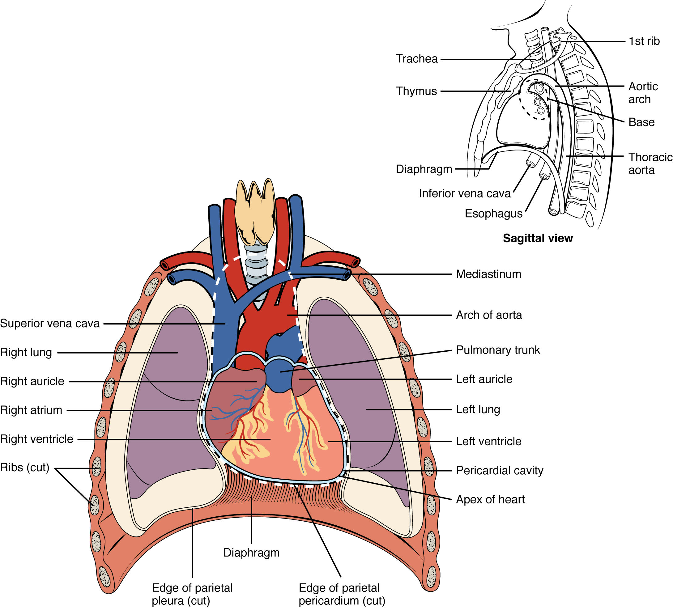
The human heart is located within the thoracic cavity, between the lungs in the space known as the mediastinum. Figure 12.1 shows the position of the heart within the thoracic cavity. Within the mediastinum, the heart is separated from the other mediastinal structures by a tough membrane known as the pericardium, or pericardial sac, and sits in its own space called the pericardial cavity. The great vessels, which carry blood to and from the heart, are attached to the superior surface of the heart, which is called the base. The base of the heart is located at the level of the third costal cartilage. The inferior tip of the heart, the apex, lies just to the left of the sternum between the junction of the fourth and fifth ribs.
Concept Check
- On the diagram below (Figure 1), locate the mediastinum, the pericardial cavity, the base of the heart and the apex of the heart.
- Locate the largest vein in the body superior vena cava.
Membranes and Layers of the Heart Walls
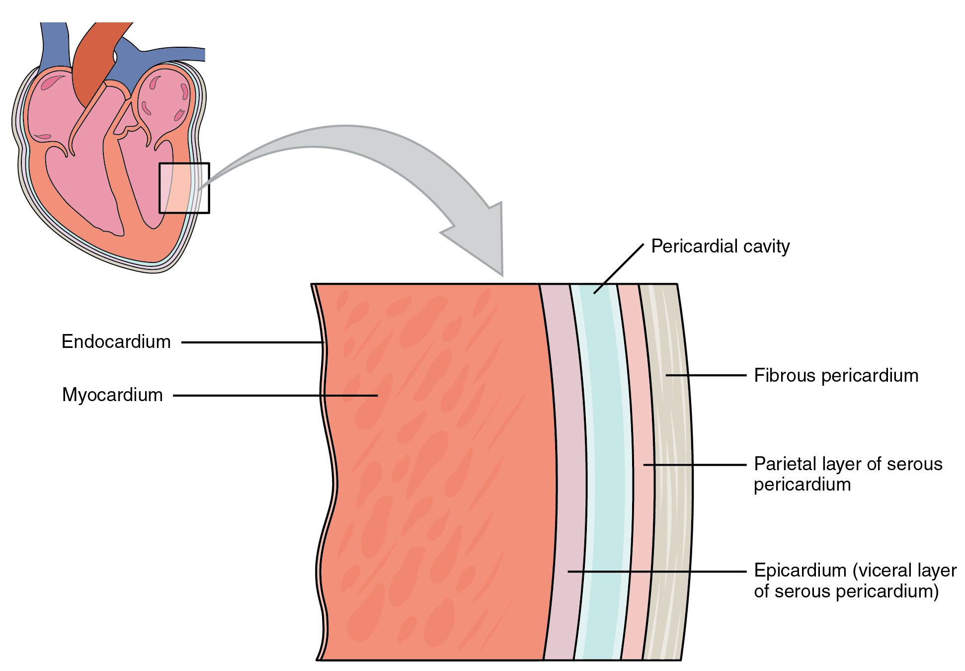
The heart and the roots of the great vessels are surrounded by a membrane known as the pericardium or pericardial sac. The pericardium consists of two distinct sub layers:
- The sturdy outer fibrous pericardium is made of tough, dense connective tissue that protects the heart and holds it in position.
- Separated by the pericardial cavity and containing pericardial fluid the inner serous pericardium consists of two layers:
- the outer parietal pericardium, which is fused to the fibrous pericardium.
- the inner visceral pericardium, or epicardium, which is fused to the heart and forms the outer layer of the heart wall.
The walls of the heart consist of three layers:
- The outer epicardium, which is another name for the visceral pericardium mentioned above.
- The thick, middle myocardium, which is made of muscle tissue and gives the heart its ability to contract.
- The inner endocardium, which lines the heart chambers and is the main component of the heart valves.
Concept Check
- Look at Figure 12.2 below, and name the layers of the heart wall and surrounding membranes, starting with the innermost layer.
- As shown on the diagram, suggest why is the myocardium layer is thicker than the endocardium layer?
Internal Structures of the Heart
The heart consists of four chambers:
- The upper chambers are the right and left atria (singular: atrium).
- The lower chambers are the right and left ventricles.
The interventricular septum is a muscular wall that separates the right and left ventricles. The interatrial septum separates the right and left atria.
The atrium and ventricle on each side of the heart are separated by an atrioventricular (AV) valve:
- The right AV valve, or tricuspid valve, separates the right atrium and right ventricle.
- The left AV valve, or bicuspid valve, separates the left ventricle and the left atrium. This valve is also called the mitral valve.
There are also two semilunar valves:
- The pulmonary valve separates the right ventricle from the pulmonary trunk.
- The aortic valve separates the left ventricle from the aorta (De Saix, et al., 2013).
Anatomy Labeling Activity
Cardiovascular System – Heart Vocabulary
5.25 liters of blood
The volume of blood ejected by the ventricle in one minute is called the cardiac output.
70 mL blood per contraction
The amount of blood ejected from the ventricle in one contraction is called the stroke volume.
AV
Atrioventricular: the area of the heart where the atria and ventricles meet.
AV Valves
Atrioventricular valves: mitral (bicuspid) valve allows blood to flow from left atrium to left ventricle, tricuspid valve allows blood to flow from right atrium to right ventricle.
Great Vessels
The great vessels include the superior vena cava, inferior vena cava, aorta and pulmonary trunk.
HDL
High-density lipoprotein, often referred to as ‘good’ cholesterol.
Heart Rate
The number of times the heart contracts in one minute.
Inferior Vena Cava
One of the two largest veins in the body. It carries deoxygenated blood from the torso and legs back to the heart.
Interatrial Septum
The wall separating the right and left atria.
Interventricular Septum
The wall of myocardium that separates the right and left ventricles.
Mitral Valve
Also known as the bicuspid valve.
Pericardial fluid
Pericardial fluid is a serous fluid which allow the 2 layers of serous pericardium to slide smoothly against each other as the heart beats.
Pulmonary Trunk
Very large artery referred to as a trunk, a term indicating that the vessel gives rise to several smaller arteries.
Roots of the Great Vessels
The part of each great vessel (aorta, pulmonary trunk, inferior vena cava, superior vena cava) that connects to the base of the heart.
Serous
You may recall that serous membranes throughout the body are folded back on themselves, which results in a double-layered membrane separated by serous fluid. The serous membrane surrounding the lungs is called pleura. The serous membrane surrounding the abdominopelvic organs is called peritoneum.
Superior Vena Cava
One of the two largest veins in the body. It carries deoxygenated blood from the head and upper extremities back to the heart.
Test Yourself
References
Canadian Medical Association. (2018). Canadian Specialty Profiles. https://www.cma.ca/canadian-specialty-profiles
Canadian Society of Cardiology Technologists. (n.d.). Becoming a registered cardiology technologist. https://www.csct.ca/education/about-being-rct
Centers for Disease Control and Prevention. (2019). Cardiomyopathy. CDC. https://www.cdc.gov/heartdisease/cardiomyopathy.htm
Centers for Disease Control and Prevention. (2019a). Valvular heart disease. CDC. https://www.cdc.gov/heartdisease/valvular_disease.htm
Centers for Disease Control and Prevention. (2019b). Aortic aneurysm. CDC. https://www.cdc.gov/heartdisease/aortic_aneurysm.htm
[CrashCourse]. (2015, July 6). The heart, part 1 – under pressure: Crash course A&P #25 [Video]. YouTube. https://youtu.be/X9ZZ6tcxArI
[CrashCourse]. (2015, July 13). The heart, part 2 – heart throbs: Crash course A&P #26 [Video]. YouTube. https://youtu.be/FLBMwcvOaEo
Heart & Stroke. (n.d.). Heart failure. Heart and Stroke Foundation. https://www.heartandstroke.ca/heart/conditions/heart-failure
Mitchener Institute for Education. (n.d.). Cardiovascular perfusion. Michener Institute of Education at UHN. https://michener.ca/program/cardiovascular-perfusion/
Tittley, J. G. (n.d.). Thoracic aortic aneurysms (TAA). Retrieved from Canadian Society for Vascular Surgery: https://canadianvascular.ca/Thoracic-Aortic-Aneurysms-(TAA)
Image Descriptions
Figure 12.1 image description: This diagram shows the location of the heart in the thorax (sagittal and anterior views). The sagittal view labels read (from top, clockwise): first rib, aortic arch, thoracic arch, esophagus, inferior vena cava, diaphragm, thymus, trachea. The anterior view lables read (from top, clockwise): mediastinum, arch of aorta, pulmonary trunk, left auricle, left lung, left ventricle, pericardial cavity, apex of heart, edge of parietal pericardium, diaphgragm, edge of parietal pleura, ribs, right ventricle, right atrium, right auricle, right lung, superior vena cava. [Return to Figure 12.1].
Figure 12.2 image description: This image shows a magnified view of the structure of the heart wall. Labels read (from top, clockwise): pericardial cavity, fibrous pericardium, parietal layer of serous pericardium, epicardium (visceral layer of serous pericardium), myocardium,
Unless otherwise indicated, this chapter contains material adapted from Anatomy and Physiology (on OpenStax), by Betts, et al. and is used under a a CC BY 4.0 international license. Download and access this book for free at https://openstax.org/books/anatomy-and-physiology/pages/1-introduction.
The number of times the heart contracts in one minute.
The amount of blood ejected from the ventricle in one contraction is called the stroke volume.
The volume of blood ejected by the ventricle in one minute is called the cardiac output
The great vessels include the superior vena cava, inferior vena cava, aorta and pulmonary trunk.
The part of each great vessel (aorta, pulmonary trunk, inferior vena cava, superior vena cava) that connects to the base of the heart
You may recall that serous membranes throughout the body are folded back on themselves, which results in a double-layered membrane separated by serous fluid. The serous membrane surrounding the lungs is called pleura, The serous membrane surrounding the abdominopelvic organs is called peritoneum.
