2 Blood Vessels and Blood
Blood Vessels and Blood Word Parts
Click on prefixes, combining forms, and suffixes to reveal a list of word parts to memorize for the Cardiovascular System – Blood.
Blood pumped by the heart flows through a series of vessels known as arteries, arterioles, capillaries, venules, and veins before returning to the heart.
- Arteries transport blood away from the heart and branch into smaller vessels, forming arterioles.
- Arterioles distribute blood to capillary beds, the sites of exchange with the body tissues.
- A capillary is a microscopic channel that supplies blood to the tissues themselves, a process called perfusion.
- Exchange of gases and other substances occurs in the capillaries between the blood and the surrounding cells and their tissue fluid (interstitial fluid).
- For capillaries to function, their walls must be leaky, allowing substances to pass through.
- Capillaries lead back to small vessels known as venules.
- Venules are small veins that converge into larger veins.
- A vein is a blood vessel that conducts blood toward the heart
- Compared to arteries, veins are thin-walled vessels with large and irregular lumens
- Larger veins are commonly equipped with valves that promote the unidirectional flow of blood toward the heart and prevent backflow toward the capillaries caused by the inherent low blood pressure in veins as well as the pull of gravity
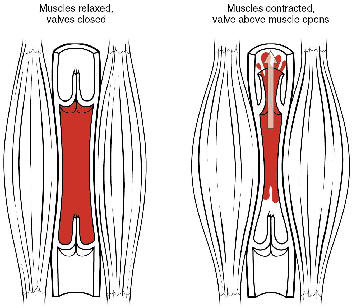
- Other ways in which the body assists the transport of venous blood back to the heart involve contractions of skeletal muscles in the extremities (see figure below), as well as pressure variations caused by breathing motion in the chest.
Concept Check
- Select the correct bolded word: Arteries always carry blood away from/towards the heart
- Select the correct bolded word: Veins always carry blood Away from/towards the heart.
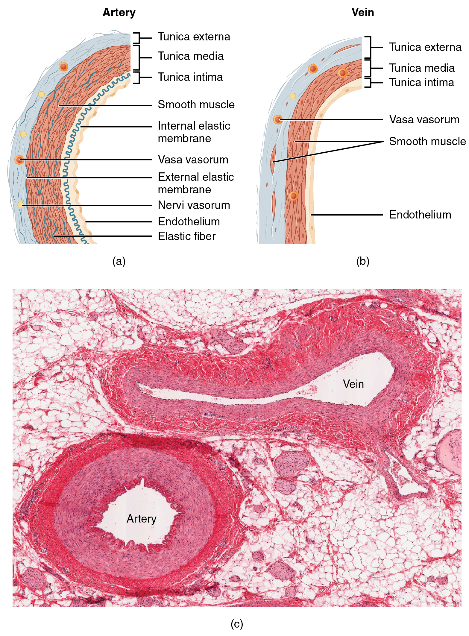
Both arteries and veins have the same three distinct tissue layers, called tunics, for the garments first worn by ancient Romans. From the most interior layer to the outer, these tunics are the tunica intima, the tunica media, and the tunica externa (see Figure 13.3). The smooth muscle in the middle layer, the tunica media, provides the vessel with the ability to vasoconstrict and vasodilate as needed to ensure sufficient blood flow.
| CHARACTERISTIC | ARTERIES | VEINS |
|---|---|---|
| Direction of blood flow | Conducts blood away from the heart | Conducts blood toward the heart |
| General appearance | Rounded | Irregular, often collapsed |
| Pressure | High | Low |
| Wall thickness | Thick | Thin |
| Relative oxygen concentration | Higher in systemic arteries
Lower in pulmonary arteries |
Lower in systemic veins
Higher in pulmonary veins |
| Valves | Not present | Present most commonly in limbs and in veins inferior to the heart |
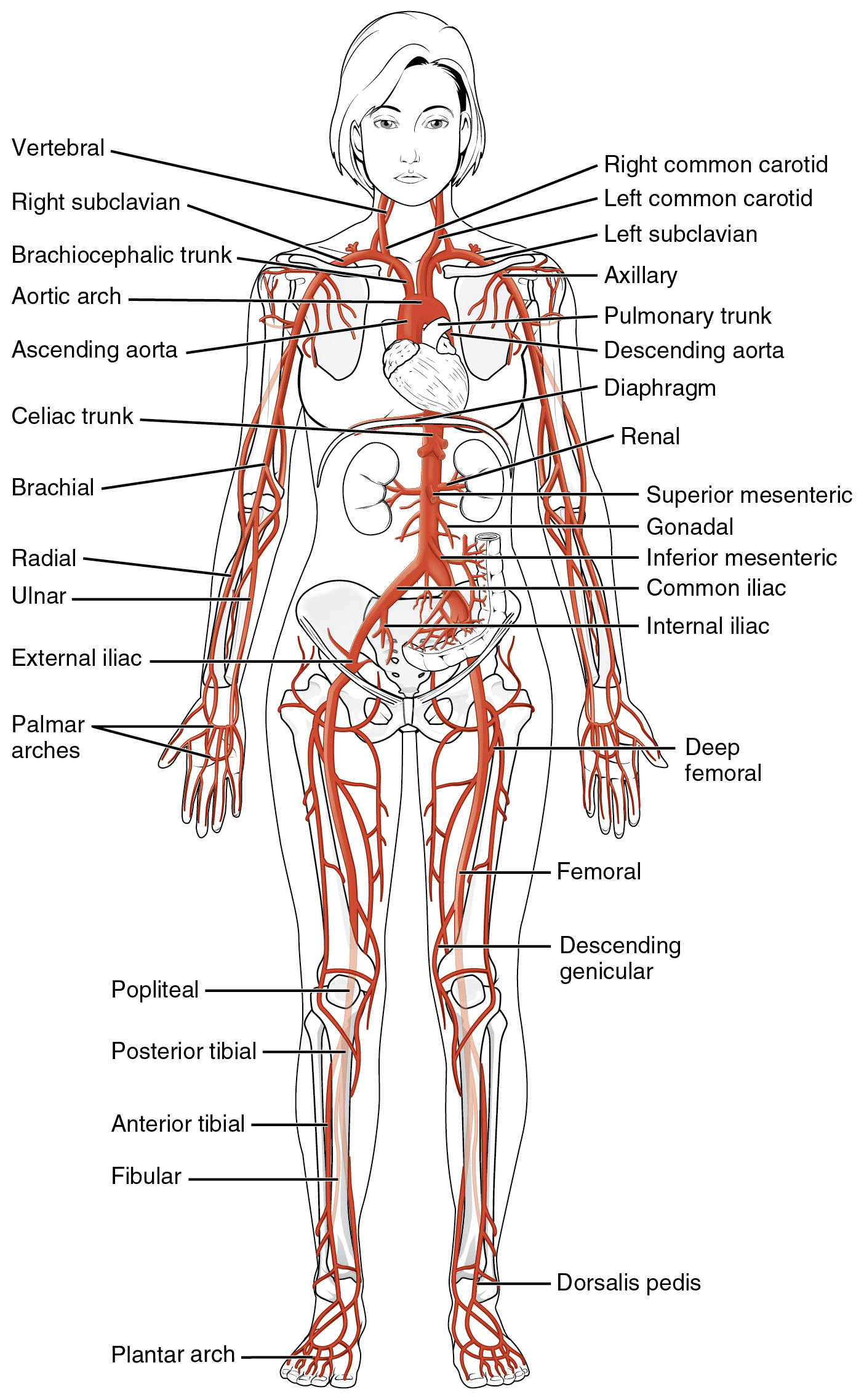
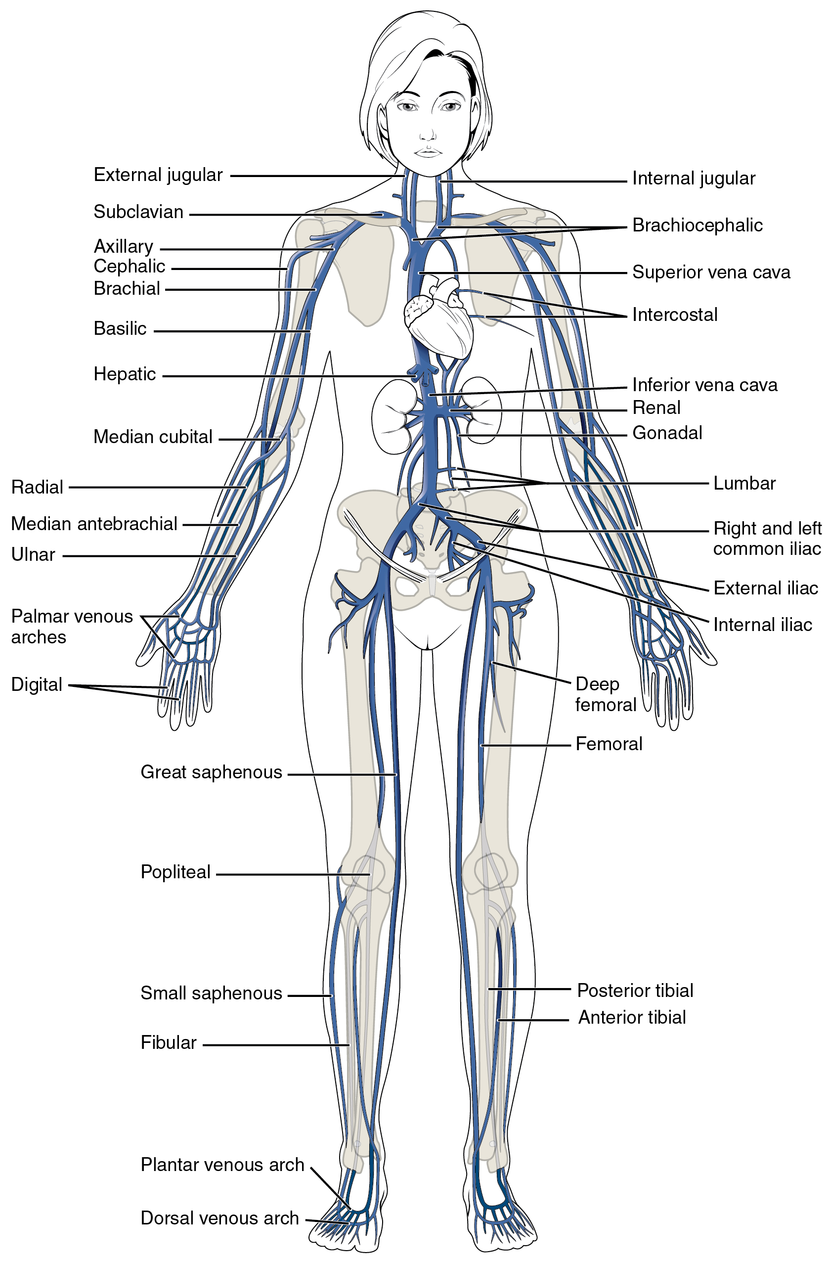
The Major Arteries and Veins in the Human Body
Concept Check
- Without looking back at the images of the main arteries and veins of the body, can you name and locate 3 arteries and 3 veins in your body?
Cardiovascular System-Blood Vocabulary
Arteries
Blood vessels that transport blood away from the heart.
Arterioles
A very small artery that leads to a capillary.
Brachial Artery
Large artery in the upper arm near the biceps muscle.
Capillary
A microscopic channel that supplies blood to the tissues through perfusion.
Carotid Artery
A large artery in the neck.
Endothelium
The lining of the lumen of a blood vessel.
Perfusion
The delivery of blood to an area/tissue/organ.
Vasoconstrict
The smooth muscle layer in the blood vessel wall contracts, causing the vessel diameter to narrow. This increases blood pressure in the vessel.
Vasodilate
The smooth muscle layer in the wall of the blood vessel relaxes, allowing the vessel to widen. This decreases blood pressure in the vessel.
Vein
Blood vessels that carry blood back to the heart.
Venules
Extremely small veins.
Vessel Compliance
The ability of any compartment to expand to accommodate increased content. The greater the compliance of an artery, the more effectively it is able to expand to accommodate surges in blood flow without increased resistance or blood pressure.
Viscosity
The thickness of fluids that affects their ability to flow.
Test Yourself
References
Canadian Medical Association. (2018, August). Hematology profile. CMA Specialty Profiles. https://www.cma.ca/sites/default/files/2019-01/hematology-e.pdf
Canadian Society for Medical Laboratory Science. (n.d.). Who are lab professionals. https://www.csmls.org/Medical-Laboratory-Professionals/About/Who-are-Lab-Professionals.aspx
Canadian Society for Vascular Surgery. (2020). Patients: What is vascular surgery? https://canadianvascular.ca/Patients
[CrashCourse]. (2015, July 20). Blood vessels, part 1 – form and function: Crash course A&P #27 [Video]. YouTube. https://youtu.be/v43ej5lCeBo
Sonography Canada. (2020). Credentials. https://sonographycanada.ca/certification/credentials
Image Descriptions
Figure 13.1 image description: The left panel shows the structure of a skeletal muscle vein pump when the muscle is relaxed, and the right panel shows the structure of a skeletal muscle vein pump when the muscle is contracted.[Return to Figure 13.1].
Figure 13.2 image description: The top left panel of this figure shows the ultrastructure of an artery (labels read from top: tunica externa, tunica media, tunica intima, smooth muscle, internal esastic membrane, vasa vasorum, external elastic membrane, nervi vasorum, endothelium, elastic fiber), and the top right panel shows the ultrastructure of a vein (labels read from top: tunica exerna, tunica media, tunica intima, vasa vasorum, smooth muscle, endothelium). The bottom panel shows a micrograph with the cross sections of an artery and a vein. [Return to Figure 13.2].
Figure 13.3 image description: The major arteries in the human body. Labels read (from top, clockwise) right common carotid, left common carotid, axillary, pulmonary trunk, descending aorta, diaphragm, renal, superior mesenteric, gonadal, inferior mesenteric, common iliac, internal iliac, deep femoral, femoral, descending genicular, doraslis pedis, plantar arch, fibular, anterior tibial, posterial tibial, popliteal, palmer arches, exernal iliac, ulnar, radial, brachial, celiac trunk, ascending aorta, aortic arch, brachiocephalic trunk, right subclavian, vertebral. [Return to Figure 13.3].
Figure 13.4 image description: The major veins in the human body. Labels read (from top, clockwise) internal jugular, brachiocephalic, superior vena cava, intercostal, inferior vena cava, gonadal, lumbar, right and left common iliac, external iliac, internal iliac, deep femoral, femoral, posterior tibial, anterior tibial, dorsal venous arch, plantar venous arch, fibular, small saphenous, popliteal, great saphenous, digital, palmar venous arches, ulnar, median antebrachial, medial cubital, hepatic, basilic, brachial, cephalic, axillary, subclavian, externa jugular. [Return to Figure 13.4].
Unless otherwise indicated, this chapter contains material adapted from Anatomy and Physiology (on OpenStax), by Betts, et al. and is used under a a CC BY 4.0 international license. Download and access this book for free at https://openstax.org/books/anatomy-and-physiology/pages/1-introduction.
definition
Blood vessels that transport blood away from the heart.
A very small artery that leads to a capillary
A microscopic channel that supplies blood to the tissues through perfusion
The delivery of blood to an area/tissue/organ
Extremely small vein
Blood vessels that carry blood back to the heart.
The smooth muscle layer in the blood vessel wall contracts, causing the vessel diameter to narrow. This increases blood pressure in the vessel.
The smooth muscle layer in the wall of the blood vessel relaxes, allowing the vessel to widen. This decreases blood pressure in the vessel.
