The Retina
Jim Hutchins
Objective 2: Discuss the microscopic anatomy of the retina.
The retina extends from the edges of the ciliary body, lining the interior of the eye.
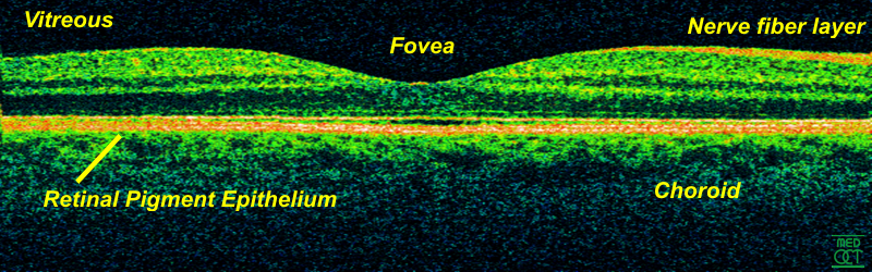
In the direct path of the photons which pass through the pupil without being refracted (i.e. without the light rays being bent) is a pit called the fovea centralis (“central pit”). This region has a high concentration of yellowish visual pigment and so is also called the macula lutea (“yellow spot”).
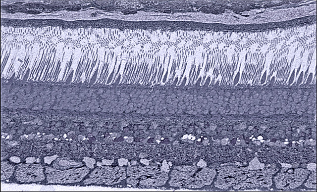
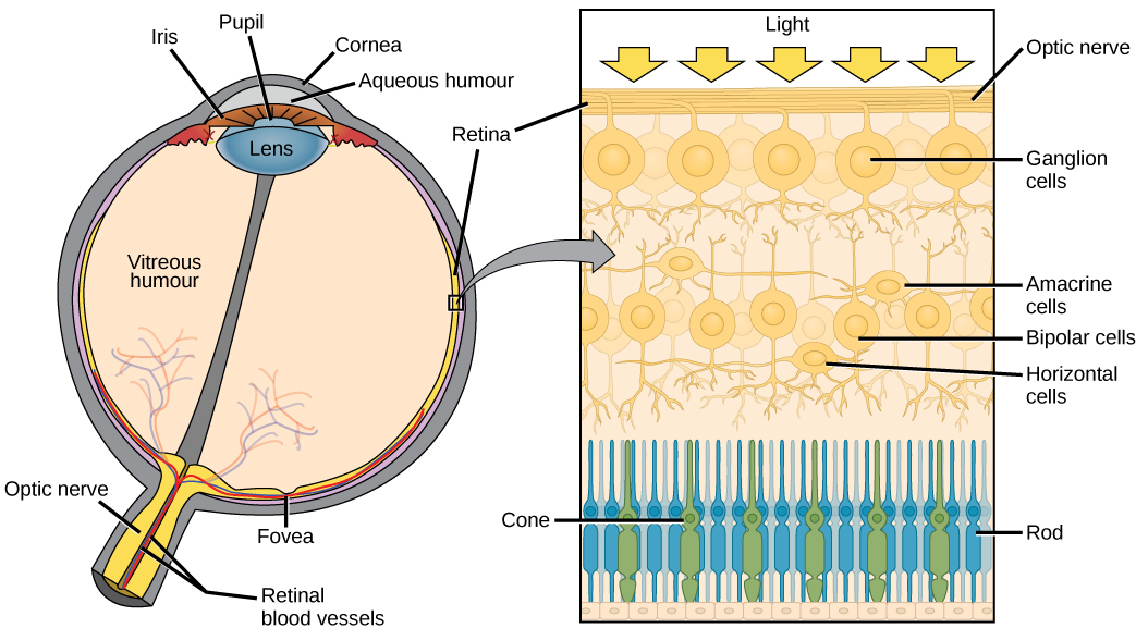
There are two types of photoreceptors, named after the distinctive shape of their modified cilia. In each case, the photoreceptor outer segments consist of stacks of membranes like a roll of pennies. The outer segment is connected to the cell body via a thin stalk.
Media Attributions
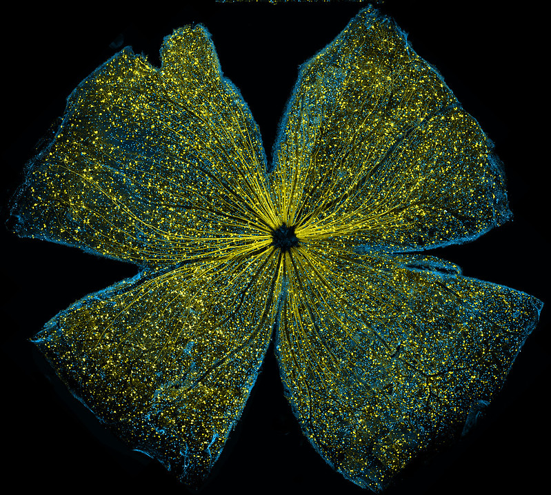
- Mouse retina © Kenyoung Kim, Wonkyu Ju, and Mark Ellisman National Center for Microscopy and Imaging Research, University of California, San Diego is licensed under a CC BY-NC (Attribution NonCommercial) license
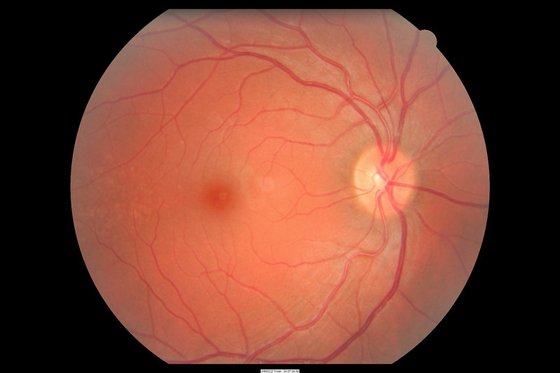
- Human retina fundus © Manuel Pinot is licensed under a CC BY-NC-ND (Attribution NonCommercial NoDerivatives) license
- Retina-OCT800 © Maksim is licensed under a CC BY (Attribution) license
- Human retina histology © Bryan Jones is licensed under a CC BY-NC-ND (Attribution NonCommercial NoDerivatives) license
- Cross section eye with detail of retina © Mary Ann Clark, Matthew Douglas, and Jung Choi is licensed under a CC BY (Attribution) license
