The Cranial Fossa
Rachel Jessop and Jim Hutchins
Objective
1. Identify the cranial fossae and pathophysiology’s associated with it.
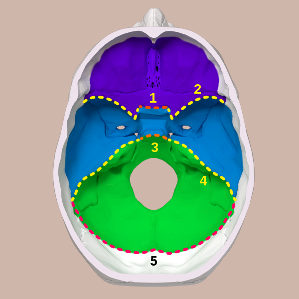
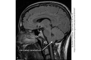
One important phenomenon to discuss in the world of brain disorders is the cranial fossae – the unique depression seen on the edge of the skull, where the brain sits. In the image above, the anterior, medial, and posterior fossae (purple, blue, and green, respectively) are seen. The posterior fossa is an important location to note when it comes to brain tumors, as it is the most common spot for tumors in children. Also seen in this image is the foramen magnum, or the large hole in the posterior fossa at the base of the skull. This is where the spinal cord comes up to connect to the brain. An Arnold-Chiari malformation occurs when the cerebellum or other ventral part of the brain pushes through the foramen (seen below).