The Embryonic Brain
Rachel Jessop and Jim Hutchins
Objective
1. Understand how the human brain is formed in utero.
2. Know how those preliminary subdivisions become fully mature adult structures.

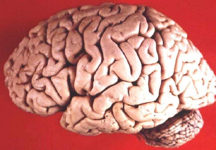
This is the human brain in lateral view. In order to understand its anatomy, we have to understand how such a complex and elaborate structure develops.
The central nervous system of the human begins to develop shortly after gastrulation, the formation of three primordial layers that occurs 16 days post-conception (30 days since the last period). A part of the future skin (ectoderm) begins to thicken, forming a neural plate. The plate then develops a shallow groove that deepens into a neural fold over the next few days.
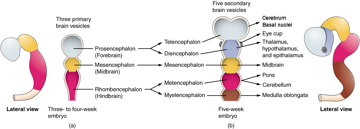
 The edges of this groove fold up and seal together forming a neural tube. This tube then begins to change shape and develop empty rooms called vesicles. The first set of three vesicles are called primary brain vesicles. The second set of vesicles are called secondary brain vesicles and we need to learn the names of these because those will help us understand the anatomy of the adult nervous system.

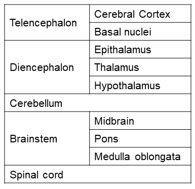
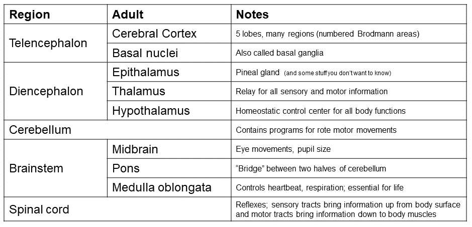
 These subdivisions of the developing brain become adult structures, as shown in the table. Because each of these areas has a different anatomy and different strategy of “brain wiring”, it’s important to understand just a little bit about the development of the brain to understand the anatomy and function of the brain.
For example, the cerebral cortex and basal nuclei, which represent the vast majority of the weight of the human brain, are extensively interconnected and control much of human behavior. The basal nuclei smooth out the activity of the part of cerebral cortex that controls muscles; this smoothing goes wrong in Parkinson disease. Changes in the activity of another part of the basal nuclei is implicated in obsessive-compulsive disorder. We will examine some of the signaling molecules and wiring of this system in Unit 12, and we will describe what goes wrong with signaling in the basal nuclei in the Pathophysiology course.


