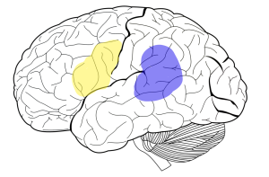
Speech Areas of the Human Brain
Rachel Jessop and Jim Hutchins
Objective
1. Identify the locations and functions of the major speech areas of the brain.
There are many portions of the brain in play when someone is speaking. The prefrontal cortex assists with mouth movement and the auditory cortex helps produce meaningful, understandable language. However, there are two other specialized speech areas that are important to understand when we look at clinical aphasia (language disorders).

The first is Broca’s area, highlighted above in yellow. It was named after the physician who studied its capabilities, Pierre Paul Broca. It is located at the posterior inferior frontal gyrus on the left hemisphere (in most of the population, a small percentage of the population has both speech areas located in the right hemisphere). Broca observed that many of his patients who suffered damage to this area exhibited expressive aphasia – meaning that the patient may know exactly what they want to say, but take great effort to mutter only a few broken keywords of their message.
The second is Wernicke’s area, highlighted in purple. Like Broca’s area, it was also named after the physician who studied it (Carl Wernicke). As seen in the diagram, this functional area is generally located at the left posterior superior temporal cortex. Though this area is also important for speech production, its contribution differs from Broca’s area. Wernicke noted that patients suffering damage to this area exhibited receptive aphasia – meaning that their intended message comes out as a jumbled “word salad”. The patient typically has no idea their speech makes no sense, and though their motor speaking ability is intact, the words lack any meaning.
The following is an example of both aphasias: Say a patient wanted to say “I need a drink of water.”
Someone with Broca’s aphasia would slowly say: “Mmrrrhph… drrrk…. wuuteeeer”
Someone with Wernicke’s aphasia would confidently say: “Why purple should we newspaper okay?”

