Meninges
Rachel Jessop and Jim Hutchins
Objective
1. Be able to identify the cerebral meninges.


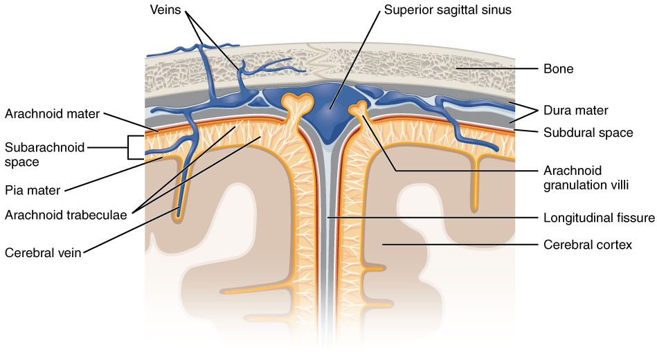
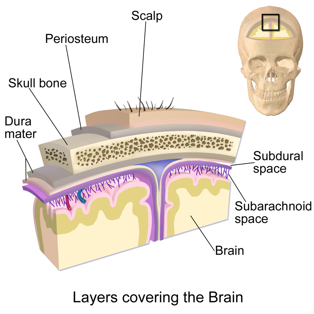
The brain is covered by a three-layered bag. These layers are collectively called the meninges (singular meninx).
From the skull to the surface of the brain, the meninges comprise:
- dura mater (Latin: “tough mother”), a leathery covering;
- arachonoid mater (“spiderweb mother”), which resembles a wet spider web and is normally filled with cerebrospinal fluid (CSF);
- pia mater (“delicate mother”), like a coating of paint on the surface of the brain, which cannot be separated from the substance (parenchyma) of the brain.
How in the world did these meninges get such strange names? They were originally named by a Persian anatomist, Ali ibn Abbas, in the 10th century. There is no Arabic word for “membrane” so he wrote al umm, “the mother”, “the womb”. The European translators rendered this literally into the Latin “mother” instead of figuratively into the original meaning of mother as a womb, swaddling or covering.
The term epidural, as in epidural anesthesia, means “on top of the dura”. Epidural anesthesia is a deadening agent applied on top of (“epi-”) the dura mater. Patients who suffer bleeding into the brain may have blood collecting in the epidural space (between the skull and dura); tearing apart the subdural region (between the dura and arachnoid); or filling the subarachnoid space (between the arachnoid and the pial surface of the brain).

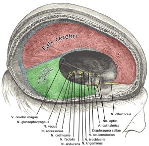
An extension of the dura mater falls in line with the longitudinal fissure to cut the brain midsaggitally and support venous drainage/CSF circulation – it is called the falx cerebri (red). It is connected posteriorly to the tentorium cerebelli (green), which sits superiorly to the cerebellum, compartmentalizing it from the cerebral structures.

