Basal Nuclei (Basal Ganglia)
Rachel Jessop and Jim Hutchins
Objective
1. Understand what major structures make up the basal ganglia, and what functions they are responsible for.
2. Identify disorders of the basal ganglia, especially Parkinson’s and Huntington’s disease.
The basal nuclei (blue structures in the diagram) are essential in the control of movement, and regulation of mood and complex behaviors.
An older name for the basal nuclei is basal ganglia. However, we’ve made a big deal of telling you that a “ganglion” is a cluster of nerve cells outside the CNS, and the basal nuclei are definitely part of the CNS, so we’d be hypocritical and confusing to call them “ganglia”, right? That doesn’t stop some neuroscientists from calling it that, but it should.
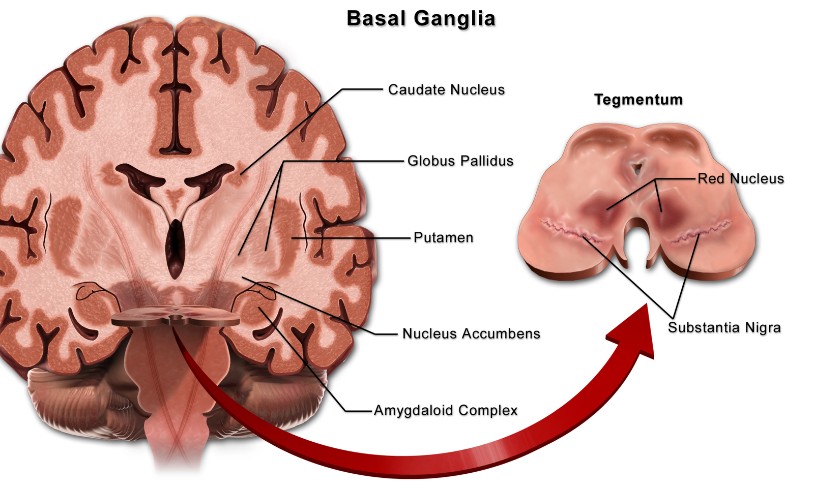
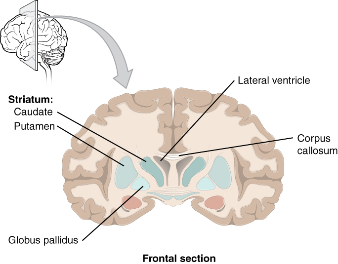 There are three structures that make up the basal nuclei:
- caudate nucleus (Latin: “tail”; remember “caudal” as an anatomical direction means “toward the tail”);
- putamen (Latin: “shell” or “crust”);
- globus pallidus (Latin: “pale globe”).
The basal nuclei receive dopamine from the substantia nigra of the midbrain; when the nigra dies, the lack of dopamine causes the movement disorders of Parkinson Disease. In about half of Parkinson patients, mental disorders also result. Another degenerative disease of the basal nuclei, Huntington Disease, also results in mental disorders. Obsessive-compulsive patients show abnormalities in the caudate nucleus.
