Visual Pathways
Rachel Jessop and Jim Hutchins
Objective
1. Trace the visual system from perception to processing.
Both your right and left eye receive photons emanating from a single point in visual space, in most cases. There is a small area of vision that only activates photoreceptors in the right eye because it is blocked by your nose and a small area of vision that only activates receptors in the left eye for the same reason. These two symmetrical areas, which activate only one eye, are called the monocular crescents because of their shape and characteristics.
For every other location in the visual world, any point is represented twice. You can confirm this by looking at something more-or-less in front of you and covering one eye at a time. Its position seems to shift slightly (more about this later) but it’s still there.
The retina nearest the nose is called the nasal retina and the retina nearest the temples is called the temporal retina. The dividing line between nasal and temporal retina is the midpoint, or bottom, of the foveal pit.
Points in visual space to the left of the midline are represented in the nasal retina of the left eye and in the temporal retina of the right eye.
Points in visual space to the right of the midline are represented in the temporal retina of the left eye and in the nasal retina of the right eye.
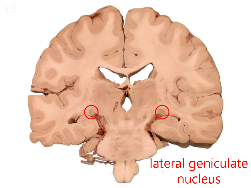
For reasons that will soon become clear, it’s important that the LGN and visual cortex on one side contains a complete representation of the opposite side of the world. Thus, we need to join the axons coming from the left nasal retina with axons coming from the right temporal retina. These axons will all go to the right LGN.
Similarly, we need to join axons coming from the left temporal retina with axons coming from the right nasal retina and send them all to the left LGN.
This sorting process occurs at the optic chiasm. Axons from the left nasal retina cross the midline. Axons from the right nasal retina cross the midline. Axons from the temporal retina stay on the same side.
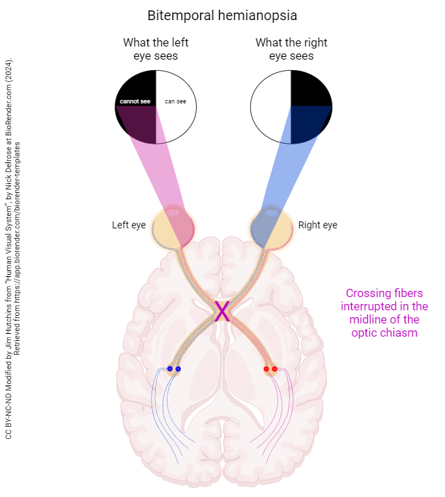
If a lesion, such as a tumor of the pituitary gland, disrupts only the crossing fibers (i.e. if it lies in the center of the optic chiasm), then only the temporal retinal axons can send information to the brain. These retinal axons convey information from the nasal world on both sides. Because we can only observe what is going on with the visual fields, not what is going on with the fibers themselves, a phenomenon known as bitemporal hemianopsia (“two temples half not seeing”) results. Information from the temporal world, which is carried by nasal ganglion cell axons, is no longer able to be transmitted to the brain and is not consciously perceived.
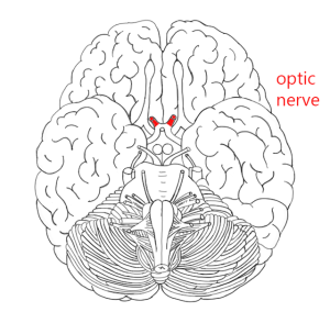 The optic nerve carries all the axons from the retinal ganglion cells. Recall that we saw this structure as the optic nerve head in the retina producing the blind spot in visual perception.
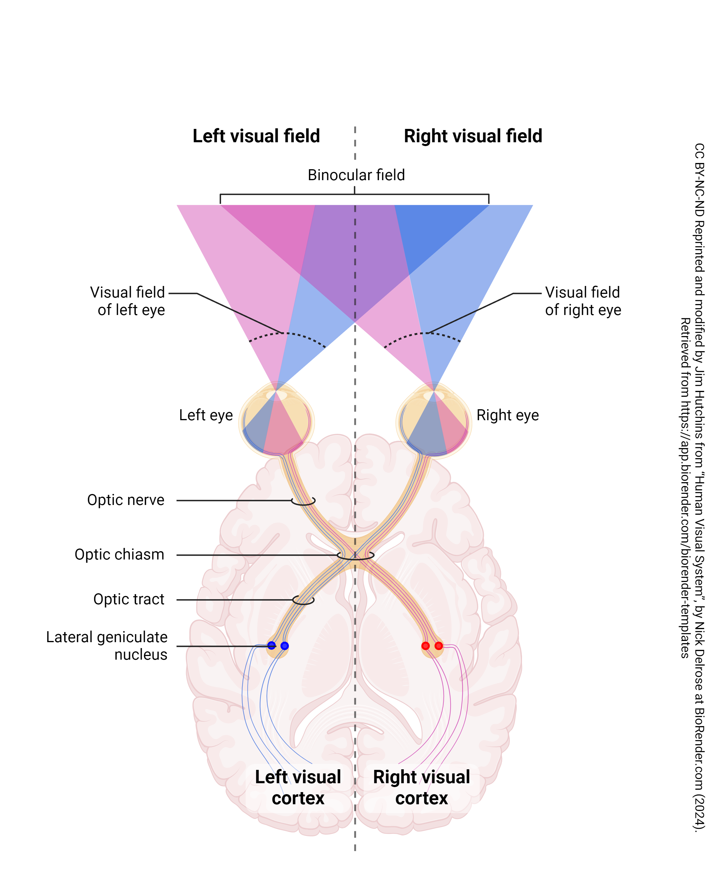
Half of the axons from each eye, representing the right temporal retina and the left nasal retina, are picking up information from the left visual world. These are represented by pink shading and pink axons.
Half of the axons from each eye, representing the right nasal retina and the left temporal retina, are picking up information from the right visual world. These are represented by blue shading and blue axons.
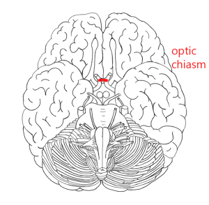
Thus, at the optic chiasm (the Greek letter chi, χ, looks like this structure), the axons from the nasal half of each retina cross to join those that carry information from the same part of the visual field.
When something is on the same side in neuroscience, we call it ipsilateral. When it’s on the opposite side, we call it contralateral.
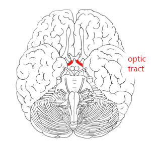 The optic tract, then, contains two complete representations of the contralateral visual field, one from ganglion cell axons originating in the right eye and one from ganglion cell axons originating in the left eye.
For example, the right optic tract contains axons from the left (contralateral) nasal retina ganglion cells and the right (ipsilateral) temporal retina ganglion cells.