The Endomembrane System of Neurons
Exocytosis in Neurons
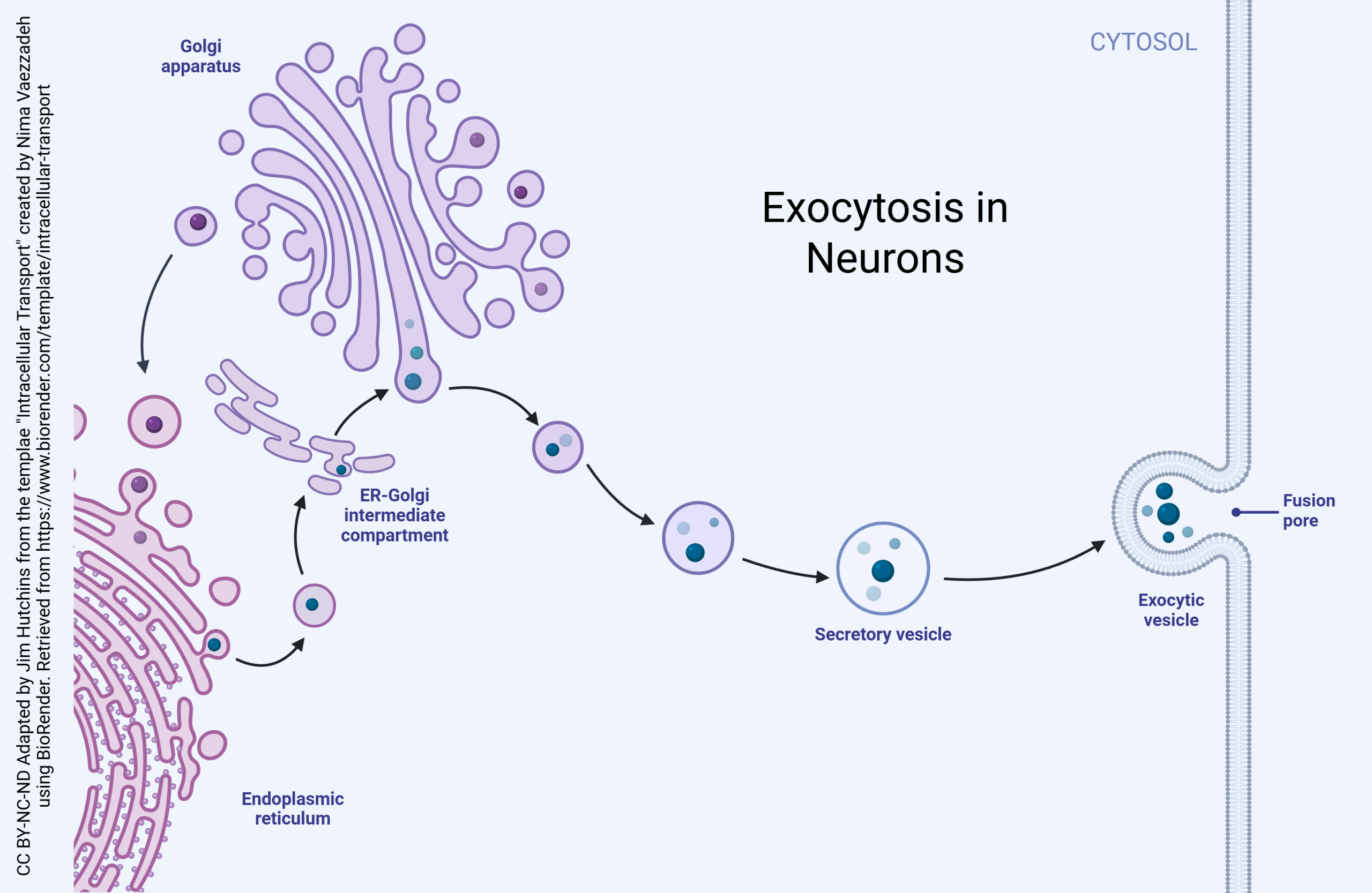
Exocytosis in neurons takes on a critical role because most neurons communicate with each other using chemical extracellular signals called neurotransmitters. The mechanism of chemical neurotransmitter release is covered elsewhere. This section will discuss exocytosis of proteins.
Proteins are made in a macromolecular machine called a ribosome. Ribosomes stitch together amino acids, the monomers of proteins, using the messenger RNA code as a template. The details of this process are explained elsewhere.
Proteins for export or proteins that will be expressed on the cell surface (such as pumps, channels, or neurotransmitter receptors) are made in the rough endoplasmic reticulum (rough ER), an organelle consisting of ribosomes attached to the endomembrane system. Protein modification then begins.
First, proteins travel into the ER-Golgi intermediate complex (ERGIC). Vesicles containing proteins bud off of the rough endoplasmic reticulum and take on an outer layer (coat) of coat protein complex II (COPII). These vesicles then fuse to form the cis face of the Golgi.
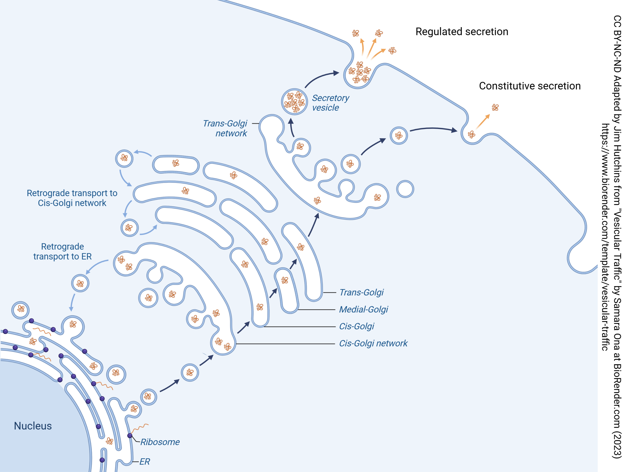
(The Golgi apparatus, also called the Golgi complex, forms at the cis face and breaks apart at the trans face. These faces are named for their relationship to the ER, with the cis face closer to the ER and the trans face further away.)
When there is a need to move proteins in the opposite direction, from the cis face of the Golgi to the rough ER, they are coated with coat protein complex Ia.
-
Carbohydrate Synthesis Stage: A transition occurs when a cis cisterna loses the ability to receive COPII vesicles while acquiring the ability to receive glycosylation enzymes and sugar nucleotide transporters in COPIb vesicles. These processes convert the cis cisterna into a medial cisterna. Medial cisternae mature into trans cisternae. Both medial and trans cisternae are involved in carbohydrate synthesis, with early-acting enzymes concentrated in medial cisternae and late-acting enzymes in trans cisternae. Cisternae at the carbohydrate synthesis stage exchange material with one another via COPIb vesicles.
-
Carrier Formation Stage: A transition occurs when a trans cisterna loses the ability to receive COPIb vesicles, and subsequently loses the ability to produce COPIb vesicles while acquiring the ability to produce clathrin-coated vesicles. The timing of secretory vesicle formation and the ultimate fate of carrier formation cisternae are still poorly understood. ER membranes attached to trans and/or TGN cisternae probably function in direct lipid transfer between the organelles (Hanada et al. 2009). This diagram depicts the mammalian TGN as the last cisterna in the stack.
Regulated and Constitutive Secretion in Neurons
Pellentesque sed venenatis purus. Ut ullamcorper, dolor ut lobortis condimentum, turpis lectus porta ipsum, vel mattis felis sem vitae turpis. Donec aliquam non nisi sagittis ultricies. Duis pretium dictum metus et scelerisque. Duis eu urna iaculis nisl eleifend cursus a quis augue. Proin vel massa purus. Interdum et malesuada fames ac ante ipsum primis in faucibus. Phasellus fermentum est at enim auctor tristique. Curabitur vel efficitur metus. Quisque quis turpis augue. Ut vel erat erat. Fusce quis malesuada sapien, et tincidunt neque. In diam tellus, vulputate vel auctor eu, vestibulum in mauris. Sed et velit orci. Mauris facilisis tellus a placerat finibus. Nullam scelerisque, velit eget blandit mollis, eros erat dignissim arcu, eu luctus dui mi id est.
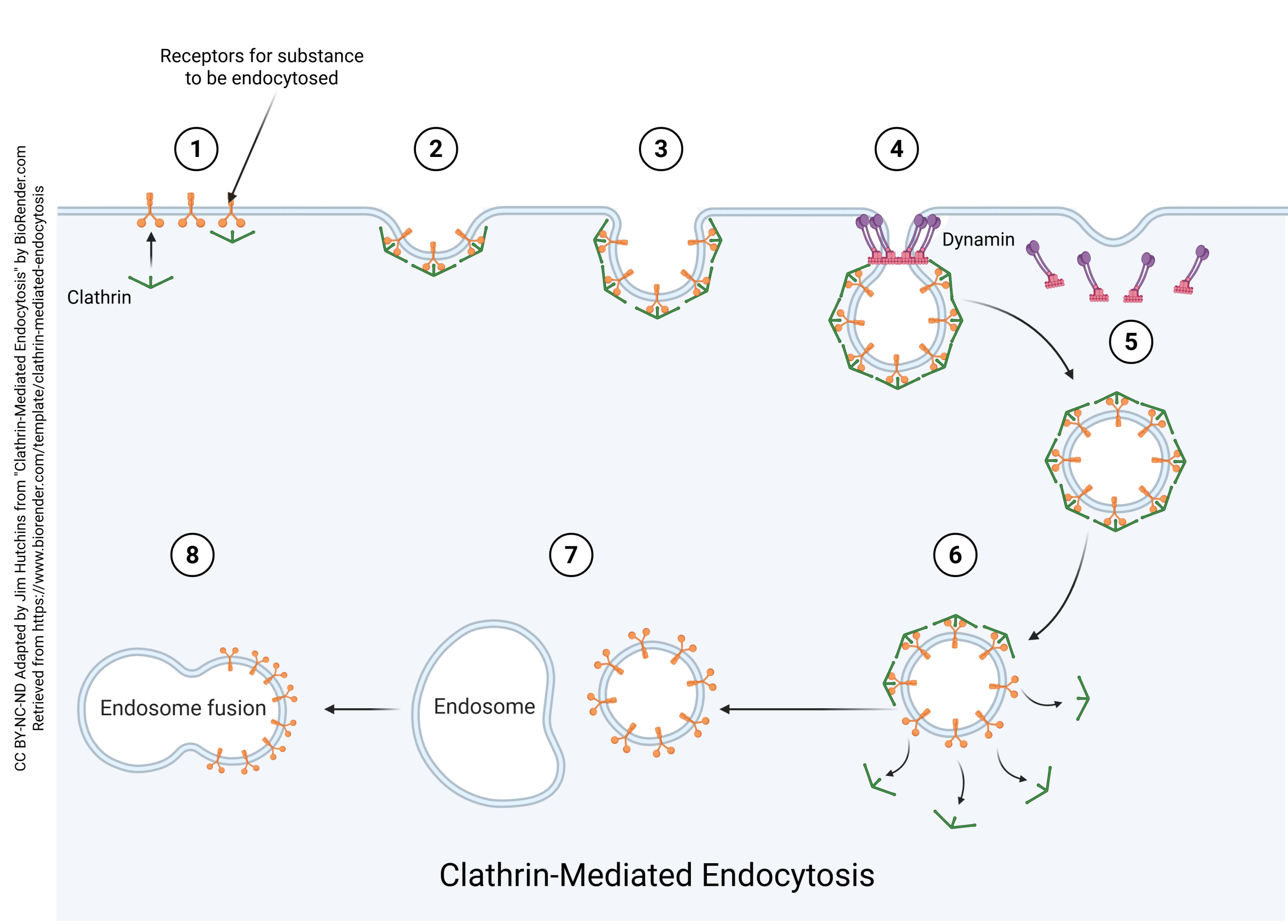
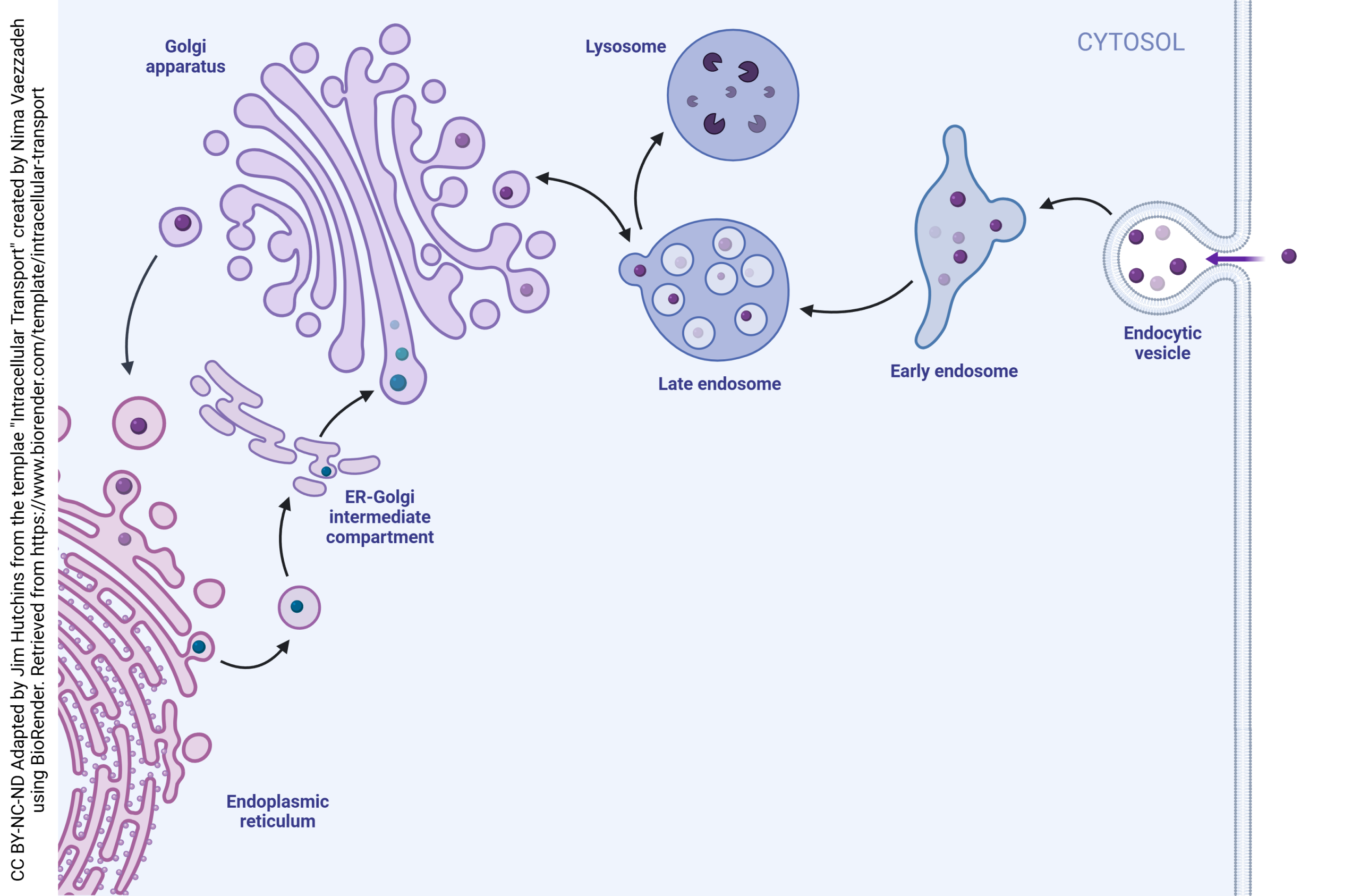
Endocytosis in Neurons
Sed quis libero ut ex luctus dapibus. Donec nisl leo, cursus sagittis nisl non, bibendum placerat dui. Cras ut nulla ac nulla condimentum tempus quis ut mi. Maecenas eros eros, finibus vitae lacinia ac, sagittis sit amet turpis. Suspendisse iaculis laoreet ultrices. Praesent sollicitudin quis nunc et semper. Donec eros elit, posuere id sodales vitae, vulputate at erat. Morbi venenatis condimentum varius. Cras vitae augue varius, mollis sem in, facilisis tortor.
Curabitur luctus tempor laoreet. Donec sed libero quis ante hendrerit accumsan eu nec tellus. Aliquam erat volutpat. Nam at sapien enim. Maecenas interdum urna sit amet tempus posuere. Sed sem justo, porta vel urna a, dictum dignissim magna. Maecenas varius feugiat elit id sagittis. Nam fringilla viverra turpis eget viverra. Ut quis rhoncus leo. Donec fermentum ultricies ligula, eget dictum velit finibus in. In hac habitasse platea dictumst. Aenean commodo sollicitudin nibh vel blandit. Aenean rhoncus, risus vitae sollicitudin sodales, ligula elit aliquam ligula, ac viverra sapien nisl sit amet turpis. Proin sagittis, nulla sed porta pretium, sem purus consequat sem, quis auctor magna sapien et diam.
Pellentesque habitant morbi tristique senectus et netus et malesuada fames ac turpis egestas. Suspendisse id mauris id nisi iaculis imperdiet. Pellentesque in lacus eros. Nunc bibendum, lacus quis lacinia commodo, nibh nisi porttitor ligula, id blandit odio nunc sit amet metus. Vivamus sit amet quam vitae lectus dictum iaculis ut id ipsum. Ut non ex id nisi pulvinar tincidunt. Aenean quis massa sagittis risus efficitur vulputate. Curabitur tincidunt, est sed congue laoreet, neque enim auctor metus, in euismod tortor quam sit amet nunc. Donec feugiat porttitor sem vel interdum. Cras commodo laoreet urna non elementum. Fusce in risus vulputate, sodales ligula sed, porttitor purus. Integer aliquet in velit vel fringilla.