The Cytoskeleton of Neurons
Tess Johnson; Caleb Bevan; Jackson T. Anderson; and Jim Hutchins
Chapter under construction. This is the first draft. If you have questions, or want to help in the writing or editing process, please contact hutchins.jim@gmail.com.
Skeletons provide structure and support. Similar to humans, cells also have a skeleton called a cytoskeleton. This helps cells hold their shape and allows for proper functioning. The cytoskeleton also has many other functions, such as supporting the plasma membrane, allowing cell movement, maintaining the organization of organelles, and creating tracks for the transport of vesicles.
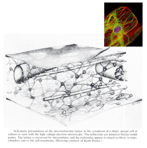
The cytoskeleton is made of three protein fibers: microfilaments, intermediate filaments, and microtubules.
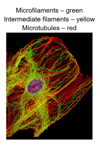
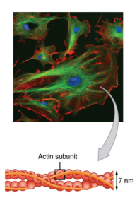
Microfilaments are very narrow and are about 7 nm (nanometers) in diameter. These are linked monomers of actin, which is a type of protein. Microfilaments have a double helix, similar to the structure of DNA, and each of the two ends is unique from each other. Another common name for microfilaments is actin filaments.
Microfilaments have many important roles. Microfilaments act as tracks for myosin, a motor protein. This allows for the movement of proteins on the surface of the cell. The transport of vesicles is also facilitated through microfilaments. The movement of the cell is created from microfilaments assembling and disassembling quickly. The structure of the cell is also provided, mainly at the edge of the cell, and connector proteins link microfilaments to the plasma membrane. This helps maintain the shape and structure of the cell.
Intermediate filaments are 8-10 nm in diameter and are made of fibrous protein strands that are woven together. There is a variety of proteins that make up these filaments, and as a result of this woven structure, they can withstand tension. Intermediate filaments also help to maintain the shape of the cell as well as provide structure. The nucleus and organelles are also anchored into place with intermediate filaments.
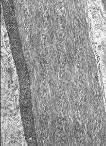
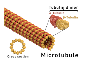 Microtubules are the largest of the protein fibers that make up the cytoskeleton. These are about 25 nm in diameter and are made of tubulin proteins. These proteins form hollow tubes, similar to a hose. Each microtubule has two unique ends, as well as two subunits (α and β). Microtubules are able to grow and shrink and are able to resist compression forces. This helps maintain the structure of the cell. Microtubules can also be tracks for the motor proteins kinesin and dynein. These are used to transport vesicles in the cell, which is important for releasing neurotransmitters.
There are also some structures of the cytoskeleton that extend from the main area of the cell. These are mostly used for cell movement and support.
Flagella are long, hair-like structures that originate on the surface of the cell and project outwards.
Cilia are similar to flagella but are much shorter and are found on main surfaces for the movement of material. The force that creates cilia movement is created by dynein protein moving on microtubules.
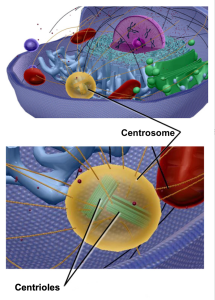
 Centrioles are held together by proteins and organize microtubules into cylinders of nine triplets. When two centrioles meet at a 90° angle, pericentriolar material surrounds them, creating microtubule anchoring sites.
Centrosomes are the main structure that organizes organelles within the cell.

Media Attributions
- Cytoskeleton 1 © Keith R. Porter is licensed under a All Rights Reserved license
- Cytoskeleton 2 © NIH Image Gallery is licensed under a CC BY-NC (Attribution NonCommercial) license
- Cytoskeleton 3
- GFAP intermediate filaments © Don W. Fawcett is licensed under a All Rights Reserved license
- Cytoskeleton 5
- Cytoskeleton 6
- Cytoskeleton 7
- Cytoskeleton 8
- Cytoskeleton 9
