Synaptic Pruning
Aliyah Grijalva and Jim Hutchins
Chapter under construction. This is the first draft. If you have questions, or want to help in the writing or editing process, please contact hutchins.jim@gmail.com.
Synaptic Pruning

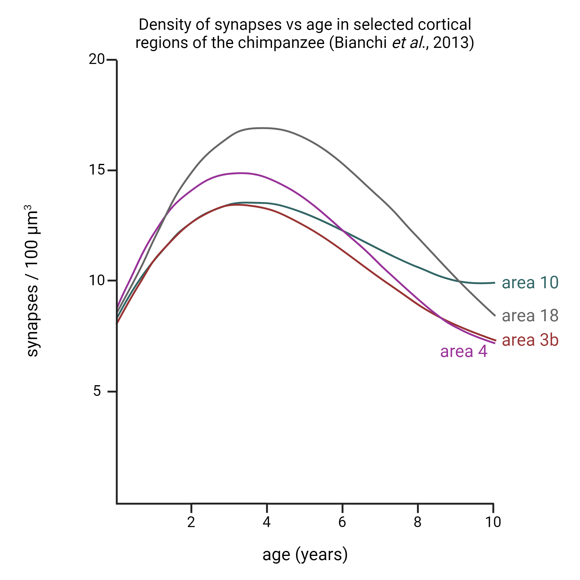
All areas of the brain overproduce synapses. In general, between 1/6 and 1/3 of the synapses in the brain are removed during normal development in a process called synaptic pruning.
The process of synaptic pruning involves removing “weak” or inappropriately formed synapses. In general, about one and half times the number of synapses reflected in the adult are made; therefore, about 1/3 of the inchoate synapses are eliminated during the process of development. Synaptic pruning mechanisms play a large role in this process. Just as pruning or removing fruit from a tree can, in the end, help the tree be more effective and produce more fruit, removing synapses will make the brain more effective as well.
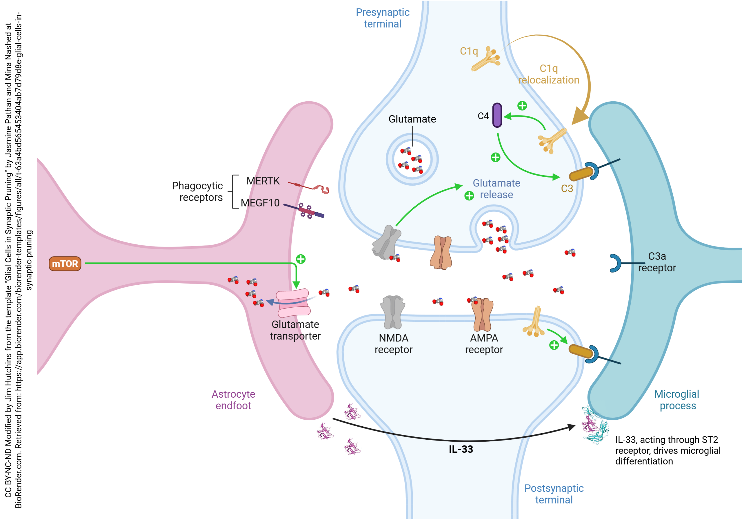
 This diagram shows the currently hypothesized role of astrocytes and microglia in synaptic pruning.
Signal Transducer and Activator of Transcription-3 (STAT3) is a transcription factor which triggers metabolic switching in astrocytes. When STAT3 is turned on in astrocytes, they shut down neuronal mitochondria and destroy glutamatergic synapses. STAT3 also up-regulates the glutamate transporter, as shown.

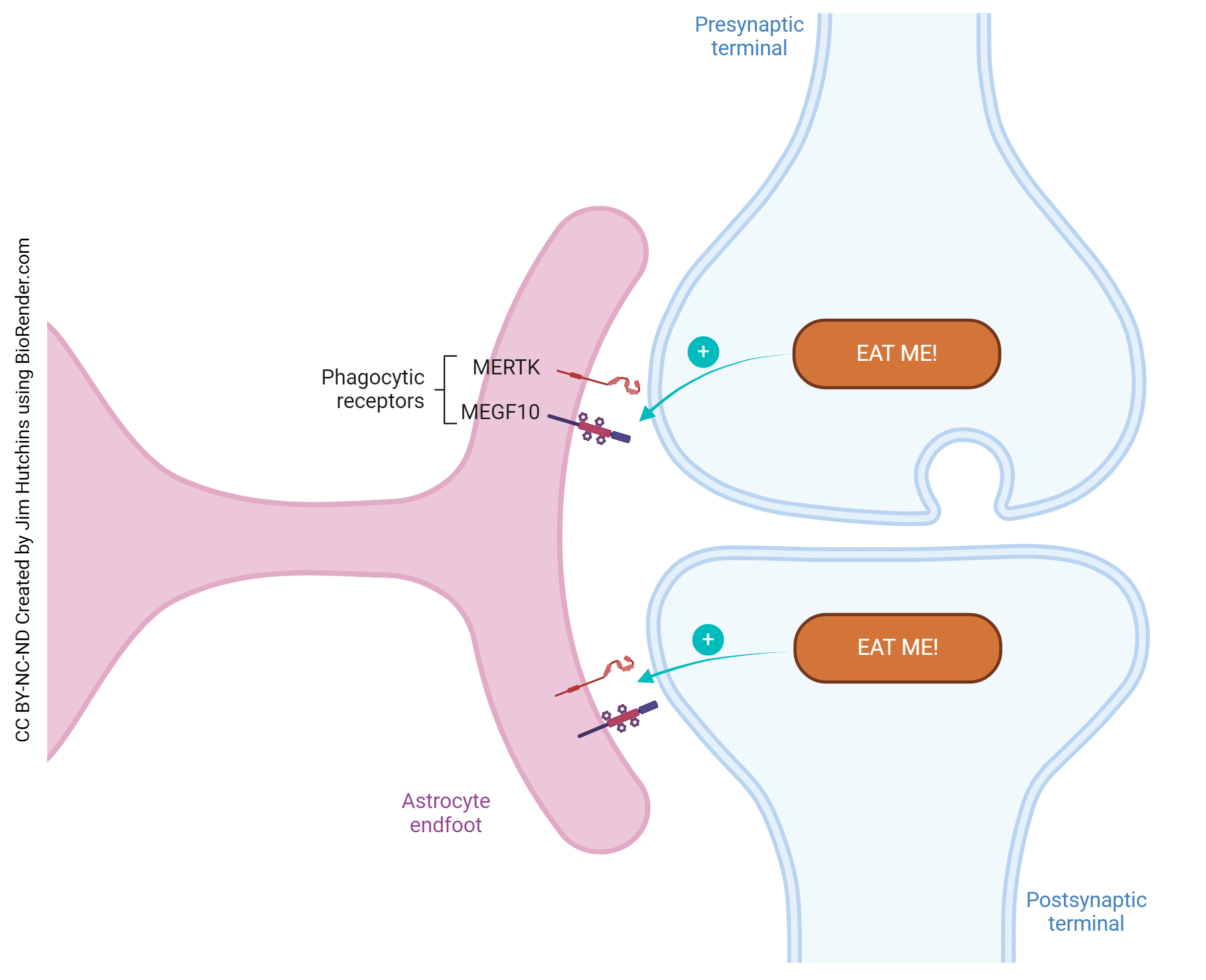
MERTK and MEGF10 act as phagocytic receptors: with their help, the astrocyte engulfs the soon-to-be-pruned synapse and begins the process of phagocytosis.
Astrocytes release transforming growth factor beta (TGF-β). TGF-β, in turn, triggers the complement cascade, particularly complement C1q and C3 in neurons.
Complement C1q, when relocated in neurons, induces the neuron to express complement C3 on the cell surface. C3 labeling of neurons, like C3 labeling of targeted bacteria, is called opsonization. Opsonization is the process by which cells are tagged for destruction. Neurons which express complement C3 are targets for the microglia, which act as macrophages in the brain.
Astrocytes also release interleukin 33 (IL-33), which activates phagocytosis, migration, and inflammatory responses in microglia.
It is now clear that in the 1988 tree shrew study, GFAP was staining the astrocytes, which might have been participating in the pruning of synapses in the developing tree shrew LGN, while vimentin may have been staining the microglia involved in the process of synaptic pruning.
Media Attributions
- Synaptogenesis © Serena Bianchi, Cheryl D. Stimpson, Tetyana Duka, and Chet C. Sherwood adapted by Jim Hutchins is licensed under a All Rights Reserved license
- Glial Cells in Synaptic Pruning © Jasmine Pathan, & Mina Nashed adapted by Jim Hutchins is licensed under a CC BY-NC-ND (Attribution NonCommercial NoDerivatives) license
- Astrocyte phagocyte receptors in synaptic pruning © Jim Hutchins is licensed under a CC BY-NC-ND (Attribution NonCommercial NoDerivatives) license

