Formation of the Neuromuscular Junction
Caleb Bevan; Tess Johnson; Jackson T. Anderson; and Jim Hutchins
Chapter under construction. This is the first draft. If you have questions, or want to help in the writing or editing process, please contact hutchins.jim@gmail.com.
What is a Neuromuscular Junction?
The term neuromuscular junction refers to the synapse between a motor neuron and a skeletal muscle fiber. When this junction gets sent a signal from your lower motor neuron, it will contract or tense up. This is how you contract every skeletal muscle in your body. This is needed for any voluntary muscular movements in your body, so without these junctions, you wouldn’t be able to control your movement at all. This junction releases a neurotransmitter called Acetylcholine which has the primary use of muscle contraction. Each time you relax your muscle, Acetylcholinase is released to help your muscles stop contracting.
Axon Growth
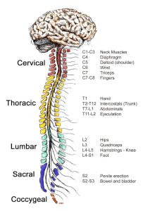
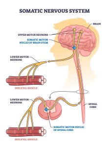
Your brain sends signals down your spine from your upper motor neurons located in the cerebral cortex. Down your spine each segment has a certain area to be controlled. These segments are called lower motor neurons. During muscle development, motor neurons extend from the spinal cord toward developing muscle fibers in a vertebrate organism. These developing muscle fibers are being grown throughout the whole body. The motor neurons are guided by chemical cues and proteins such as netrins, ephrins, and semaphorins, which help them navigate to the correct muscle or area. The axon also uses a growth cone made up of several membranous organelles on the tip of the axon to help guide it through the environment. This helps the axon find that muscle target and connect to right place.
Initial Contact
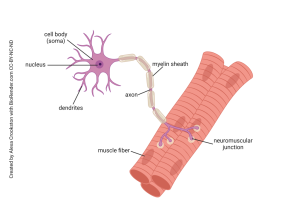
When the motor neuron’s growth cone reaches the muscle fiber this is called initial contact. The muscle secretes basal lamina proteins into the synaptic cleft. Then it binds to its receptor, MuSK (Muscle-Specific Kinase), on the surface of the muscle cell. This triggers intracellular signaling, which is essential for synapse formation. Activation of MuSK leads to the clustering of nicotinic acetylcholine receptors (AChRs) at the contact site. Every muscle has to have this receptor in order for the muscle to be able to function with the help of the brain. This is used for the contraction and release of muscles using an electric current using the acetylcholine. Another protein, rapsyn, helps stabilize these AChR clusters in the muscle membrane. Rapsyn also helps to link the receptors to the cytoskeletal complex as a sort of anchor to keep all of the AChR attached to the muscle cell. The areas on the muscle membrane that are not in contact with the neuron overtime lose their AChRs which means that most of the receptors are concentrated at the synapse for the highest effect.
Maturation
One the muscle fiber is connected and in position it can start to develop and mature. One of the first things they do is form junctional fold to increase the surface area for more contact of the neuron and more space for the AChRs. The neuron starts to release ACh to allow the muscle to start responding to those signals send from the lower motor neurons. At the start motor neurons may be connected to multiple muscle fibers but overtime the ones with the weaker connections will be eliminated because of a process called synaptic pruning. The end result of the pruning is leaving just a single motor neuron per muscle fiber. This helps with energy efficiency and higher control over the muscles in your body. When you see baby’s spending hours and hours just moving their muscles or working on motor movements this is what is happening in their body. This motor control only happens over hours and hours of practice with the muscles in your body.
Functionality
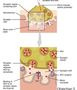
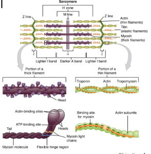
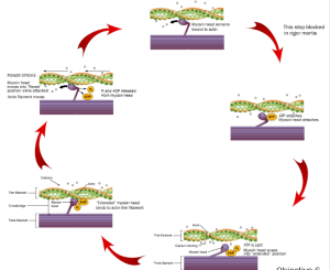
The final function of the neuromuscular junction is to provide voluntary movement to the body. This is something we use all day everyday because of the daily use of the muscles in our body. Your brain sends a signal down to your spine which then connects to a motor neuron directly attached to the muscle that reaches your synapse. This causes a nerve impulse to go into the synapse to cause vesicles to release Acetylcholine. Acetylcholine then goes and binds to the nicotinic acetylcholine receptors where there is an electrical change in the muscle using a tube through a muscle called a T-Tubule. The T-Tubule connects to sarcoplasmic reticulum which is a calcium storage for your body. It releases the calcium because of the change in voltage. The calcium then goes on to bind to troponin to create an area for the myosin head to bind called the tropomyosin. The contraction of your muscle is within something called a sarcomere it uses fibers called actin and myosin which are proteins. The Sarcomere pulls the fibers in to provide the muscles with contraction. Myosin reaches up and grabs actin to shorten the muscle. To actually contract the myosin head binds to the tropomyosin inside of the sarcomere to pull the sarcomere closer together which causes the contraction. The only way to release the contraction is to use ATP (Adenosine Triphosphate) to use its stored energy. This then splits the ATP into ADP (Adenosine Diphosphate) with an extra phosphate ion near the site. These ions are then transported where they can be combined again for future use. This is a cycle labeled in the image below of the muscle contraction and the use of ATP with the reaction. Every single time we contract any of our muscles this is what happens. There is tons of things that go on in our body even though it is a very simple task.
Media Attributions
- BrainSpinalCordlabld
- Somatic nervous system with human brain impulse to muscle outlin
- Motor neuron © Alexa Crookston is licensed under a CC BY-NC-ND (Attribution NonCommercial NoDerivatives) license
- Screenshot 2024-11-18 105740
- Thick and Thin Filaments © Betts, J. Gordon; Young, Kelly A.; Wise, James A.; Johnson, Eddie; Poe, Brandon; Kruse, Dean H. Korol, Oksana; Johnson, Jody E.; Womble, Mark & DeSaix, Peter is licensed under a CC BY (Attribution) license
- Cross-bridge cycle © Betts, J. Gordon; Young, Kelly A.; Wise, James A.; Johnson, Eddie; Poe, Brandon; Kruse, Dean H. Korol, Oksana; Johnson, Jody E.; Womble, Mark & DeSaix, Peter adapted by Jim Hutchins is licensed under a CC BY (Attribution) license
