Determining Orientation in the Development of the Neural Tube
Jim Hutchins
Chapter under construction. This is the first draft. If you have questions, or want to help in the writing or editing process, please contact hutchins.jim@gmail.com.
The role of Hedgehog signaling
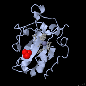
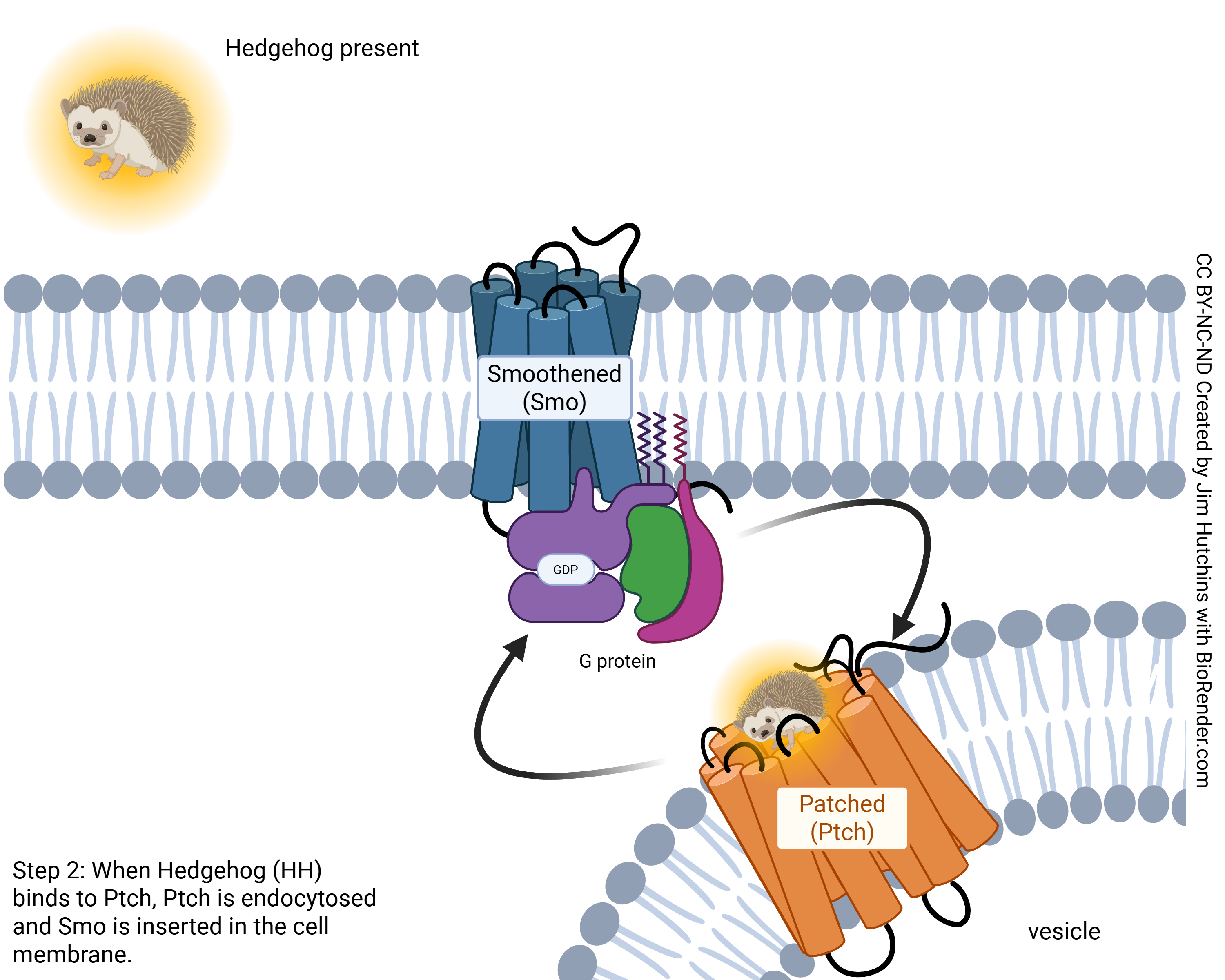
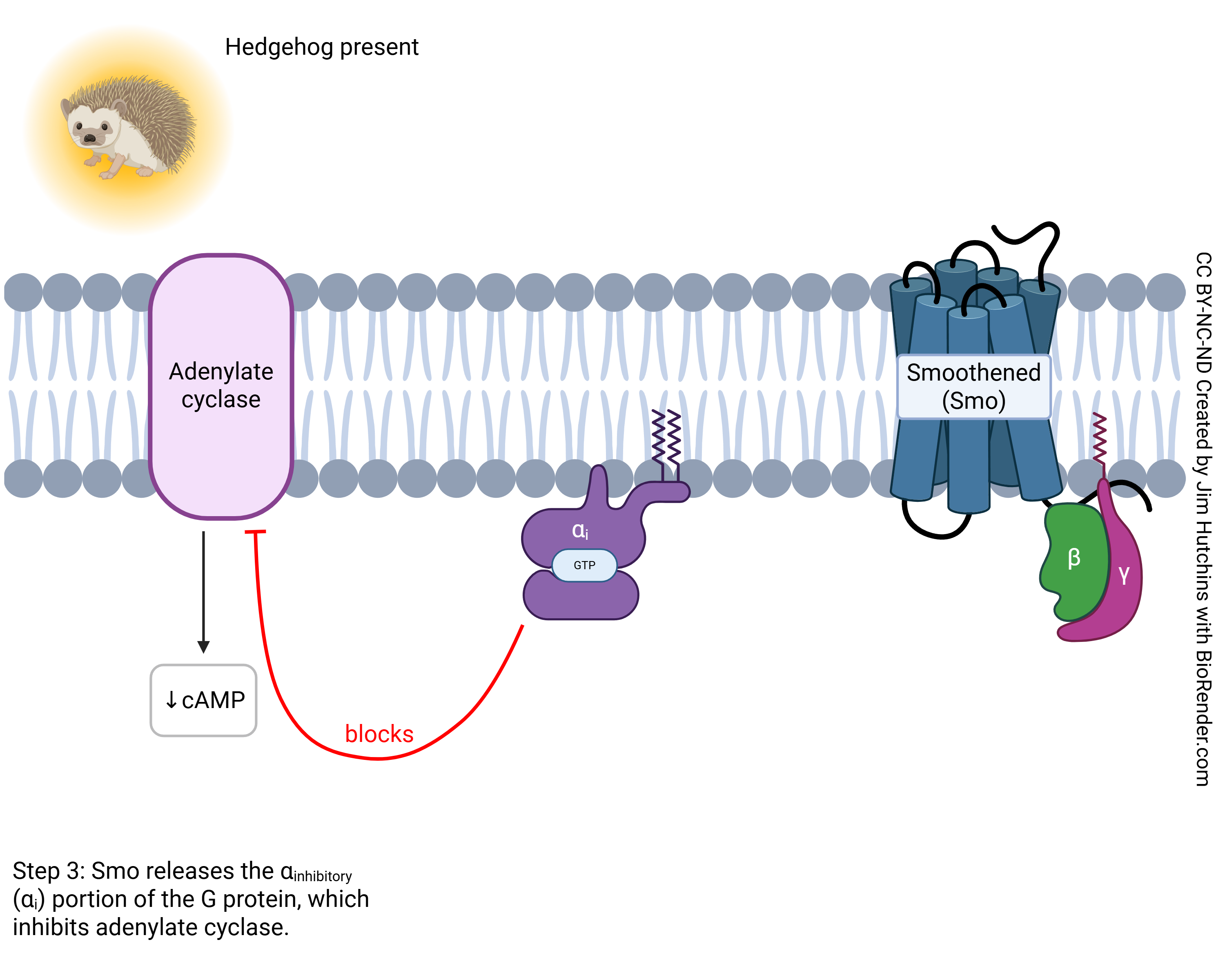
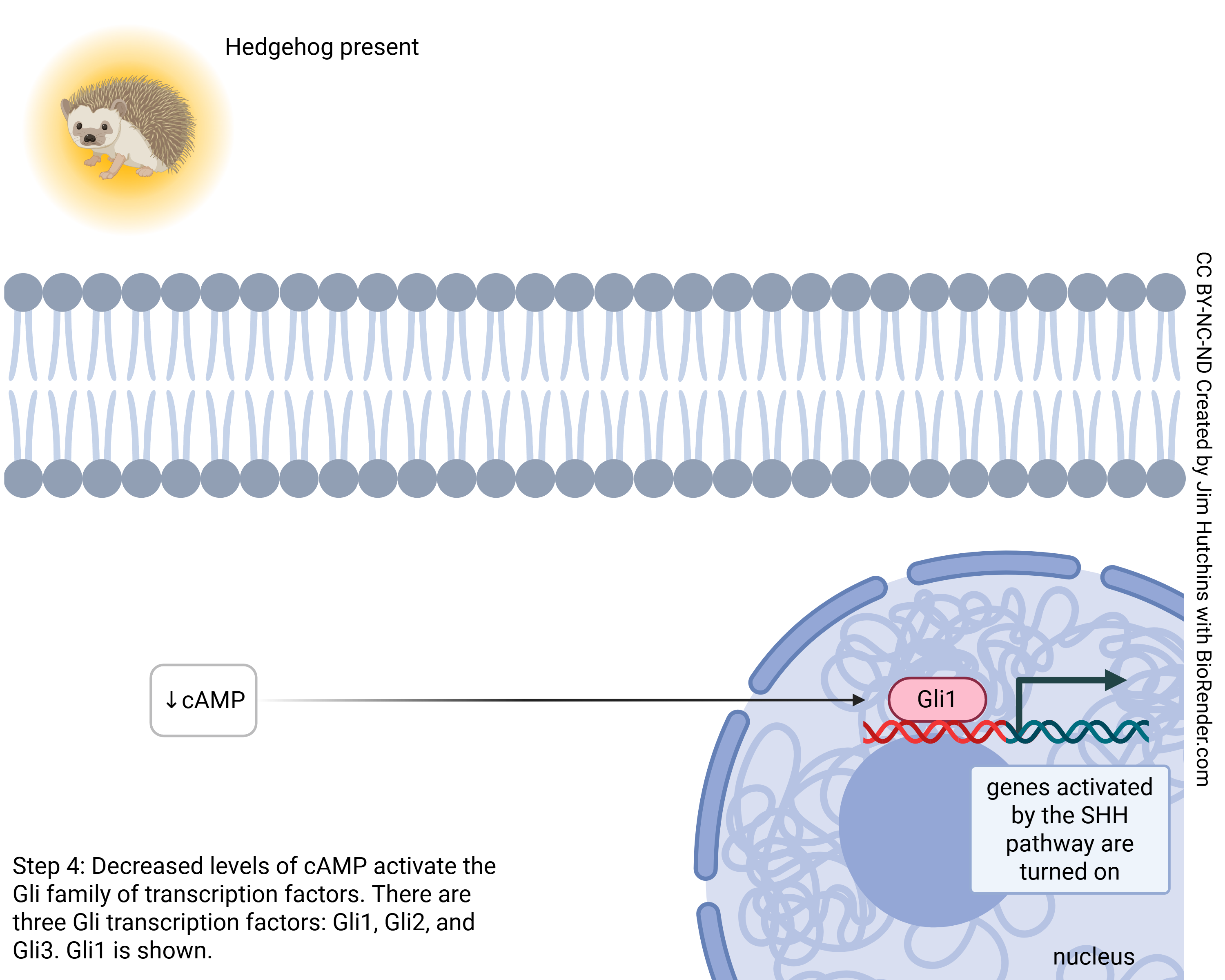
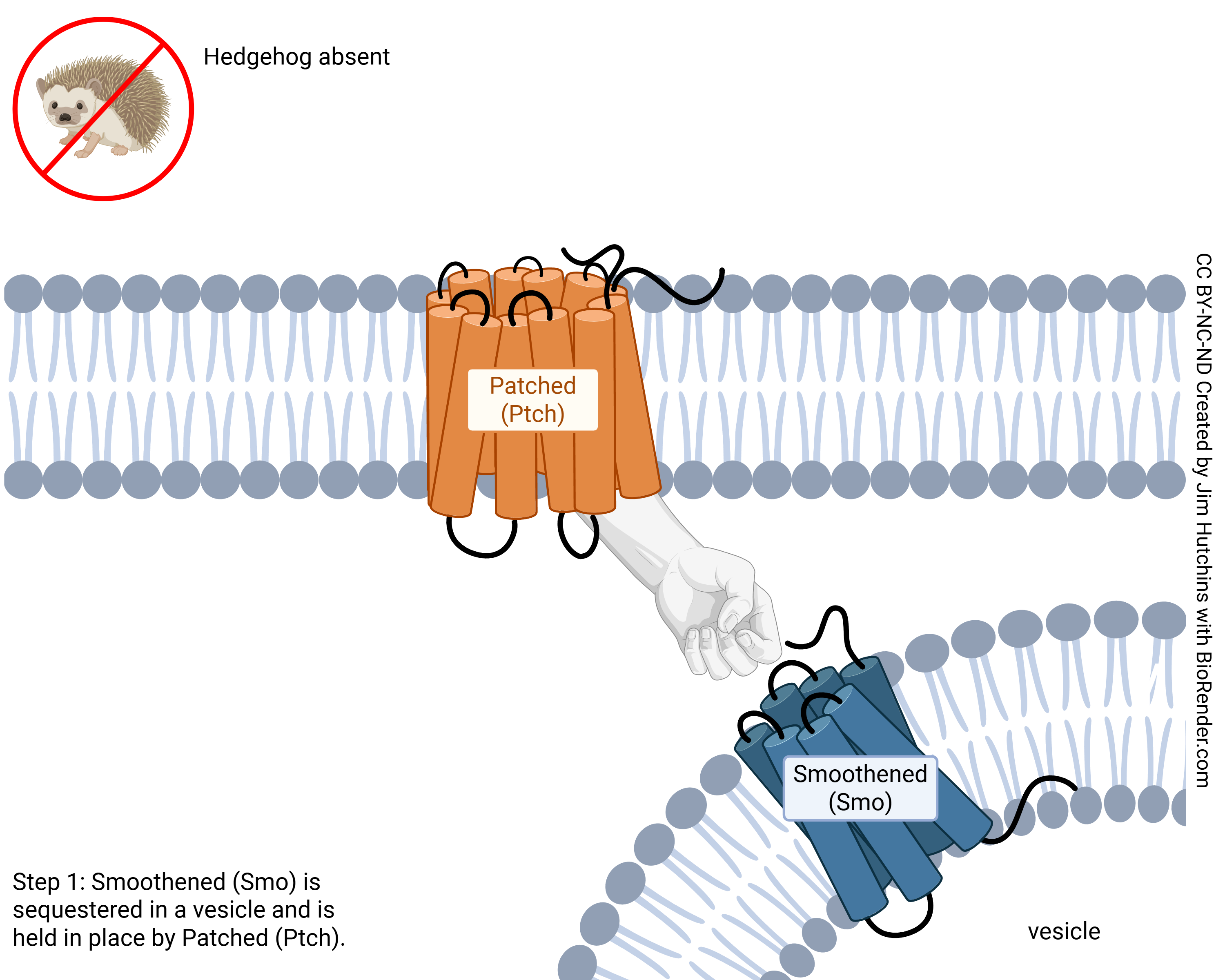 In the absence of a Shh signal, a 12 transmembrane receptor protein called Patched blocks the function of Smoothened (Smo), a seven-pass transmembrane protein, by keeping it sequestered in an intracellular vesicle (Figure 3)[16]. When Shh binds to Patched, inhibition of Smo by Patched is relieved. Patched becomes endocytosed, and Smo translocates to the cell surface. In vertebrates, Smo localizes to the surface of the primary cilium, initiating a signaling cascade that leads to the activation of Gli transcription factors [8]. Present in both the nucleus and cytoplasm, there are three of these regulatory proteins (Gli1, Gli2, and Gli3). Following Shh signaling, all three proteins can act as transcriptional activators of Shh target genes. Gli3, however, can act as both an activator and repressor; in the absence of Shh signaling, Gli3 is cleaved by the proteasome, and its truncated form accumulates in the nucleus where it represses transcription of Shh-responsive genes (Figure 3) [16].
https://proteopedia.org/wiki/index.php/Sonic_Hedgehog
Randi Woodbeck, Michal Harel, Joel L. Sussman, David Canner, Riley Hicks, Andrea Gorrell, Alexander Berchansky
Media Attributions
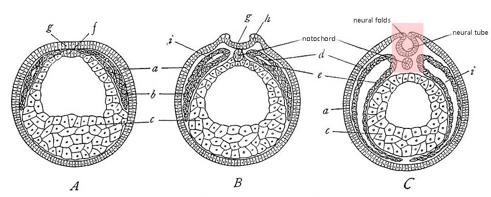
- Amphibia formation of the neural tube © Libbie Henrietta Hyman is licensed under a Public Domain license
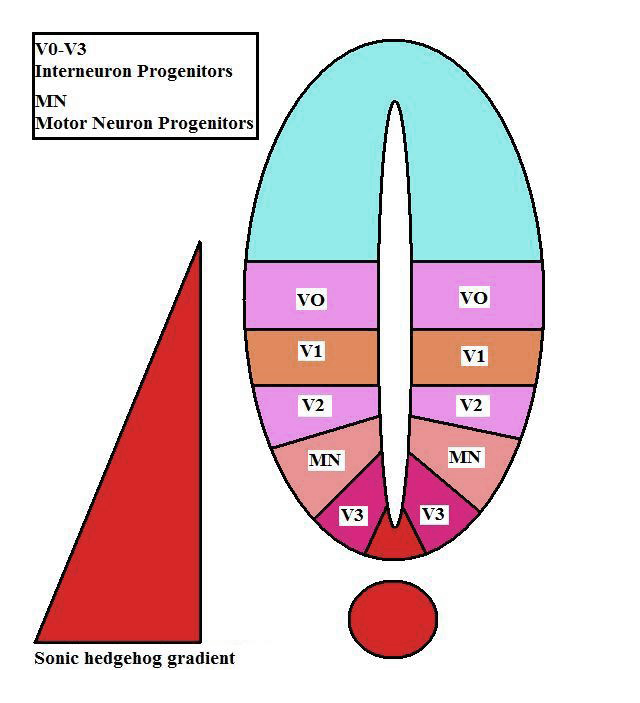
- Neural Tube Progenitor Domains desaturated © Catcasillas is licensed under a CC BY (Attribution) license
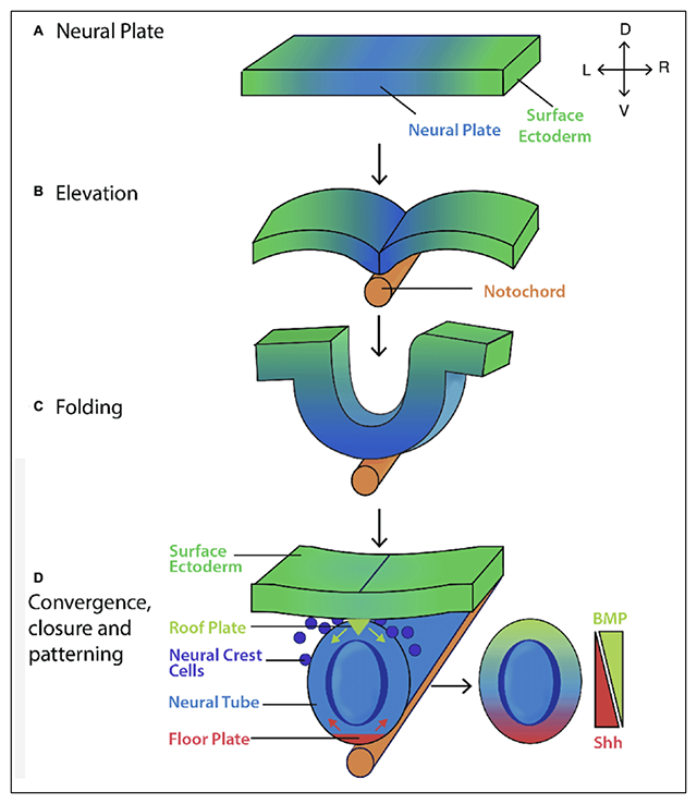
- Formation and patterning of the mouse neural tube desaturated © Rachel A. Shparberg, Hannah J. Glover, Michael B. Morris is licensed under a CC BY (Attribution) license
- Sonic Hedgehog © Randi Woodbeck, Michal Harel, Joel L. Sussman, David Canner, Riley Hicks, Andrea Gorrell, Alexander Berchansky is licensed under a CC BY-SA (Attribution ShareAlike) license
- Hedgehog signaling step 1 © Jim Hutchins is licensed under a CC BY-NC-ND (Attribution NonCommercial NoDerivatives) license
- Hedgehog signaling step 2 © Jim Hutchins is licensed under a CC BY-NC-ND (Attribution NonCommercial NoDerivatives) license
- Hedgehog signaling step 3 © Jim Hutchins is licensed under a CC BY-NC-ND (Attribution NonCommercial NoDerivatives) license
- Hedgehog signaling step 4 © Jim Hutchins is licensed under a CC BY-NC-ND (Attribution NonCommercial NoDerivatives) license
