11
Diagnostic Urinalysis and Respiratory Gases Lab
Introduction
Urine is typically a sterile liquid by-product of the body which is excreted by the kidney. A urinalysis is a common clinical test because urine can be used as a diagnostic tool to detect disease conditions. We will learn and measure the various characteristics of urine and analyze these findings to determine any possible abnormalities.
Pre-Lab Questions
1. What disorders can be identified using a diagnostic urinalysis?
2. What is the principal nitrogenous waste in mammals?
3. How much urine volume is excreted in a day?
4. Are proteins or blood normally found in urine? If they are present, how might they get there?
5. If the urine sample tests positive for ketones and glucose, for what disease should the patient be checked?
Activity 1: Urinalysis
Materials
- Urine cups
- Urine dipstick
- Prepared “unknown” urine sample
- Microscope Slides
- Slide cover
- Microscope
- Test tubes
- Wooden Sticks
- Centrifuge
Visual Exam
Physical characteristics can be applied to urine including color, turbidity, smell, pH, and density.
|
 |
| Characteristic | Normal | Sample |
| Color and turbidity | Pale yellow to deep yellow/clear | |
| Odor | Sweet, ammonia, sulfur |
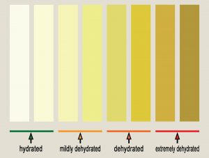
How many liquids have you drank today? How much of that was water?
What observations can you make on the color of your urine?
Chemical Exam
- pH: Normal pH range of urine is 4-8 but much variation comes from diet. High protein diets result in more acidic urine while vegetarian urine diets result in more basic urine. People experiencing burning with urination with no indication of a UTI are given suggestion to raise the pH of their urine by changing their diet and drinking more water.
- Density: The specific gravity of urine is the ratio of the weight of volume of the urine compared with the weight of the same volume of DI water. The SG of DI water is 0.001 and normal urine ranges from 0.001-0.035.
- Leukocytes: WBC’s
- Nitrites: Nitrites aren’t usually found in urine. They are associated with the presence of gram-negative bacteria that can convert nitrate into nitrite. The presence of nitrites can be suggestive of a UTI.
- Protein: Protein molecules are too large to pass through the glomerular filtration barrier, the first part of urine formation process of blood filtration in the kidney, so protein is undetectable in a healthy person. When protein can pass through this barrier, it is known as proteinuria. Proteinuria can be caused by many things like damage or disease to the glomerular filtration barrier, hypertension, kidney damage or stones, and diabetes.
- Glucose – Glycosuria is glucose in the urine which can occur in pregnancy or patients taking corticosteroids. It can also be indicative of diabetes but is not normally in urine.
- Ketones – Ketones are chemicals that are formed during the abnormal breakdown of fat and are not normally in urine. Ketones may result from prolonged vomiting, fasting or starvation, individuals on a diet, or individuals with poorly controlled diabetes resulting in diabetic ketoacidosis.
- Bilirubin and Urobilinogen: Bilirubin is produced when red blood cells are broken down.
- It’s transported in the blood to the liver, where it’s processed and excreted into the gut and makes up part of bile. In the gut, bacteria transform the bilirubin into urobiligen. It is normal for urine to contain urobiligen but not bilirubin. Bilirubin in the urine may be an indicator of a breakdown of red blood cells, indicating liver disease or a problem with drainage of bile into the gut, like gall stones.
- Hemoglobin: Hematuria, blood in the urine, can be macroscopic or microscopic. Macroscopic being large volumes of blood in the urine that changes the color or microscopic being undetectable to the naked eye so chemical strips are used to identify it. Blood in the urine can be the result of kidney disease, kidney stones, anticoagulation disease, menstrual blood, or UTI.
- While wearing gloves, dip the chemstrip into your urine and wait one minute before reading the results.
- Read the results of the chemstrip and input into the table.
| Test | Result | Significance (normal or if abnormal, possible cause) |
| Acidity (4.5 – 8.0) | ||
| Specific gravity (1.001 – 1.035) | ||
| PH | ||
| Protein | ||
| Glucose | ||
| Ketones | ||
| Urobilinogen | ||
| Bilirubin | ||
| Blood |
Microscopic
- Red Blood Cells: kidney disease, urinary tract infection, a drug reaction, or cancer.
- White Blood Cells: infection or inflammation in your urinary tract.
- Epithelial Cells: Normal to see a few, but an excessive amount can indicate UTI
- Bacteria: UTI
- Pour 1 mL of your urine into a test tube, place in centrifuge and spin urine for 5 minutes at 1,500 rpm
- Decant off supernatant and use the wooden stick to stir the pellet
- Drag the pellet down the test tube and place a drop onto the slide.
- Place a slide cover on top of the drop of sediment and tilt the slide until the sides are dried
- Observe the slide under the microscope at low and high power and note observations
| Cell types | Average number seen per movement of frame |
| RBC | |
| WBC | |
| Epithelial | |
| Bacteria | |
| Other (sediment/crystals) |
Activity 2: Diagnostic Urinalysis
Perform the same tests as Activity 2 using an unknown urine sample
Which sample will you be analyzing?
Visual Exam
- Obtain a specimen jar and collect a sample of the unknown urine.
- Note the color of the urine and the smell.
| Characteristic | Normal | Sample |
| Color and turbidity | Pale yellow to deep yellow/clear | |
| Odor | Sweet, ammonia, sulfur |
Chemical Exam
- While wearing gloves, dip the chemstrip into your urine and wait one minute before reading the results.
- Read the results of the chemstrip and input into the table.
| Test | Result | Significance (normal or if abnormal, possible cause) |
| Acidity (4.5 – 8.0) | ||
| Specific gravity (1.001 – 1.035) | ||
| PH | ||
| Protein | ||
| Glucose | ||
| Ketones | ||
| Urobilinogen | ||
| Bilirubin | ||
| Blood |
Microscopic Exam
- Pour 1 mL of your urine into a test tube, place in centrifuge and spin urine for 5 minutes at 1,500 rpm
- Decant off supernatant and use the wooden stick to stir the pellet
- Drag the pellet down the test tube and place a drop onto the slide.
- Place a slide cover on top of the drop of sediment and tilt the slide until the sides are dried
- Observe the slide under the microscope at low and high power and note observations
| Cell types | Average number seen per movement of frame |
| RBC | |
| WBC | |
| Epithelial | |
| Bacteria | |
| Other (sediment/crystals) |
Post Lab Questions
1. What did you notice about the color of your urine? How does this relate to the amount of water you’ve had today?
2. How would you diagnose the patient with the unknown urine sample? What is your reasoning?
3. What would most likely be the cause of a urine sample with a positive test for nitrites, leukocytes and a slightly higher than normal pH?
4. What dietary habits may cause an acidic urine sample (more acidic than normal)? What would cause a basic urine sample?
5. Elevated levels of urobilinogen and bilirubin may indicate problems with what organ?
| Sample | Specific gravity | pH | Leukocytes | Nitrite | Protein | Glucose | Ketones | Urobilinogen | Bilirubin | Blood | Color & Transparency |
| Control | Normal = 1.01 – 1.040 | Normal = pH 5-9
Avg. = 6 |
Normally Negative Positive = >25 cells/μL | Normal = <0.05 mg/dL Positive = >0.05 mg/dL | Normal = <30 mg/dL Positive = >30 mg/dL | Normal = Negative Positive = >90 mg/dL | Normal = <10 mg/100 mL Positive = >10 mg/100 mL | Normal = <0.4 mg/dL Positive = >0.4 mg/dL | Normal = <0.05 mg/dL Positive = >0.05 mg/dL | Normal = <5 cells/μL Positive = >5 cells/μL | Straw, yellow, amber |
| A | |||||||||||
| B | |||||||||||
| C | |||||||||||
| D | |||||||||||
| Yours | |||||||||||
| Causes |

