15.1 Features of the Animal Kingdom
Learning Objectives
By the end of this section, you will be able to:
- List the features that distinguish the animal kingdom from other kingdoms
- Explain the processes of animal reproduction and embryonic development
- Describe the hierarchy of basic animal classification
- Compare and contrast the embryonic development of protostomes and deuterostomes
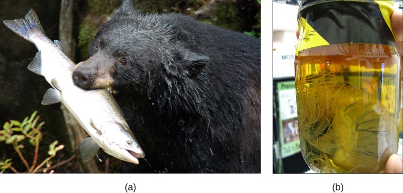
Even though members of the animal kingdom are incredibly diverse, animals share common features that distinguish them from organisms in other kingdoms. All animals are eukaryotic, multicellular organisms, and almost all animals have specialized tissues. Most animals are motile, at least during certain life stages. Animals require a source of food to grow and develop. All animals are heterot rophic, ingesting living or dead organic matter. This form of obtaining energy distinguishes them from autotrophic organisms, such as most plants, which make their own nutrients through photosynthesis and from fungi that digest their food externally. Animals may be carnivores, herbivores, omnivores, or parasites (Figure 15.2). Most animals reproduce sexually: The offspring pass through a series of developmental stages that establish a determined body plan, unlike plants, for example, in which the exact shape of the body is indeterminate. The body plan refers to the shape of an animal.
Complex Tissue Structure
A hallmark trait of animals is specialized structures that are differentiated to perform unique functions. As multicellular organisms, most animals develop specialized cells that group together into tissues with specialized functions. A tissue is a collection of similar cells that had a common embryonic origin. There are four main types of animal tissues: nervous, muscle, connective, and epithelial. Nervous tissue contains neurons, or nerve cells, which transmit nerve impulses. Muscle tissue contracts to cause all types of body movement from locomotion of the organism to movements within the body itself. Animals also have specialized connective tissues that provide many functions, including transport and structural support. Examples of connective tissues include blood and bone. Connective tissue is comprised of cells separated by extracellular material made of organic and inorganic materials, such as the protein and mineral deposits of bone. Epithelial tissue covers the internal and external surfaces of organs inside the animal body and the external surface of the body of the organism.
Concept in Action

View this video to watch a presentation by biologist E.O. Wilson on the importance of animal diversity.
Animal Reproduction and Development
Most animals have diploid body (somatic) cells and a small number of haploid reproductive (gamete) cells produced through meiosis. Some exceptions exist: For example, in bees, wasps, and ants, the male is haploid because it develops from an unfertilized egg. Most animals undergo sexual reproduction, while many also have mechanisms of asexual reproduction.
Sexual Reproduction and Embryonic Development
Almost all animal species are capable of reproducing sexually; for many, this is the only mode of reproduction possible. This distinguishes animals from fungi, protists, and bacteria, where asexual reproduction is common or exclusive. During sexual reproduction, the male and female gametes of a species combine in a process called fertilization. Typically, the small, motile male sperm travels to the much larger, sessile female egg. Sperm form is diverse and includes cells with flagella or amoeboid cells to facilitate motility. Fertilization and fusion of the gamete nuclei produce a zygote. Fertilization may be internal, especially in land animals, or external, as is common in many aquatic species.
After fertilization, a developmental sequence ensues as cells divide and differentiate. Many of the events in development are shared in groups of related animal species, and these events are one of the main ways scientists classify high-level groups of animals. During development, animal cells specialize and form tissues, determining their future morphology and physiology. In many animals, such as mammals, the young resemble the adult. Other animals, such as some insects and amphibians, undergo complete metamorphosis in which individuals enter one or more larval stages. For these animals, the young and the adult have different diets and sometimes habitats. In other species, a process of incomplete metamorphosis occurs in which the young somewhat resemble the adults and go through a series of stages separated by molts (shedding of the skin) until they reach the final adult form.
Asexual Reproduction
Asexual reproduction, unlike sexual reproduction, produces offspring genetically identical to each other and to the parent. A number of animal species—especially those without backbones, but even some fish, amphibians, and reptiles—are capable of asexual reproduction. Asexual reproduction, except for occasional identical twinning, is absent in birds and mammals. The most common forms of asexual reproduction for stationary aquatic animals include budding and fragmentation, in which part of a parent individual can separate and grow into a new individual. In contrast, a form of asexual reproduction found in certain invertebrates and rare vertebrates is called parthenogenesis (or “virgin beginning”), in which unfertilized eggs develop into new offspring.
Classification Features of Animals
Animals are classified according to morphological and developmental characteristics, such as a body plan. With the exception of sponges, the animal body plan is symmetrical. This means that their distribution of body parts is balanced along an axis. Additional characteristics that contribute to animal classification include the number of tissue layers formed during development, the presence or absence of an internal body cavity, and other features of embryological development.
Visual Connection
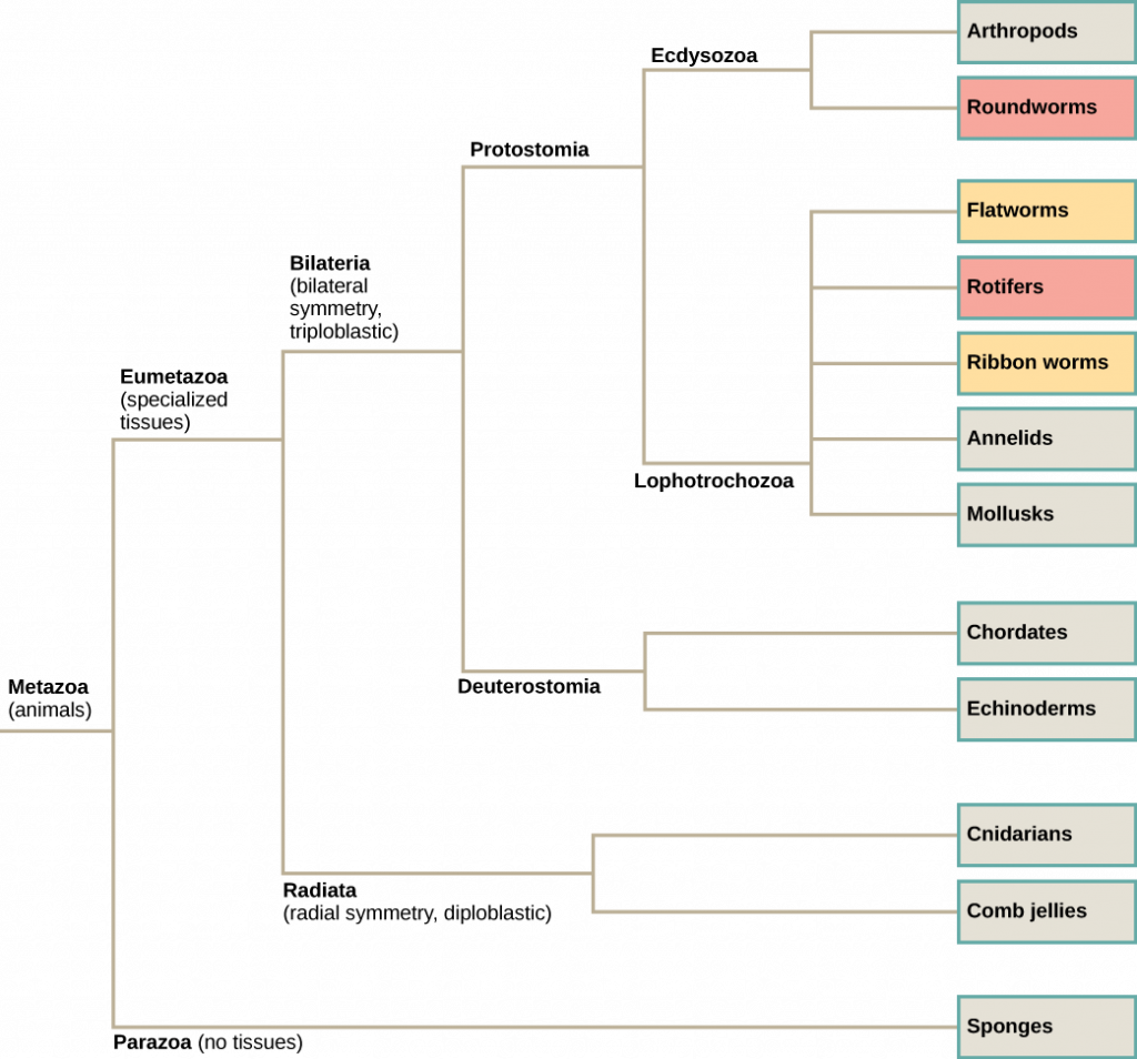
Which of the following statements is false?
- Eumetazoa have specialized tissues and Parazoa do not.
- Both acoelomates and pseudocoelomates have a body cavity.
- Chordates are more closely related to echinoderms than to rotifers according to the figure.
- Some animals have radial symmetry, and some animals have bilateral symmetry.
(Answer: 2)
Body Symmetry
Animals may be asymmetrical, radial, or bilateral in form (Figure 15.4). Asymmetrical animals are animals with no pattern or symmetry; an example of an asymmetrical animal is a sponge (Figure 15.4a). An organism with radial symmetry (Figure 15.4b) has a longitudinal (up-and-down) orientation: Any plane cut along this up–down axis produces roughly mirror-image halves. An example of an organism with radial symmetry is a sea anemone.
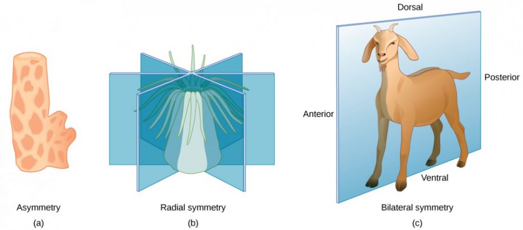
Bilateral symmetry is illustrated in Figure 15.4c using a goat. The goat also has upper and lower sides to it, but they are not symmetrical. A vertical plane cut from front to back separates the animal into roughly mirror-image right and left sides. Animals with bilateral symmetry also have a “head” and “tail” (anterior versus posterior) and a back and underside (dorsal versus ventral).
Concept in Action

Watch this video to see a quick sketch of the different types of body symmetry.
Layers of Tissues
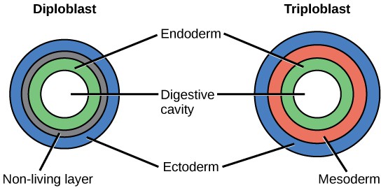
Most animal species undergo a layering of early tissues during embryonic development. These layers are called germ layers. Each layer develops into a specific set of tissues and organs. Animals develop either two or three embryonic germs layers (Figure 15.5). The animals that display radial symmetry develop two germ layers, an inner layer (endoderm) and an outer layer (ectoderm). These animals are called diploblasts. Animals with bilateral symmetry develop three germ layers: an inner layer (endoderm), an outer layer (ectoderm), and a middle layer (mesoderm). Animals with three germ layers are called triploblasts.
Presence or Absence of a Coelom
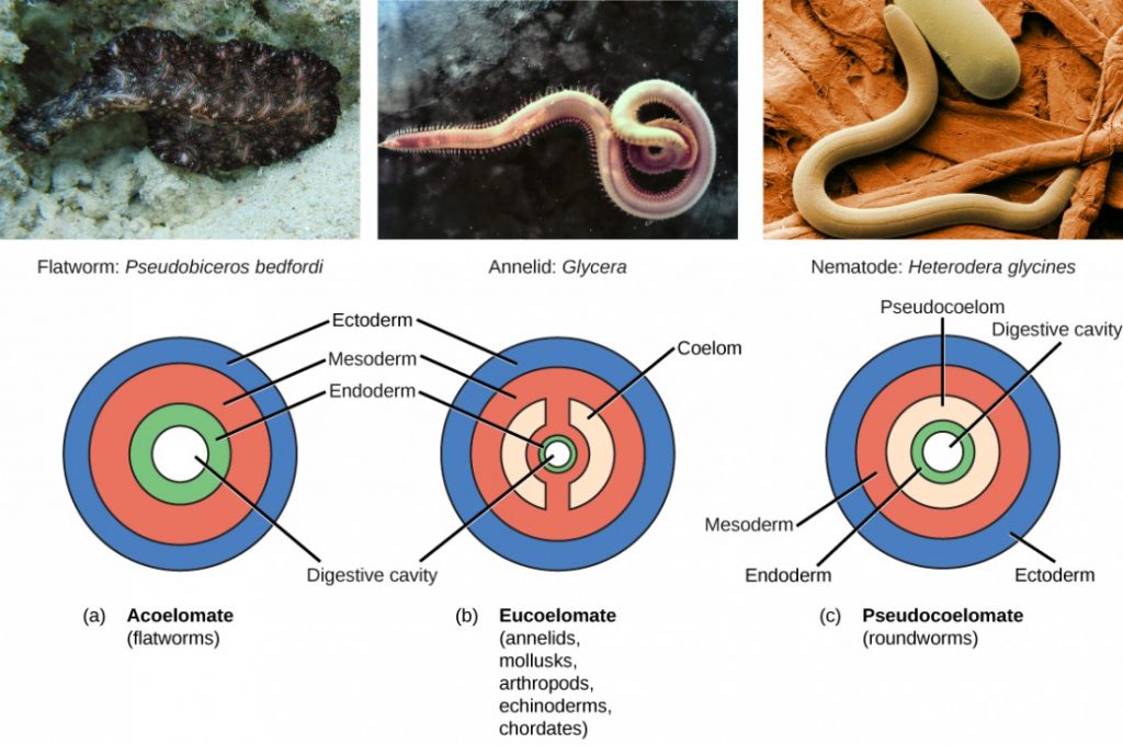
Triploblasts may develop an internal body cavity derived from mesoderm, called a coelom (pr. see-LŌM). This epithelial-lined cavity is a space, usually filled with fluid, which lies between the digestive system and the body wall. It houses organs such as the kidneys and spleen, and contains the circulatory system. Triploblasts that do not develop a coelom are called acoelomates, and their mesoderm region is completely filled with tissue, although they have a gut cavity. Examples of acoelomates include the flatworms. Animals with a true coelom are called eucoelomates (or coelomates) (Figure 15.6). A true coelom arises entirely within the mesoderm germ layer. Animals such as earthworms, snails, insects, starfish, and vertebrates are all eucoelomates. A third group of triploblasts has a body cavity that is derived partly from mesoderm and partly from endoderm tissue. These animals are called pseudocoelomates. Roundworms are examples of pseudocoelomates. New data on the relationships of pseudocoelomates suggest that these phyla are not closely related and so the evolution of the pseudocoelom must have occurred more than once (Figure 15.3). True coelomates can be further characterized based on features of their early embryological development.
Protostomes and Deuterostomes
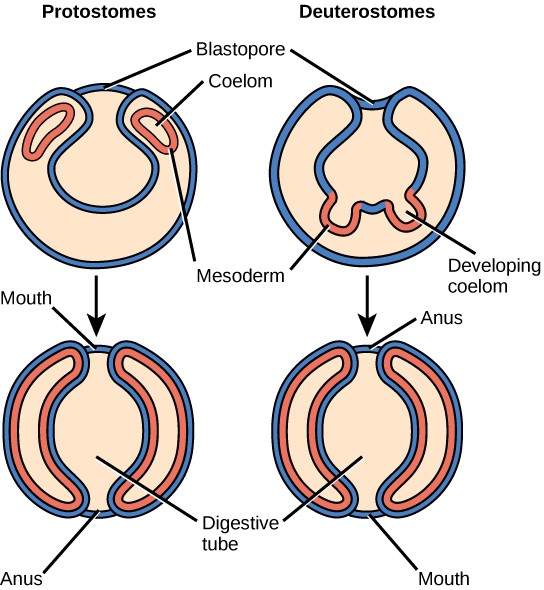
Bilaterally symmetrical, triploblastic eucoelomates can be divided into two groups based on differences in their early embryonic development. Protostomes include phyla such as arthropods, mollusks, and annelids. Deuterostomes include the chordates and echinoderms. These two groups are named from which opening of the digestive cavity develops first: mouth or anus. The word protostome comes from Greek words meaning “mouth first,” and deuterostome originates from words meaning “mouth second” (in this case, the anus develops first). This difference reflects the fate of a structure called the blastopore (Figure 15.7), which becomes the mouth in protostomes and the anus in deuterostomes. Other developmental characteristics differ between protostomes and deuterostomes, including the mode of formation of the coelom and the early cell division of the embryo.
Section Summary
Animals constitute a diverse kingdom of organisms. Although animals range in complexity from simple sea sponges to human beings, most members share certain features. Animals are eukaryotic, multicellular, heterotrophic organisms that ingest their food and usually develop into motile creatures with a fixed body plan. Most members of the animal kingdom have differentiated tissues of four main classes—nervous, muscular, connective, and epithelial—that are specialized to perform different functions. Most animals reproduce sexually, leading to a developmental sequence that is relatively similar across the animal kingdom.
Organisms in the animal kingdom are classified based on their body morphology and development. True animals are divided into those with radial versus bilateral symmetry. Animals with three germ layers, called triploblasts, are further characterized by the presence or absence of an internal body cavity called a coelom. Animals with a body cavity may be either coelomates or pseudocoelomates, depending on which tissue gives rise to the coelom. Coelomates are further divided into two groups called protostomes and deuterostomes, based on a number of developmental characteristics.
Exercises
Glossary
- acoelomate: without a body cavity
- asymmetrical: having no plane of symmetry
- bilateral symmetry: a type of symmetry in which there is only one plane of symmetry that creates two mirror-image sides
- body plan: the shape and symmetry of an organism
- coelom: a lined body cavity derived from mesodermal embryonic tissue
- deuterostome: describing an animal in which the blastopore develops into the anus, with the second opening developing into the mouth
- diploblast: an animal that develops from two embryonic germ layers
- eucoelomate: describing animals with a body cavity completely lined with mesodermal tissue
- germ layer: a collection of cells formed during embryogenesis that will give rise to future body tissues
- protostome: describing an animal in which the mouth develops first during embryogenesis and a second opening developing into the anus
- pseudocoelomate: an animal with a coelom that is not completely lined with tissues derived from the mesoderm as in eucoelomate animals
- radial symmetry: a type of symmetry with multiple planes of symmetry all cross at an axis through the center of the organism
- triploblast: an animal that develops from three germ layers
Media Attributions
- Figure 15.2 © (a) Modification of work by USDA Forest Service; (b) Modification of work by Clyde Robinson; OpenStax is licensed under a CC BY (Attribution) license
- 15.1aQR
- Figure 15.3 © OpenStax is licensed under a CC BY (Attribution) license
- Figure 15.4 © OpenStax is licensed under a CC BY (Attribution) license
- 15.1QR
- Figure 15.5 © OpenStax is licensed under a CC BY (Attribution) license
- Figure 15.6 © (a) Modification of work by Jan Derk; (b) Modification of work by NOAA; (c) Modification of work by USDA, ARS; OpenStax is licensed under a CC BY (Attribution) license
- Figure 15.7 © OpenStax is licensed under a CC BY (Attribution) license

