Water Balance
Objective 9
Describe the fluid compartments of the body and state the relative volumes for the intra- and extracellular compartments. Compare the sources for daily water gain and loss. Describe the mechanism to regulate daily water gain. Relate changes in intracellular and interstitial osmolarity to water movement.
Body Fluids
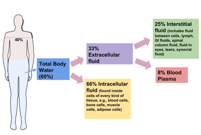
The fluids in the body account for about 60% of the overall body mass. This varies based on age, sex, and body size. Infants are leaner, so they have a little more fluid. The elderly typically have a lower percentage of total water. Differences in fat (hydrophobic) and muscle (hydrophilic) mass account for differences between sexes.

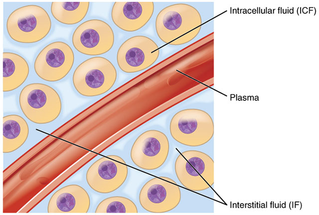
Two-thirds of the total fluid in the body is intracellular, the remaining one-third is extracellular. The extracellular environment can be divided into a number of compartments. The main extracellular space for fluid is the interstitial compartment. Essentially, this is fluid that is within tissue, but is bathing the cells. Other extracellular fluids are the plasma, urine, CSF, aqueous and vitreous humors, endolymph, perilymph, and the lymphatic, synovial, pleural, pericardial, and peritoneal fluids.
Fluid balance doesn’t mean that the same amount of fluid is in each compartment. It simply means that the fluids are present in the correct proportions. The body is constantly moving fluids by various mechanisms, but the compartmental volumes stay relatively constant.
Moving water between the intracellular and interstitial compartments is accomplished by osmosis, so the solute concentrations become very important. Most of these solutes are electrolytes.
Water Loss & Gain

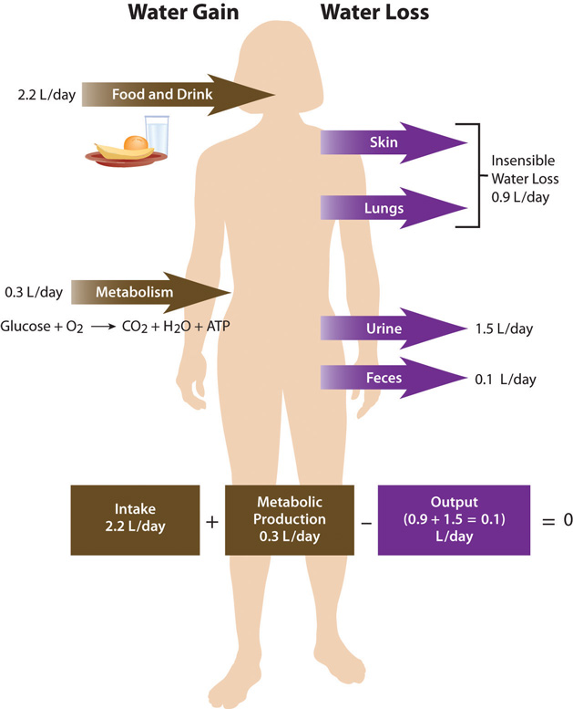
Most of the water gained by the body on a daily basis (2500 ml) comes from ingestion. People consume foods containing water and other miscellaneous liquids throughout the day. A small volume of water is gained through ATP synthesis and is called metabolic water.
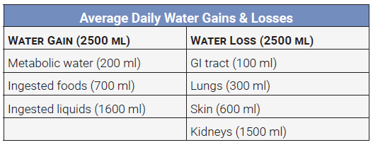
The body loses about 2500 ml of water per day from the kidneys, skin, lungs and GI tract. To maintain normal fluid proportions, the water gained should be equal the water lost.
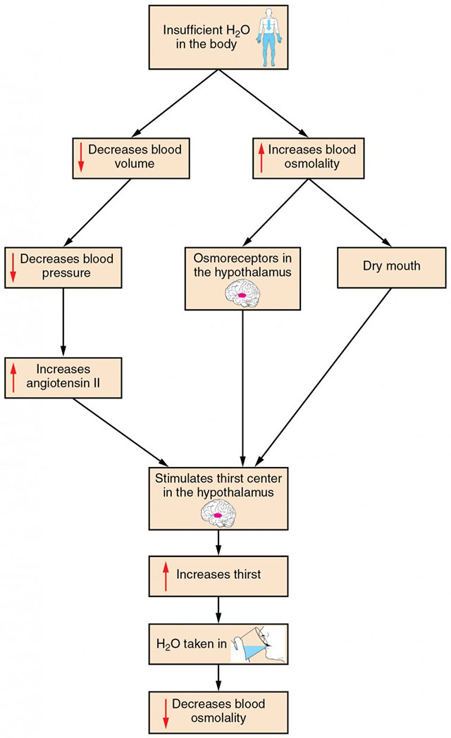 The body’s thirst center is located in the hypothalamus. Dehydration is a condition that occurs when water loss exceeds water gain. This results in a decrease in blood pressure and an increase in blood osmolarity (concentration). The hypothalamus detects the increase in blood osmolarity and triggers the thirst response. Other receptors for dehydration include the kidneys, baroreceptors in the arteries, and neurons in the mouth that detect dryness.
Changes in water quantity and availability throughout the body have a dramatic effect on cells. Remember learning about tonicity and how the solute environment around a cell can cause it to shrink or swell? To avoid this, the body carefully regulates water volume and solute concentration, using the kidneys to excrete excess water and solutes. When blood volume gets too low, the body must pull water from wherever it can to maintain adequate blood pressure to vital organs. Clinically, we can give a patient fluid if they need it, but what is infused into the patient is typically isotonic saline or something similar to it, like lactated Ringer’s solution. The composition of these solutions varies, but their purpose is to add to the volume while maintaining the balance of solutes.
Media Attributions
- U19-036 Fluid Compartments © Betts, J. Gordon; Young, Kelly A.; Wise, James A.; Johnson, Eddie; Poe, Brandon; Kruse, Dean H. Korol, Oksana; Johnson, Jody E.; Womble, Mark & DeSaix, Peter is licensed under a CC BY (Attribution) license
- U19-037 Distribution of Body Water © Calabrese, Allison is licensed under a CC BY (Attribution) license
- U19-038 Regulation of Water Balance © University of Hawai‘i at Mānoa Food Science and Human Nutrition Program is licensed under a CC BY-NC-SA (Attribution NonCommercial ShareAlike) license
- U19-039 Table of Daily Water Gains and Losses © Price, Travis is licensed under a CC BY-SA (Attribution ShareAlike) license
- U19-040 Flowchart of Thirst Response © Betts, J. Gordon; Young, Kelly A.; Wise, James A.; Johnson, Eddie; Poe, Brandon; Kruse, Dean H. Korol, Oksana; Johnson, Jody E.; Womble, Mark & DeSaix, Peter is licensed under a CC BY (Attribution) license
