Veins
Objective 7
Name and label the major veins of the body.
Veins are, by their very nature, much more variable and inconsistent than arteries. (If you’re missing an artery, major structures don’t get supplied by blood, but there are many pathways to drain deoxygenated blood from a structure.)
Veins of the Lower Extremity
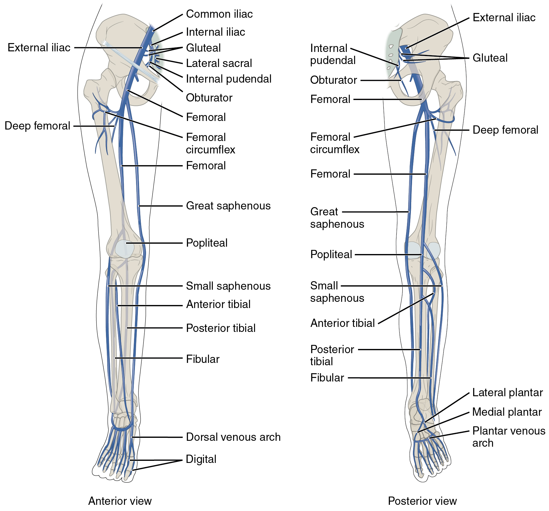 The names of the veins draining the lower extremity parallel those of the arteries. There is a notable exception in the saphenous vein, the longest vein of the body. The saphenous vein is often used as a source for a coronary artery bypass graft.
Veins of the Lower Extremity
Venous drainage of the foot
- Digital veins
- Dorsal venous arch
- Plantar venous arch
- Medial plantar vein
- Lateral plantar vein
Venous drainage of the leg
- Fibular vein
- Anterior tibial vein
- Posterior tibial vein
- Small saphenous vein
- Great saphenous vein
Venous drainage of the knee
- Popliteal vein
Venous drainage of the thigh
- Deep femoral veins
- Femoral circumflex veins
Venous drainage of the lower extremity
- Femoral vein = External iliac vein
Venous drainage of the buttocks
- Gluteal vein
Venous drainage of the groin
- Obturator vein
- Internal pudendal vein
- Lateral sacral vein
Common iliac vein
How Pulmonary Emboli Form in Veins
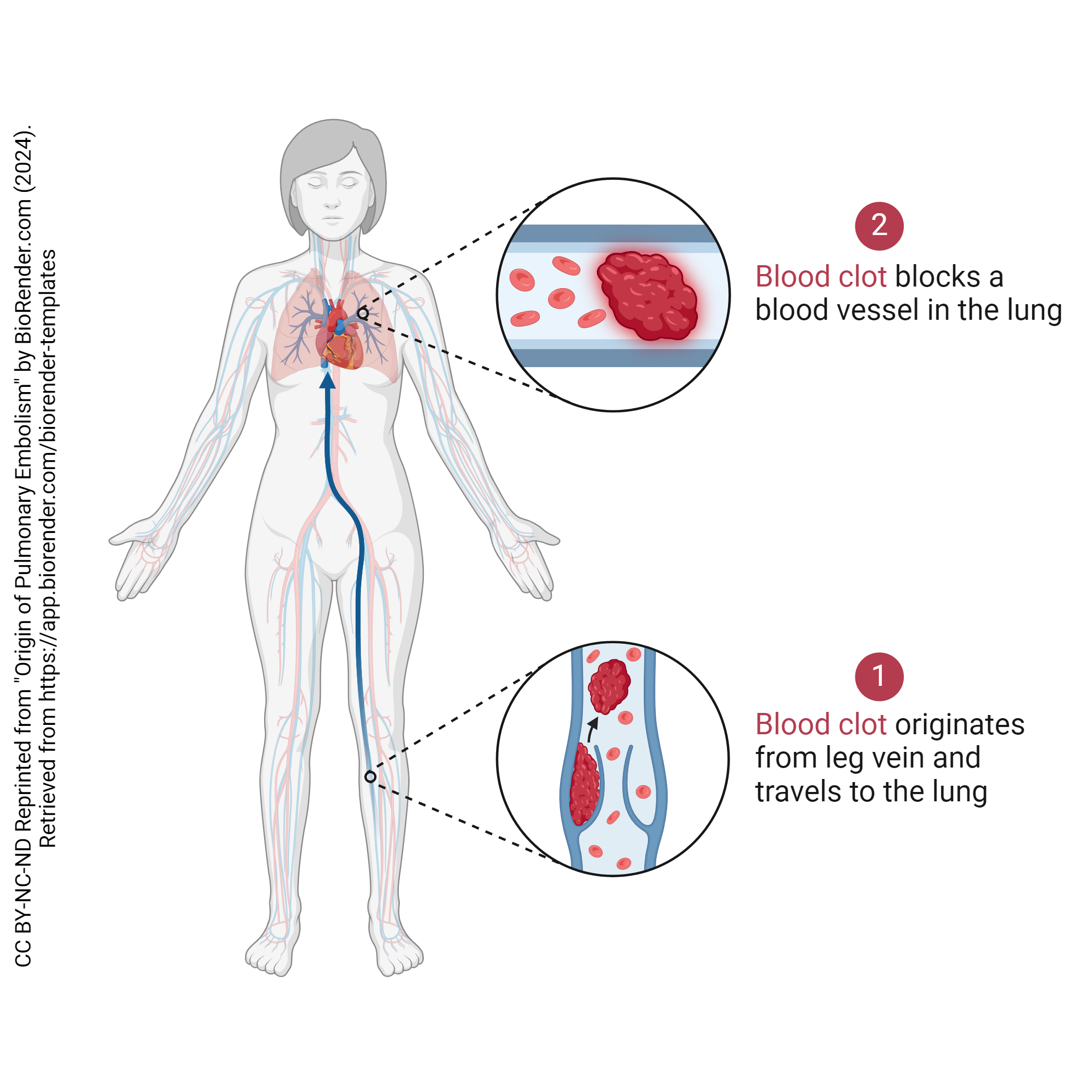 It is quite common for blood clots to arise from the veins of the leg. Blood flow is often restricted as patients get older; the veins become flabby and distended (varicose), which creates turbulence, which promotes clot formation. If a clot forms and breaks free (a thromboembolus), it travels through successively larger veins (femoral vein, common iliac vein, inferior vena cava) to the heart. The right atrium and right ventricle are similarly large structures. Next, the thromboembolus will pass through the pulmonary valve into the pulmonary artery. The first small vessels or capillary bed it will encounter is in the lung, where it becomes a pulmonary embolism.
Veins of the Upper Extremity
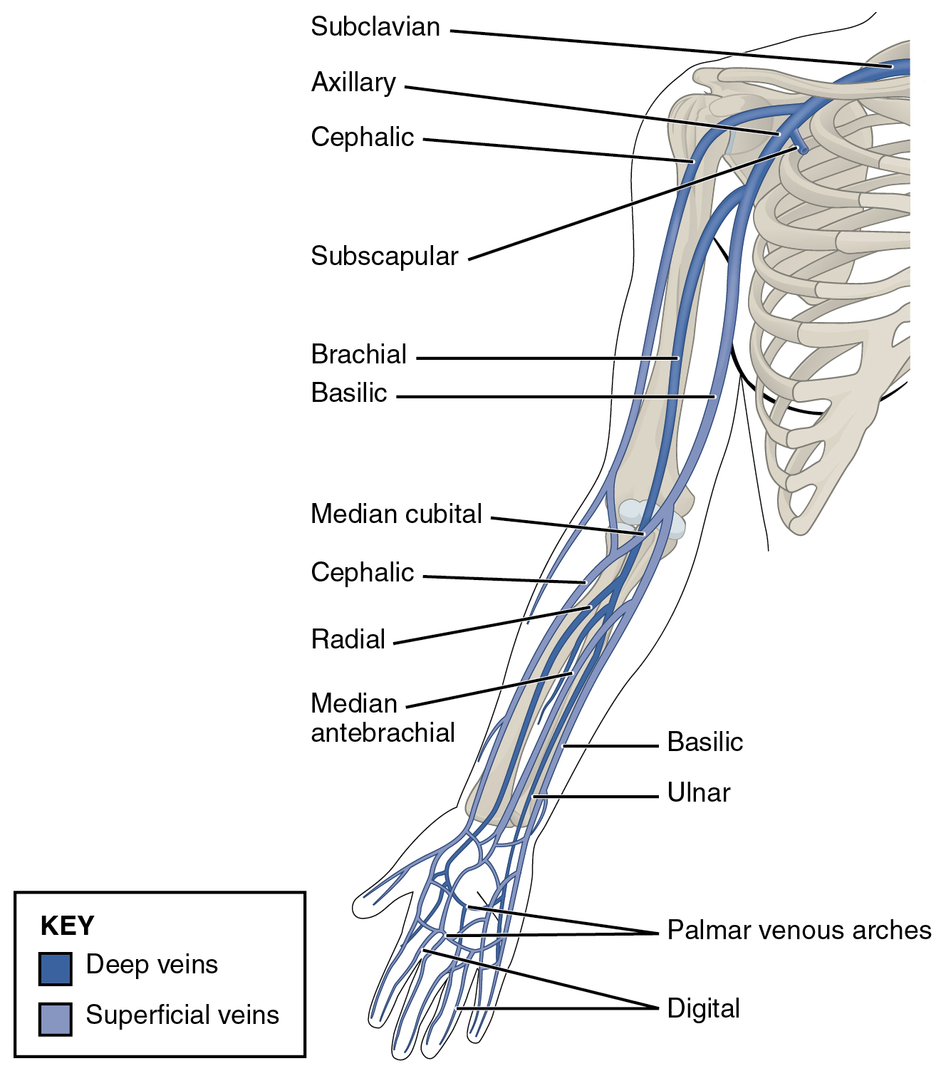 Like the foot, the hand has an arching system of veins (palmar venous arches and dorsal venous arches, not shown) that receive drainage from the digital veins.
The basilic and ulnar veins are found on the medial side of the forearm; the median antebrachial vein is medial, as we’d expect from the name; and the radial and cephalic veins are found laterally.
The median cubital vein, in the anterior elbow (cubital) region, is where phlebotomists take blood from most patients.
The basilic vein continues into the arm proper on the medial side. The ulnar, median antebrachial, and radial veins have joined and anastomosed with each other and with the other veins of the forearm to form a larger brachial vein. The cephalic vein continues on the lateral side of the arm.
Like the arterial supply, the venous drainage of the upper extremity is carried by a large vessel with three names. The brachial artery becomes the axillary vein in the axillary region. The subscapular vein joins at this point. As the vein is covered by the clavicle, it changes name to the subclavicular vein which joins the veins of the head and neck to form the brachiocephalic vein. The brachiocephalic vein drains into the superior vena cava. (Note that while the brachiocephalic artery is typically found on only one side, the brachiocephalic veins are generally on both sides.)
Veins of the Upper Extremity
Venous drainage of the hand
- Digital veins
- Palmar venous arches
Venous drainage of the forearm
- Basilic vein
- Ulnar vein
- Median antebrachial vein
- Radial vein
- Cephalic vein
Venous drainage of the elbow (cubital)
- Median cubital vein
Venous drainage of the arm
- Basilic vein
- Brachial vein
- Cephalic vein
Venous drainage of the shoulder
- Subscapular vein
Venous drainage of the upper extremity
Brachial vein = Axillary vein = Subclavian vein
Veins of the Head and Neck
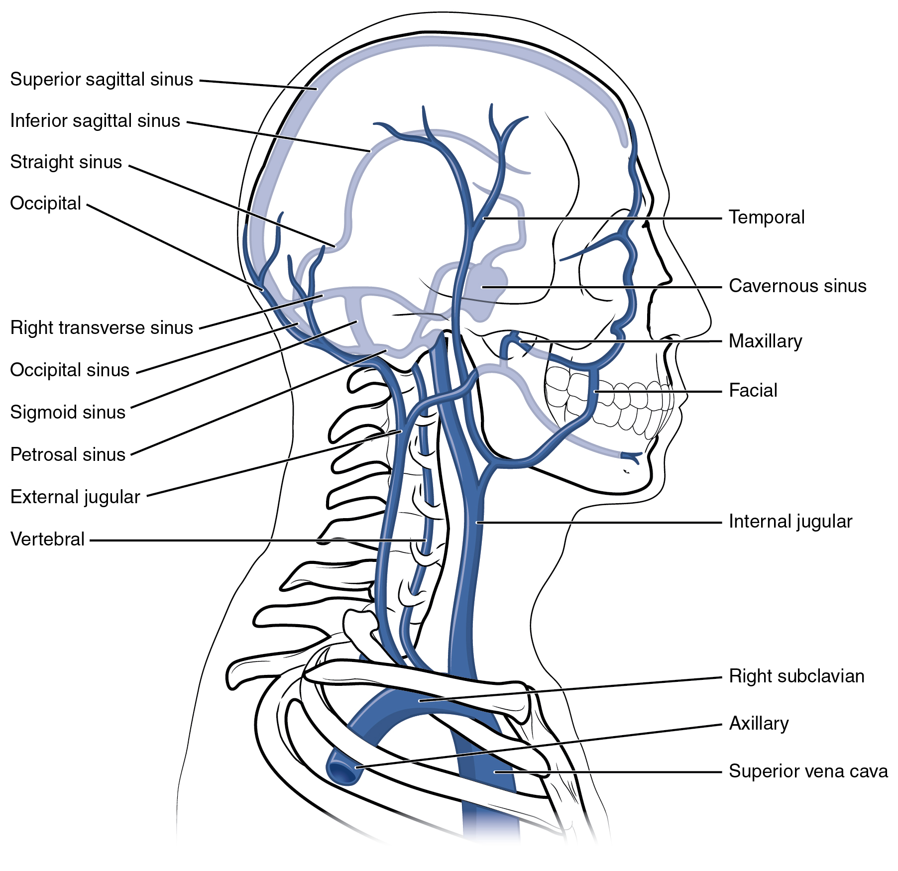 The venous drainage of the brain is carried by a complicated system of venous sinuses (i.e. open spaces not clearly bounded by vessel walls). We have already studied one of these: the cavernous sinus. All these gather together to form the internal jugular vein and the vertebral vein, which are responsible for returning venous blood to the heart.
For the scalp and face, the occipital and temporal veins drain the posterior and lateral scalp, respectively. The maxillary and inferior alveolar (not shown) veins join with the superficial veins of the head to form the external jugular vein. The internal jugular vein, external jugular vein, and vertebral vein join the subclavian vein from the upper extremity to form the brachiocephalic vein. The two brachiocephalic veins (right and left) then join to form the superior vena cava, returning blood to the right atrium of the heart.
Veins of the Head and Neck
Venous drainage of the brain
- Superior sagittal sinus
- Inferior sagittal sinus
- Straight sinus
- Transverse sinus
- Cavernous sinus
- Sigmoid sinus
- Occipital sinus
- Petrosal sinus
- Internal jugular vein
- Vertebral vein
Venous drainage of the scalp and face
- Occipital vein
- Temporal vein
- Maxillary vein
- Facial vein
- External jugular vein
Veins of the Trunk
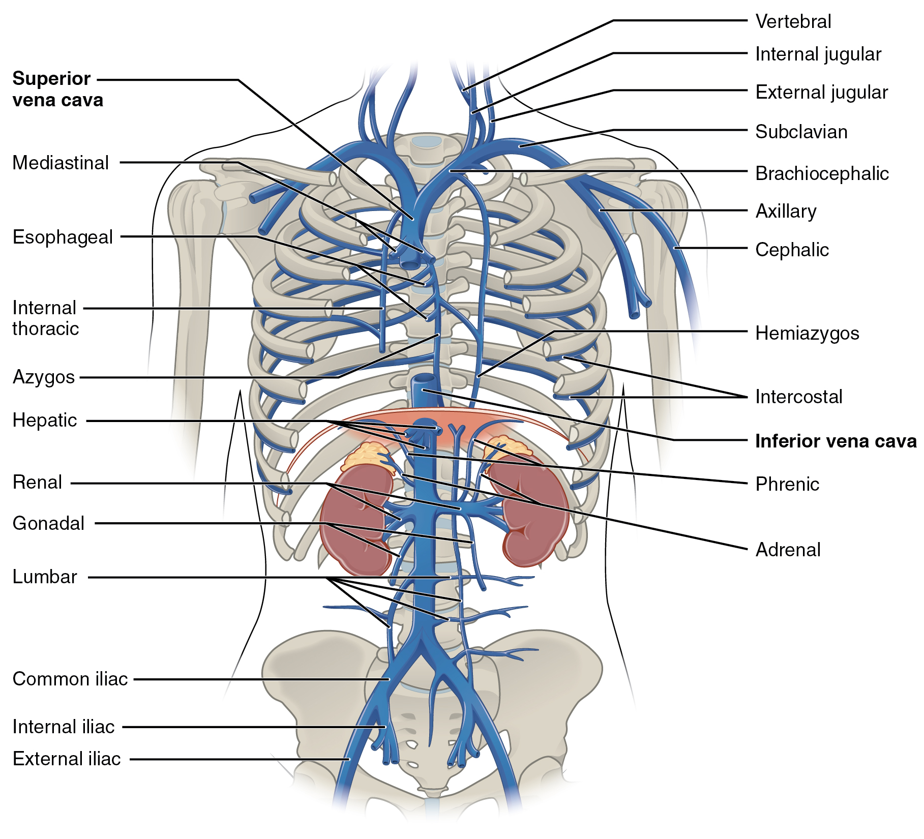 We have already seen how the veins of the head, neck, and upper extremity join to form the brachiocephalic trunk.
A set of anastomosing veins called the azygos system drains the thoracic wall and upper lumbar region. (The name azygos means “unpaired”.) Venous drainage from the chest wall (intercostal veins) and the lungs gather into this system and the hemiazygos vein and this blood then drains into the superior vena cava. The names of the other veins of the trunk are familiar to us:
- the hepatic vein from the liver
- the phrenic vein from the diaphragm
- the renal vein from the kidney
- the gonadal vein from the gonads
- the lumbar vein from the lumbar region
All these join with the inferior vena cava.
The Hepatic Portal Vein System
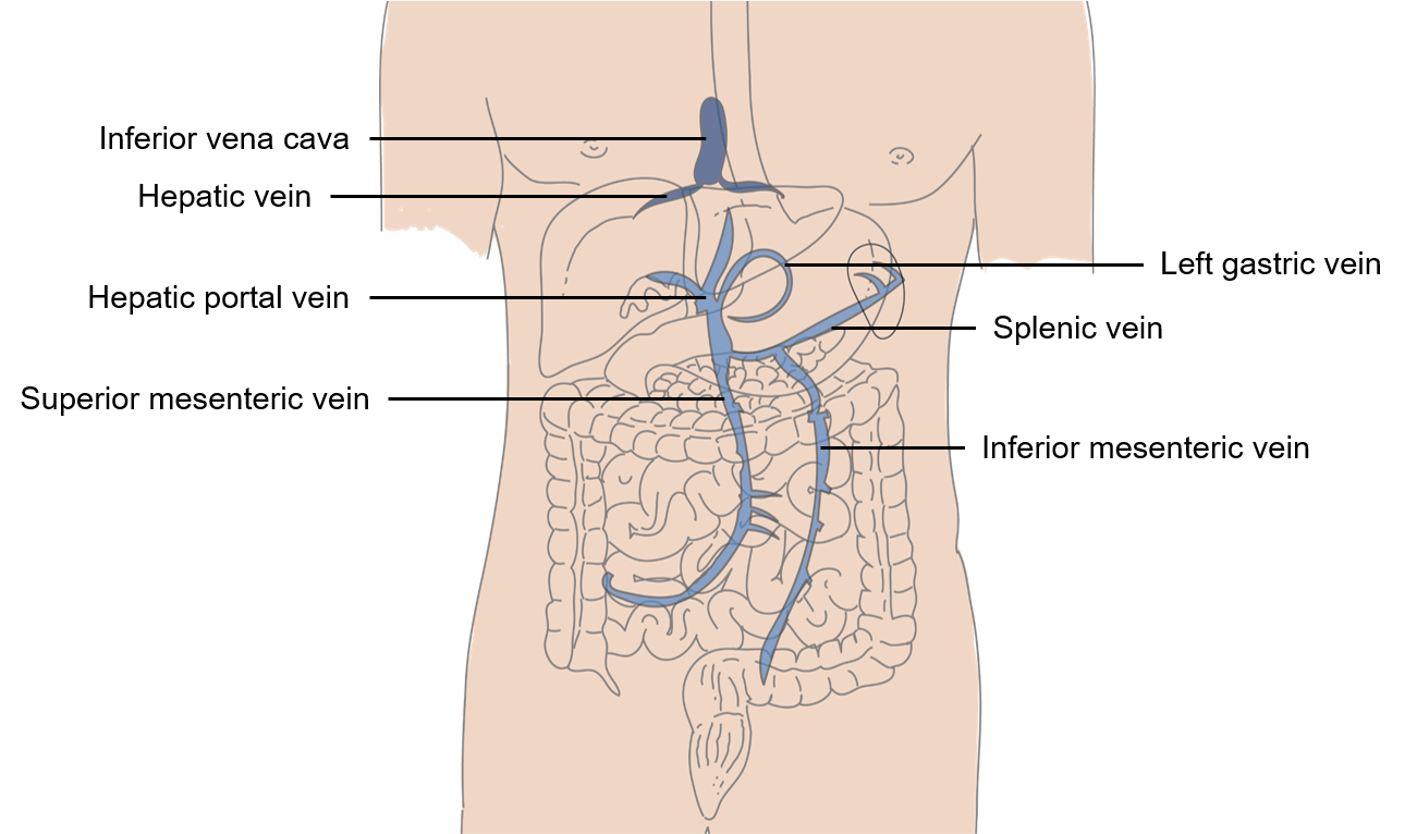 As we’ll see in Unit 18, the digestive system has a special problem: deoxygenated, nutrient-rich blood must be processed by the liver before it can be returned to the systemic circulation. In order to achieve this, there are a series of “private” veins that travel between the digestive organs and the liver: the superior mesenteric vein; the inferior mesenteric vein; and the gastric veins.
Additionally, the spleen acts as a blood reservoir. It holds about 240 mL of blood, which can be released into the systemic circulation by sympathetic stimulation. For example, after trauma, if extra blood is needed, the spleen’s reserve can be tapped.
The spleen also destroys old red blood cells. The average lifetime of a red blood cell is 90 to 120 days. “Old” red blood cells are broken down in the spleen and their molecular parts are released into the bloodstream.
The output of the spleen is carried by the splenic vein.
All these veins join the hepatic portal vein. Just as the hypophyseal portal vein carries releasing hormones and inhibiting hormones from the hypothalamus to the anterior pituitary, the hepatic portal vein carries deoxygenated, nutrient-rich blood from the stomach and intestines to the liver, as well as reserve blood or destroyed blood from the spleen.
Once this blood has been processed through the liver (Unit 18), it drains into the hepatic vein (sometimes called the “hepatic vein proper”). The hepatic vein joins the inferior vena cava to return this blood to the heart.
Media Attributions
- U16-048 Major Veins Serving the Lower Limbs © Betts, J. Gordon; Young, Kelly A.; Wise, James A.; Johnson, Eddie; Poe, Brandon; Kruse, Dean H. Korol, Oksana; Johnson, Jody E.; Womble, Mark & DeSaix, Peter is licensed under a CC BY (Attribution) license
- U16-049 Origin of Pulmonary Embolism © BioRender is licensed under a CC BY-NC-ND (Attribution NonCommercial NoDerivatives) license
- U16-050 Veins of the Upper Limb © Betts, J. Gordon; Young, Kelly A.; Wise, James A.; Johnson, Eddie; Poe, Brandon; Kruse, Dean H. Korol, Oksana; Johnson, Jody E.; Womble, Mark & DeSaix, Peter is licensed under a CC BY (Attribution) license
- U16-051 Veins of the Head and Neck © Betts, J. Gordon; Young, Kelly A.; Wise, James A.; Johnson, Eddie; Poe, Brandon; Kruse, Dean H. Korol, Oksana; Johnson, Jody E.; Womble, Mark & DeSaix, Peter is licensed under a CC BY (Attribution) license
- U16-052 Veins of the Thoracic and Abdominal Regions © Betts, J. Gordon; Young, Kelly A.; Wise, James A.; Johnson, Eddie; Poe, Brandon; Kruse, Dean H. Korol, Oksana; Johnson, Jody E.; Womble, Mark & DeSaix, Peter is licensed under a CC BY (Attribution) license
- U16-053 Hepatic Portal Vein System © Jenks, Julie and Donnelly, Daniel is licensed under a CC BY (Attribution) license
