Ureters, Bladder, Urethra, and Micturition
Objective 7
Identify and describe the structure of the ureters, and relate the structure to urine flow. Describe the structure of the bladder and explain how it is able to store varying amounts of urine. Explain the micturition reflex and how bladder muscles and the urinary sphincters play a role in that process. Compare the characteristics of the male and female urethra.
Ureters
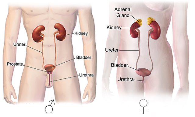
The ureters, one originating from each kidney, transport urine from the renal pelvis to the bladder. It is not purely a gravity-driven system. Hydrostatic pressure and gravity assist with urine flow, while peristaltic contractions of smooth muscle in the ureter walls push the urine towards the bladder.
The ureters are approximately 10-12 inches long and, similar to the kidneys, are retroperitoneal. Both ureters attach to the posterior base of the bladder.
There isn’t a valve present at the connection of the ureter and bladder, but as the bladder fills, the opening to the ureter closes to prevent backflow of urine back to the kidneys.
The ureters are lined by a mucous membrane containing transitional epithelium. This allows the ureter to stretch with variable volumes of urine. The secreted mucus protects the epithelium from coming into direct contact with the urine, which is often slightly acidic.
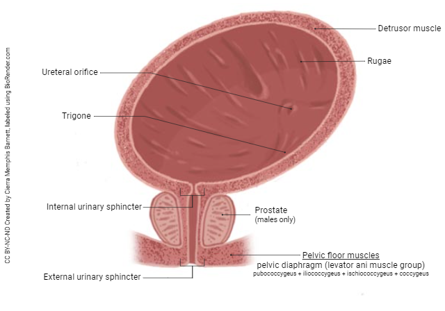
Urinary Bladder
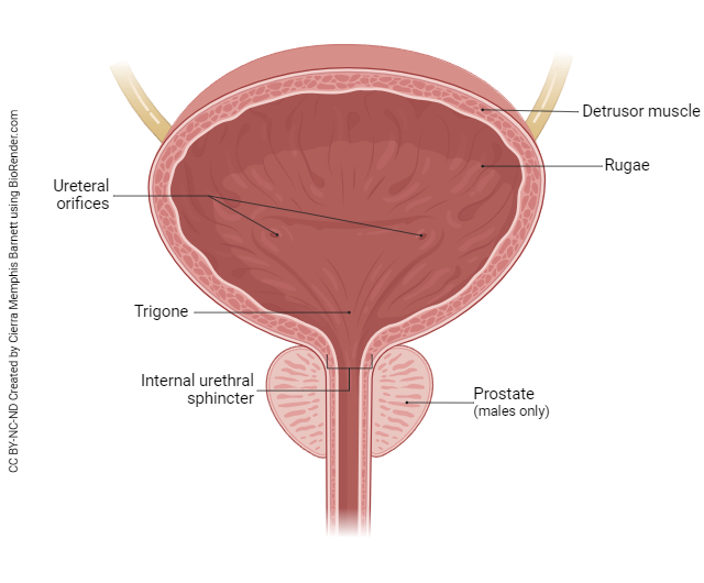
The urinary bladder is a hollow, distensible (stretchable) organ that is located posterior to the pubic symphysis in the pelvic cavity. Because the uterus also occupies space in the pelvic cavity, the female bladder is slightly smaller than the males.
The bladder can accommodate approximately 700-800 ml of urine. Folds (rugae) in the mucosal layer and the presence of transitional epithelium provide for the bladder’s distensibility.
At the base of the bladder, there is a triangular-shaped area called the trigone. The three apices of the triangle are the openings of the two ureters and the urethra (anterior)
The detrusor muscle surrounds the mucosal layer. It consists of three layers of muscle fibers:
- inner (longitudinal)
- middle (circular)
- outer (longitudinal)
Its function is to assist with the emptying of the bladder.
Finally, the superficial layers of the bladder are the adventitia and serosa. The adventitia is a layer of areolar connective tissue located on the posterior and inferior surfaces and is continuous with the ureters. The serosa is a layer of visceral peritoneum on the superior surface.
There are two sphincters that control urine release from the bladder, the internal and external urethral sphincters. The circular fibers around the opening of the urethra, in the bladder, form the internal sphincter. Deep skeletal muscles of the perineum form the external urethral sphincter. It is important understand that the internal sphincter is under involuntary control from the CNS, and once learned, the external sphincter is voluntarily controlled.
Micturition, also called urination or voiding, is the process of releasing urine from the urinary bladder. It is both a voluntary and an involuntary process.
The bladder can hold almost a liter of urine, but when the volume exceeds 200-400 ml, the distension of the bladder triggers stretch receptors that convey impulses to the micturition center in the sacral spinal cord. These signals cause a conscious sensation of bladder fullness and initiate a parasympathetic response. The parasympathetic response causes relaxation of the internal urethral sphincter and contraction of the detrusor muscle in the bladder. This response can be overridden by maintaining tension in the skeletal muscle of the external urethral sphincter. This is why we use the term “potty training.” A child is literally training their conscious control of the external urethral sphincter to be able to control when voiding occurs. Without this control, the bladder would fill, stretch receptors would signal fullness, and almost immediately your bladder would empty whether you wanted it to or not. That would be embarrassing.
Urethra
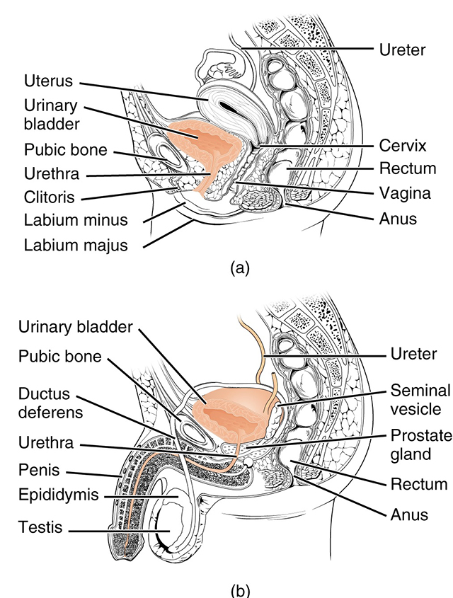
In both males and females, the urethra is the end of the urinary system. It is a small tube leading from the urinary bladder to the exterior of the body. In males, the urethra extends through the penis, is about 20 cm in length, and is divided into three regions: the prostatic, membranous and spongy urethra. In males, the urethra also functions to transport semen, formed by the testes and accessory sex glands.
In females, the urethra is only about 4 cm. Its external opening is located between the clitoris and the vaginal opening. The shorter length of the urethra and its proximity to the anus is what contributes to the higher incidence of bladder infections in females.
Media Attributions
- U19-004 Urinary System © BruceBlaus adapted by Jim Hutchins is licensed under a CC BY-SA (Attribution ShareAlike) license
- U19-028 Bladder Anatomy © Barnett, Cierra Memphis is licensed under a CC BY-NC-ND (Attribution NonCommercial NoDerivatives) license
- U19-029 Internal and External Urethral Sphincters © Barnett, Cierra Memphis is licensed under a CC BY-NC-ND (Attribution NonCommercial NoDerivatives) license
- U19-030 Female and Male Urethra © Betts, J. Gordon; Young, Kelly A.; Wise, James A.; Johnson, Eddie; Poe, Brandon; Kruse, Dean H. Korol, Oksana; Johnson, Jody E.; Womble, Mark & DeSaix, Peter adapted by Jim Hutchins is licensed under a CC BY (Attribution) license

.png and https://simple.wikipedia.org/wiki/Bladder#/media/File:Urinary_System_(Female).png)