Tolerance and Memory | Changes in the Adaptive Immune System Throughout Life
Objective 12
Describe the steps in the development of immune tolerance. Explain the differences between the primary and secondary immune response. Review the formation of memory B cells and memory T cells.
The Thymus
We’ve said this several times already: the reason T cells are called “T” is because they mature in the thymus. What we’ve left out, of course, is exactly what we mean by “mature”. It’s time to discuss that now.
Nursing students have to pass a standardized test to get licensed. Attorneys have to pass the bar exam. Immature lymphocytes have to pass two tests to become mature T cells.
The Tolerance Tests
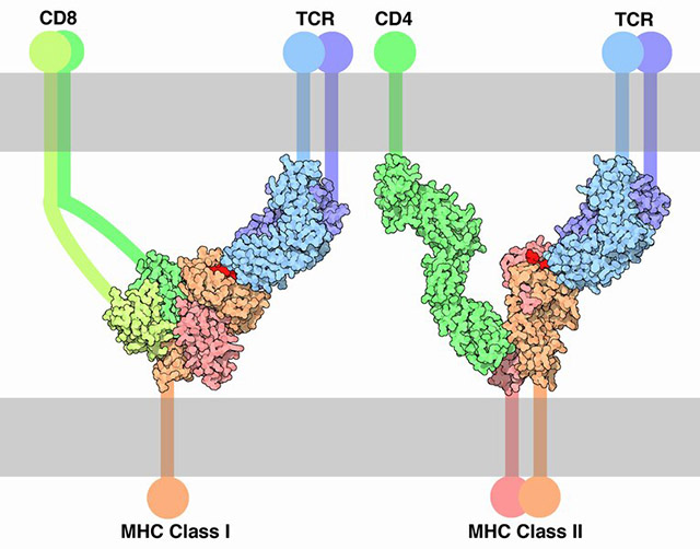
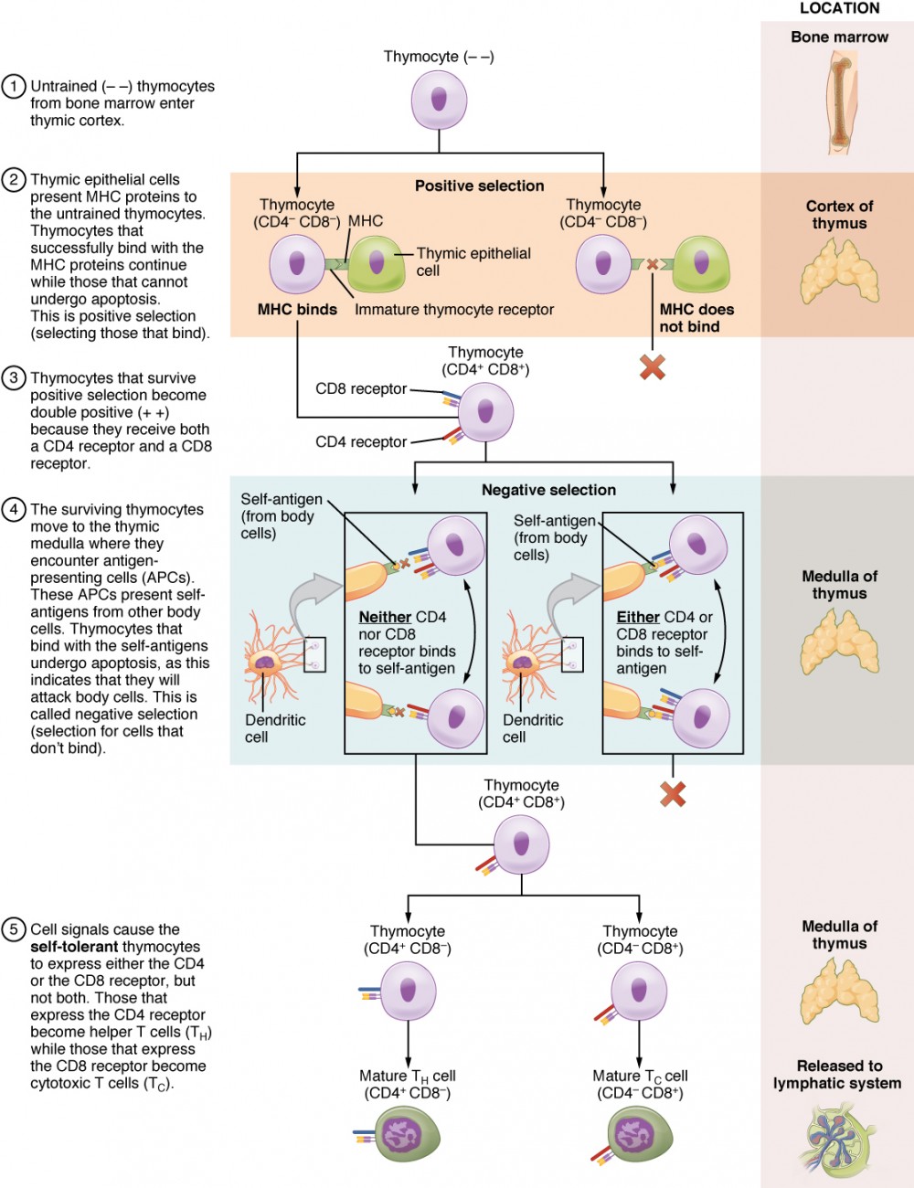
Remember from our diagram of MHC molecules that they have to bind both the CD marker and the T cell receptor. It’s of no use to have T cells that can’t bind to your body’s MHC molecules, or worse, ones that bind to someone else’s MHC molecules. Getting rid of these useless wastrels is the job of the first test.
Because immature T cells that “pass” are the ones that positively bind MHC markers, this is called a positive selection.
Going into the positive selection tests, immature T cells have neither CD4 nor CD8 markers on their surface (i.e., they are called “– –“ at the top of the diagram), with the blanks indicating what they don’t have — CD4 or CD8. They are tested by the epithelial cells of the thymus, which challenges them to bind the MHC molecules specific to that individual. If they pass, they are “rewarded” with both CD4 and CD8, so we now call them “CD4+ CD8+”.
Now it’s time for a much more rigorous, and important, test. Only 3% of cells will “pass” this test, and that’s a good thing, because the other 97% have the potential to destroy your body’s cells.
This test is adminstered by the dendritic cells, veterans of the immune wars. They bring the immature CD4+ CD8+ cells examples of antigens they’ve gathered from normal cells in your body. If you bind those, you get washed out of the program. You just deployed your weapon and shot a friend. Get outta here. Go get apoptotic, we can’t use you.
If the CD4+ CD8+ T cells fail to bind self-antigens, they have passed by doing nothing. Thus, this is called a negative selection.
Only one more step: either the CD4 or CD8 markers are lost.
Now we have selected for cells that meet the following three criteria:
- bind to your personal MHC type
- do not bind to antigens found on the surface of your body cells (i.e., “self”); they recognize only foreign antigens
- have CD4 markers (mature CD4+ TH cells) or CD8 markers (mature CD8+ TC cells)
The battle-ready soldiers are now ready to be deployed into the lymphatic system.
The Persistence of Memory

The key players in the adaptive immune response are lymphocytes. Remember we have B lymphocytes that develop and mature in the bone marrow and T lymphocytes which develop in the bone marrow but mature in the thymus. After activation by foreign antigens, B cells and T cells can stimulate the production of clones, or copies of themselves.
These clones can then differentiate into memory cells, whose job is, you guessed it, to remember! Like old folks that like to sit in their favorite recliners and tell you about “the good ol’ days”, memory B and T cells hang around for a long time, just waiting for the chance to inform newer cells of the antigen they once encountered. By doing so, new groups of lymphocytes can recognize and respond to antigens more quickly.
The cells that are not designated to become memory cells carry out their unique functions. Generally speaking, B cells have the primary responsibility of producing antibodies while T cells attack abnormal or infected cells of the body and help with the overall immune response.
Because the primary response generates memory cells and long lasting IgG antibodies, the secondary response is much faster. The antigen can be taken care of before a person even becomes symptomatic. Time to process and present antigen is not necessary. There is still an immediate increase in IgM antibodies, however, the IgG antibodies that are around from the last exposure provide a rapid response to the antigen. Titers of IgG antibodies will increase significantly after a secondary exposure. It’s almost like the first exposure makes the body think, “this thing may come back some day” while the second exposure makes it think “this thing is definitely coming back again, let’s make sure we are ready.” This is one of the main reasons to provide booster shots for various vaccinations.
Primary & Secondary Immune Responses
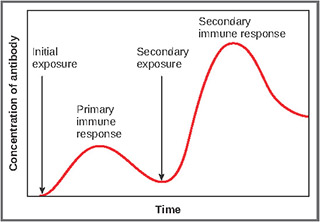
The differences between the primary and secondary immune responses illustrate the ability of the immune system to demonstrate memory. The primary immune response is the sequence of events and outcomes of the immune response with an initial exposure to an antigen. The secondary response relates to second or subsequent exposures.
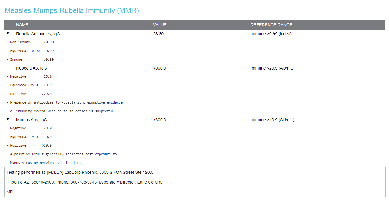
The indicator for an antibody-mediated response is an antibody titer. A titer is a test that detects the relative amount of antibody released by activated B cells. The test results shown are from a 60 year old man who was a child before the measles/mumps/rubella vaccine was developed. As a result, he (his parents, really) suffered through all three of those diseases in the early 1960s. His immune system created thousands of B cell clones that for about 60 years now have cranked out massive amounts of IgG against measles, mumps, and rubella. The levels of IgGs specific to those three diseases are shown in the test report. This patient would be unlikely to “catch” any of these diseases, even in an outbreak, because his IgGs would immediately attack and neutralize any virus particles which were able to enter his body.
There are two main differences between the primary and secondary immune responses, time and the antibody titer (antibody concentration). The first time the immune system recognizes an antigen, it takes a period of time for antigen processing, presentation, and antibody production. This period lasts approximately 5-7 days but can vary with different types of immune system challenges. Initially, the plasma cells will produce a population of IgM antibodies, followed by a population of IgG. The goal of the primary response is to provide an initial antibody-mediated response and produce a population of memory cells and IgG antibodies that can remain in the body and provide a better response the next time that same antigen is encountered. IgM is an excellent complement activator so it’s ideal for an initial response.
In time, the memory B cells undergo a process called class switching and while they keep the same antigen-binding specificity, they turn from producing IgMs to producing IgGs. The genes which make antibodies are arranged in a way that promotes an easy switch between classes while keeping the same “pocket shapes” that give the antigen-binding site its specificity.
Memory B Cells & Memory T Cells
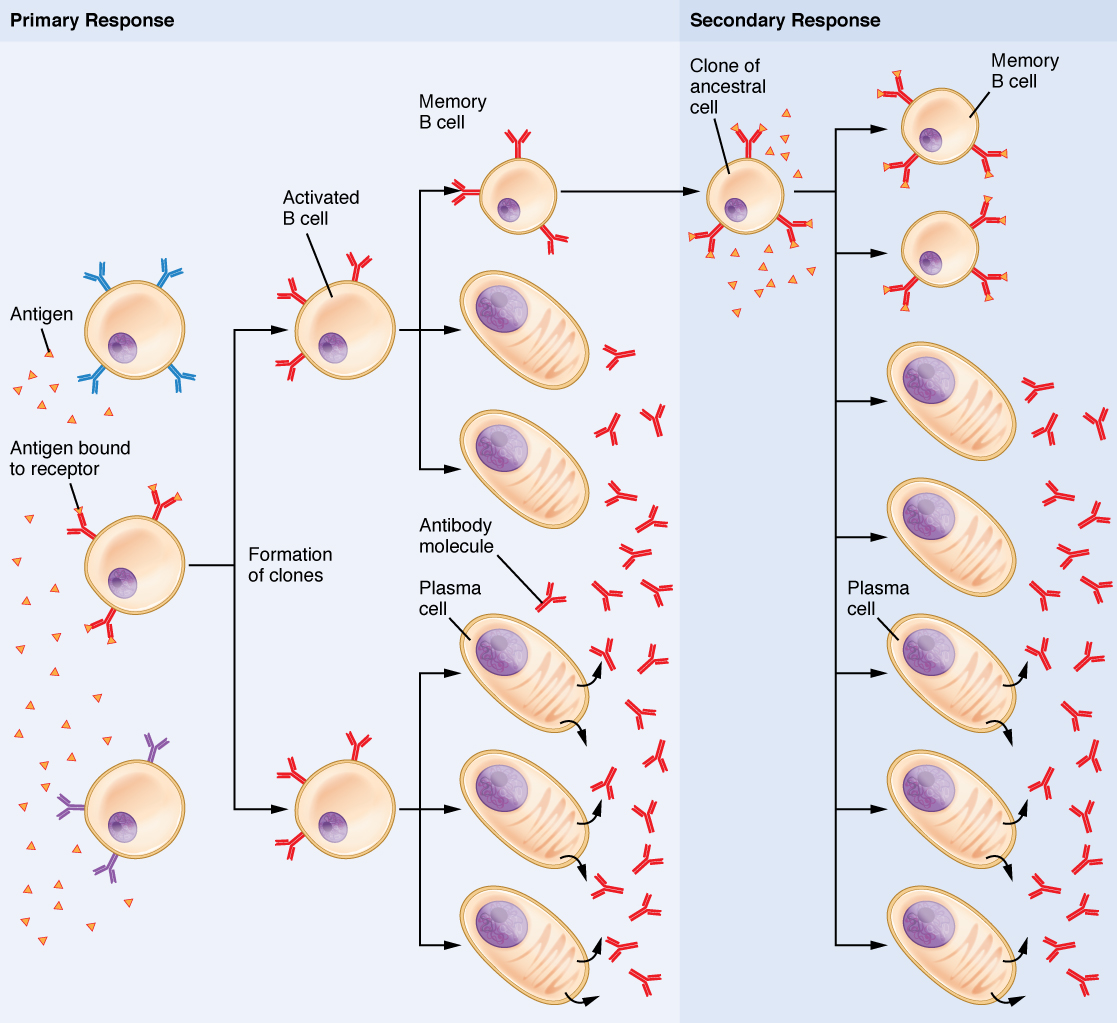
We’ve seen how one of the main cytokine signals released by TH cells promotes rapid cell division. As B cells and T cells divide under the influence of this cytokine, some of the clones (exact copies) develop into memory cells.
The first time the patient whose titers are shown above encountered measles, there was not time for the immune system to ramp up its activity before the disease got the upper hand. This means the second time he encountered the measles antigen, there will be a massive and devastating response mounted by the immune system. For example, with immunity against the measles virus, even a single virion will be immediately surrounded and destroyed by the large number of measles-specific memory B cells and memory T cells. You can only fool an effective immune system once.
Media Attributions
- U15-056 T Cell Receptor MHC © Goodsell, David is licensed under a CC BY (Attribution) license
- U15-082 Differentiation of T Cells Within the Thymus © Betts, J. Gordon; Young, Kelly A.; Wise, James A.; Johnson, Eddie; Poe, Brandon; Kruse, Dean H. Korol, Oksana; Johnson, Jody E.; Womble, Mark & DeSaix, Peter is licensed under a CC BY (Attribution) license
- U15-083 Memory © Crookston, Alexa is licensed under a CC BY-SA (Attribution ShareAlike) license
- U15-084 Primary and Secondary Antibody Response © Betts, J. Gordon; Young, Kelly A.; Wise, James A.; Johnson, Eddie; Poe, Brandon; Kruse, Dean H. Korol, Oksana; Johnson, Jody E.; Womble, Mark & DeSaix, Peter adapted by Jim Hutchins is licensed under a CC BY (Attribution) license
- U15-085 MMR Titer © Hutchins, Jim is licensed under a CC0 (Creative Commons Zero) license
- U15-086 Clonal Selection of B Cells © Betts, J. Gordon; Young, Kelly A.; Wise, James A.; Johnson, Eddie; Poe, Brandon; Kruse, Dean H. Korol, Oksana; Johnson, Jody E.; Womble, Mark & DeSaix, Peter is licensed under a CC BY (Attribution) license

