The Hypothalamus and Pituitary Gland
Objective 3
Describe the anatomical and physiological relationships between the pituitary (which includes the adenohypophysis and the neurohypophysis) and the hypothalamus. Describe hypothalamic-pituitary hormone pathways. Differentiate between releasing hormones, inhibiting hormones, and tropic hormones.
The Hypothalamus and Pituitary Gland
The hypothalamus and the pituitary gland, located just inferior to the hypothalamus, can be thought of as the “command center” of the endocrine system. Messages between the endocrine and the nervous system are coordinated through this command center. Hormones produced and released here may directly produce responses in target tissue. Other hormones may regulate the production and release of hormones in other glands.
Many of these hormone pathways act like a relay system, with one hormone targeting the release of the next, much like handing off a baton in a relay race.
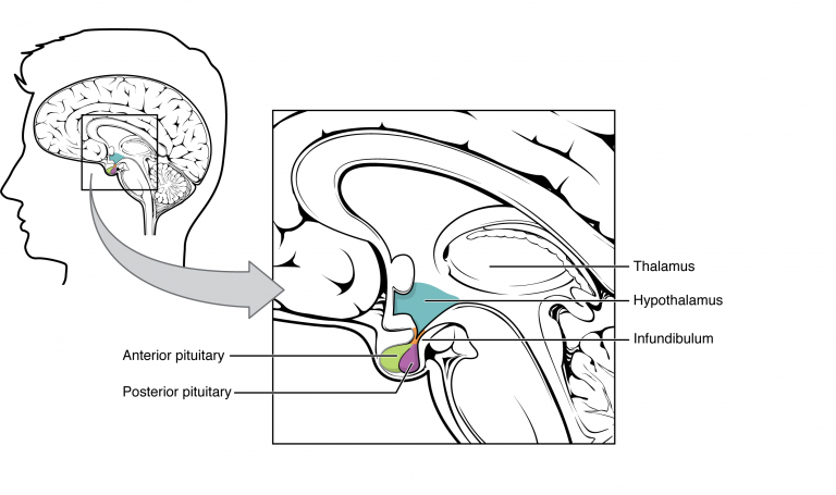
We’ve already looked at the hypothalamus, part of the diencephalon of the brain. The hypothalamus has both neural and endocrine functions. The pituitary gland (or hypophysis) is a pea-shaped gland that hangs like a boxing bag from the hypothalamus by a stalk, the infundibulum. The origin of the term hypophysis is Greek, referring to its position and growth under the brain. The pituitary is also commonly referenced as the master gland, but the truth is that it has a master, the hypothalamus. The hypothalamus is the connection between the nervous and endocrine systems.
The Posterior Pituitary (Neurohypophysis)
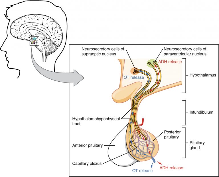
The posterior pituitary is nervous tissue; hence, it’s named the neurohypophysis. It consists of special axons and axon terminals from neurosecretory neurons. The posterior pituitary does not synthesize any hormones. The hormones from the posterior pituitary are produced by neurosecretory cells of the hypothalamus. These hormones are transported by axoplasmic transport within axons to be stored in the axon terminals.
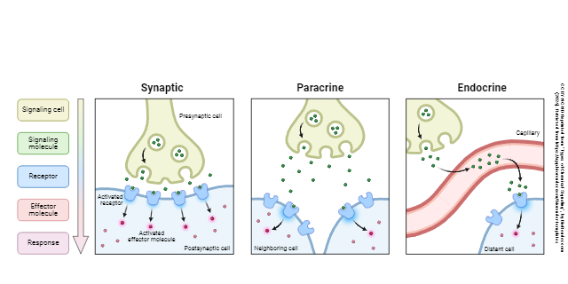
When these hypothalamic neurons send signals, hormone is released from the axon terminals housed in the posterior pituitary. In synaptic signaling, a peptide neurotransmitter is released into a synaptic cleft. In endocrine signaling, a peptide hormone is released directly into the bloodstream.
The Anterior Pituitary (Adenohypophysis)
The pituitary has two lobes that are named based on their position and functional relationship to the hypothalamus. The anterior lobe of the pituitary, also called the adenohypophysis, is about 75% of the total pituitary weight. The second lobe is the posterior pituitary or neurohypophysis.
The terms adeno- and neuro- are very important to understand their relationships with the hypothalamus. “Adeno–” means “gland”, so the relationship of the hypothalamus and anterior pituitary is a gland-to-gland relationship. The hypothalamus connects to the adenohypophysis through a vascular system, the hypophyseal portal veins. The hypothalamus produces hormones that will travel through these portal veins and then release a group of hormones from the anterior pituitary, which then target other cells or glands.
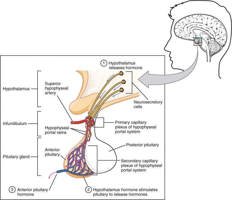
Let’s look at growth hormone as an example using the picture above:
- The hypothalamus receives a signal that more growth hormone is needed. In response, special neurosecretory cells (which respond to neural control), secrete growth hormone releasing hormone (GHRH).
- This hormone needs a route to travel to the anterior pituitary. GHRH travels through blood vessels that connect the hypothalamus to the adenohypophysis. We give these blood vessels a special name, hypophyseal portal veins.
- Specialized cells, somatotrophs, in the adenohypophysis have receptors for GHRH. When this hormone binds to the GHRH receptors, the somatotrophs secrete growth hormone (GH). Growth hormone is sometimes abbreviated with the prefix “human” (hGH).
- GH targets cells in the body, especially cells in the liver, skeletal muscle, bone, and adipose tissue. We will discuss the actions of growth hormone further in
Objective 5.
The Hypothalamic–Pituitary–Adrenal (HPA) Axis
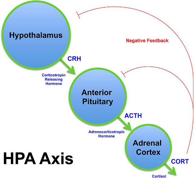
Let’s look at one more example: the hypothalamic-pituitary-adrenal (HPA) axis.
- The hypothalamus secretes corticotropin-releasing hormone (CRH) which travels through the hypophyseal portal veins to the anterior pituitary.
- The baton is passed as the anterior pituitary releases adrenocorticotropic hormone (ACTH).
- ACTH travels through the bloodstream to receptors on the superficial part of the adrenal glands, the adrenal cortex.
- The adrenal cortex will secrete other hormones; in this image, we see cortisol released. Notice that this is a negative feedback system.
You will need to understand many hormone pathways. As you are introduced to different pathways, practice drawing these until you have each of them mastered.
To understand the control of the hormones to and from the pituitary, it is important to learn some terminologies. To encourage the release of hormones from the pituitary, the hypothalamus secretes a number of releasing hormones. If “releasing” is in the name of the hormone, it comes from the hypothalamus. If the hypothalamus needs to suppress the action of the pituitary, it secretes specific inhibiting hormones. Again, this illustrates the negative-feedback responses of the endocrine system. If a hormone is a tropic hormone, it is released from one endocrine gland and targets another. The term tropic (pronounced “troh-pick”, with a long ō) may also be used in the name of the specific cells that secrete it. Thyrotrophs, corticotrophs, and gonadotrophs are all cells of the anterior pituitary that secrete a particular hormone that targets another gland.
Media Attributions
- U14-012 Hypothalamus-Pituitary Complex © Betts, J. Gordon; Young, Kelly A.; Wise, James A.; Johnson, Eddie; Poe, Brandon; Kruse, Dean H. Korol, Oksana; Johnson, Jody E.; Womble, Mark & DeSaix, Peter is licensed under a CC BY (Attribution) license
- U14-013 Posterior Pituitary Complex © Betts, J. Gordon; Young, Kelly A.; Wise, James A.; Johnson, Eddie; Poe, Brandon; Kruse, Dean H. Korol, Oksana; Johnson, Jody E.; Womble, Mark & DeSaix, Peter is licensed under a CC BY (Attribution) license
- U14-001 Types of Chemical Signaling © BioRender is licensed under a CC BY-NC-ND (Attribution NonCommercial NoDerivatives) license
- U14-013 Anterior Pituitary Complex © Betts, J. Gordon; Young, Kelly A.; Wise, James A.; Johnson, Eddie; Poe, Brandon; Kruse, Dean H. Korol, Oksana; Johnson, Jody E.; Womble, Mark & DeSaix, Peter is licensed under a CC BY (Attribution) license
- U14-014 HPA Axis Diagram © Adams, Shelley is licensed under a CC BY-SA (Attribution ShareAlike) license

.svg)