Sensory Systems | Olfactory (Smell) System
Objective 8
Explain the location and function of olfactory receptors. Describe the path taken by neural information from the olfactory receptor sheet to the brain.
The olfactory sense is probably the oldest and least modified of the senses developed in the process of human evolution. Even bacteria have a chemical sense — they move toward food sources — even though they have no nervous system. The simplest nervous systems probably had the ability to detect chemicals, and then narrowed in on some of those chemicals as neurotransmitters or hormones while others were “just” used for the sense of smell.
The Human Genome Project identified about 30,000 functional genes; about 1% of those (300 in all) are used for this single sense. When you think of all the processes going on in the body, 1% is a huge number. It would be as though this textbook devoted 16 of its 1600 pages to olfaction. (Which it won’t, thankfully.)
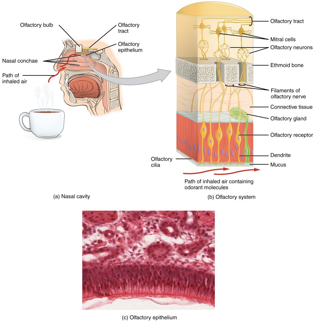
Olfactory receptors are found on the superior surface of the nasal sinuses. In humans, the olfactory receptor sheet is about the size of a postage stamp. In dogs, it’s about the size of a page in this book, which accounts, in part, for the superior sense of smell in dogs.
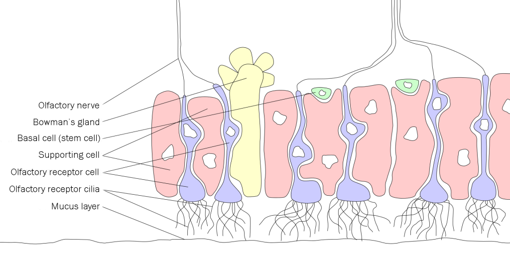
Odorants are small molecules, carried by air, that are dissolved in the mucus layer that covers the olfactory epithelium.
Olfactory transduction depends on a G protein pathway. We will discuss these pathways in more detail in Unit 13. Olfactory cells have cilia with protein receptor molecules that specifically bind a certain type of odorant. When an odorant binds to a olfactory receptor, it triggers the G protein biochemical pathway and sets into motion a cascade of chemical reactions that result in the opening of thousands of Na+ channels.
The delicate nerve axons which pass through the cribriform plate of the ethmoid bone are easily torn by head trauma. When this happens, the patient permanently loses the sense of smell, a condition called anosmia (Greek οσμη, osme, “smell”). Collectively, these nerve axons form the olfactory nerve (CN I).
The axon terminals of the olfactory nerve make synaptic contact with the dendrites of mitral cells and tufted cells in a literal ball of synapses called the olfactory glomerulus. A number of other neurons (juxtaglomerular neurons) surround the glomerulus but we won’t discuss those. The glomeruli are the processing units for olfactory information, with wiring analogous to that of the retina we studied earlier.
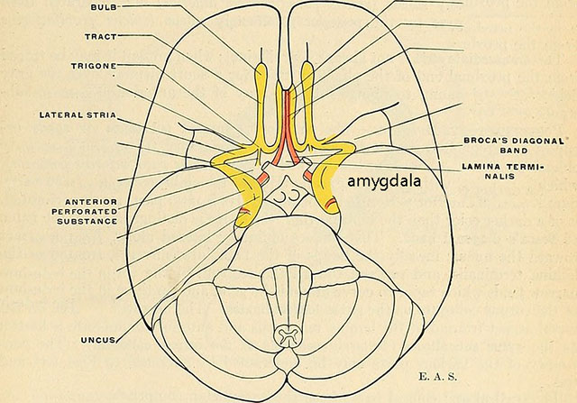
The mitral and tufted cells send axons via the olfactory tract to several areas of cortex, notably the piriform cortex but also periamygdaloid cortex, surrounding the amygdala; entorhinal cortex; the olfactory tubercle; and the anterior olfactory nucleus. These areas are shown in yellow in this diagram. You do not need to learn their names; this diagram shows their general location. The lamina terminalis is interesting, because this is the anatomical location of the border between the thalamus (diencephalon) and cortex (telencephalon, forebrain) as the forebrain grows over the thalamus like an overactive yeasty bread overlapping the edges of its container.
You might remember from Unit 11 that the tip of the temporal lobe also contains structures of the limbic system that are involved in emotional responses. You’d be right to remember that. The piriform cortex is nearby, and shares extensive connections with, the amygdala and entorhinal cortex which in turn is hooked up to the hippocampus. Being sort of a dense young man, I made the mistake of telling my then-girlfriend (who later forgave me and became my wife) that I was buying her a particular brand of perfume because my first girlfriend in high school wore it. I found out a lot of important things about the connections of the olfactory brain and the emotional brain that day.
Media Attributions
- U12-073 Olfactory © Betts, J. Gordon; Young, Kelly A.; Wise, James A.; Johnson, Eddie; Poe, Brandon; Kruse, Dean H. Korol, Oksana; Johnson, Jody E.; Womble, Mark & DeSaix, Peter is licensed under a CC BY (Attribution) license
- U12-074 Olfactory Epithelium © Sigler, Marian adapted by Jim Hutchins is licensed under a CC BY-SA (Attribution ShareAlike) license
- U12-075 Location of Olfactory Cortex in Human Brain © Gray, Henry, 1825-1861; Spitzka, Edward Anthony, 1876-1922 adapted by Jim Hutchins is licensed under a CC BY-SA (Attribution ShareAlike) license
