Sensory Systems | Gustatory (Taste) System
Objective 9
State the three types of taste papillae and five primary taste sensations. Explain the location and function of gustatory receptors. Describe the path taken by neural information from the gustatory receptors to the brain.
Like olfaction, taste (gustation) is a chemical sense, which depends on chemical substances dissolved in saliva. Taste and smell work together and are hard to separate from each other, which is why holding your nose can sometimes reduce the nasty taste of a dose of medicine.
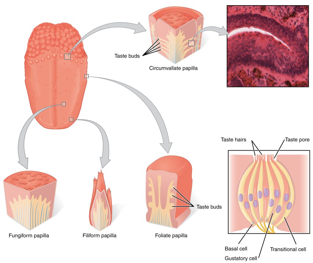
Within each papilla are found a hundred or so taste buds; each of these taste buds comprises 30-100 taste receptor cells and basal cells. Basal cells are glia-like cells which provide structural and chemical support. The apical (top) surface of a receptor cell is studded with microvilli. The microvilli are encrusted with receptor proteins which are sensitive to tastants (dissolved substances which trigger taste sensations).
The tongue has a rough surface because of bumps called papillae (sing. papilla, Latin, “nipple”). They come in four different types: filiform, fungiform, foliate, and vallate.
Filiform papillae are by far the majority of the papillae. They give the tongue its rough surface, but do not contribute to the taste function of the tongue.
The other three types of papillae have a taste function. In these papillae, gustatory receptor cells are clustered in taste buds.
- Fungiform papillae (Latin fungus, “mushroom”), as the name suggests, look like little mushrooms.
- Foliate papillae (Latin folium, “leaf”), are leaf-shaped papillae found along the lateral surface of the posterior tongue.
- Vallate (or circumvallate) papillae (Latin vallatus, “surrounded by a wall”) form a V-shaped row along the posterior tongue. There are usually 7-12 of these, and it’s easier to see them on a friend’s tongue than on your own. Go ahead and ask to see them. It’s not weird if you’re studying anatomy.
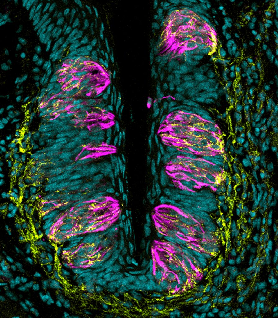
Within each papilla are found a hundred or so taste buds; each of these taste buds comprises 30-100 ciliated taste receptor cells with a glial-like cell, the basal cell, alongside for structural and chemical support.
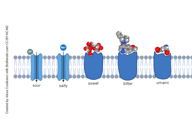 There are five primary tastes, because there are five types of taste receptors, each with a different relevant stimulus and different transduction mechanism. The five primary taste sensations are:
- sour or acid, which measures the concentration of H+ ions; HCl in water is the “index” taste that we measure other tastes against;
- salty, with an index taste of Na+Cl– (table salt) in water;
- sweet, with an index taste of sucrose (table sugar) in water;
- bitter, with an index taste of quinine (as in tonic water);
- umami, Japanese for “tasty” or “meaty” with an index taste of monosodium glutamate, as in protein-rich foods such as sushi.
Most of the taste receptors are located on the tongue, but the lips, soft palate and oropharynx contain taste receptors as well. Oddly, taste receptors are found outside the oral cavity but their wiring and therefore functions are not known.
We used to believe that different taste receptors were found in different locations, and children still do Science Fair projects which purport to show the different distribution of different taste sensations, but more recent evidence suggests that all five types of receptors are pretty much evenly spread over the surface of the tongue, lips, soft palate and oropharynx. However, there is a tendency for the sensations of salty, sweet and umami to be perceived as stronger towards the front of the tongue with sour and bitter perceived as stronger in back. This is thought to be the way that we decide what should go into our mouth (i.e. salty, sweet, and umami) and what might elicit a vomit reflex by activating the vagus (CN X: sour and bitter).
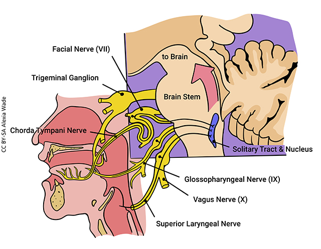
Taste information from the taste buds is transferred to afferents from three cranial nerves.
- Information from the anterior 2/3 of the tongue is carried on a branch of the facial nerve (CN VII);
- Information from the posterior 1/3 of the tongue is carried on a branch of the glossopharyngeal nerve (CN IX)
- Information from the tonsils, palate, and oropharynx is carried on a branch of the vagus nerve (CN X).
Receptors which give us a strong sense of noxious taste are more likely to be found in the oral cavity outside the tongue. Activation of the vagus, then, is most likely to trigger a gag reflex; contrariwise, damage to the vagus suppresses this reflex. Since the vagus innervates the stomach directly, this makes sense functionally.
These three nerve branches combine to form a bundle of axons called the chorda tympani.
Some authorities would add a fourth source of taste information, arriving on the trigeminal nerve (CN V). Stimulation of pain receptors by the chemical capsaicin, found in hot peppers, produces a pain response (and the subsequent release of endogenous opiates) without actual tissue damage. This is why some people find consuming spicy foods to be pleasurable.
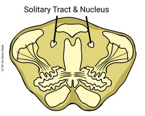
The chorda tympani carries information to the nucleus of the solitary tract in the medulla.
The nucleus of the solitary tract is — wait for it — a nucleus that surrounds the solitary tract in the medulla.
The nucleus, shaped like a long tube and appearing like a donut in cross-section, is why the solitary tract is called “solitary”: in myelin-stained sections, the solitary tract (myelin surrounding axons appears black) stands off by itself like a bullseye.
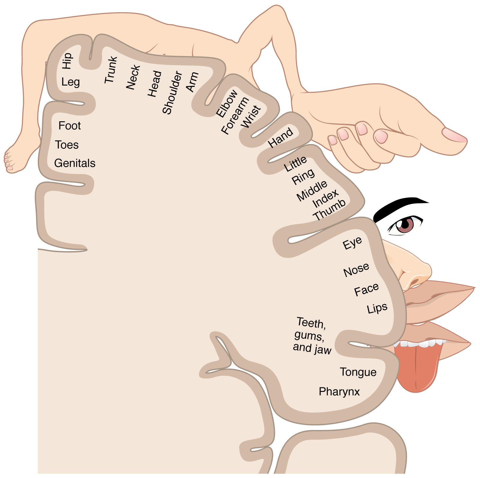
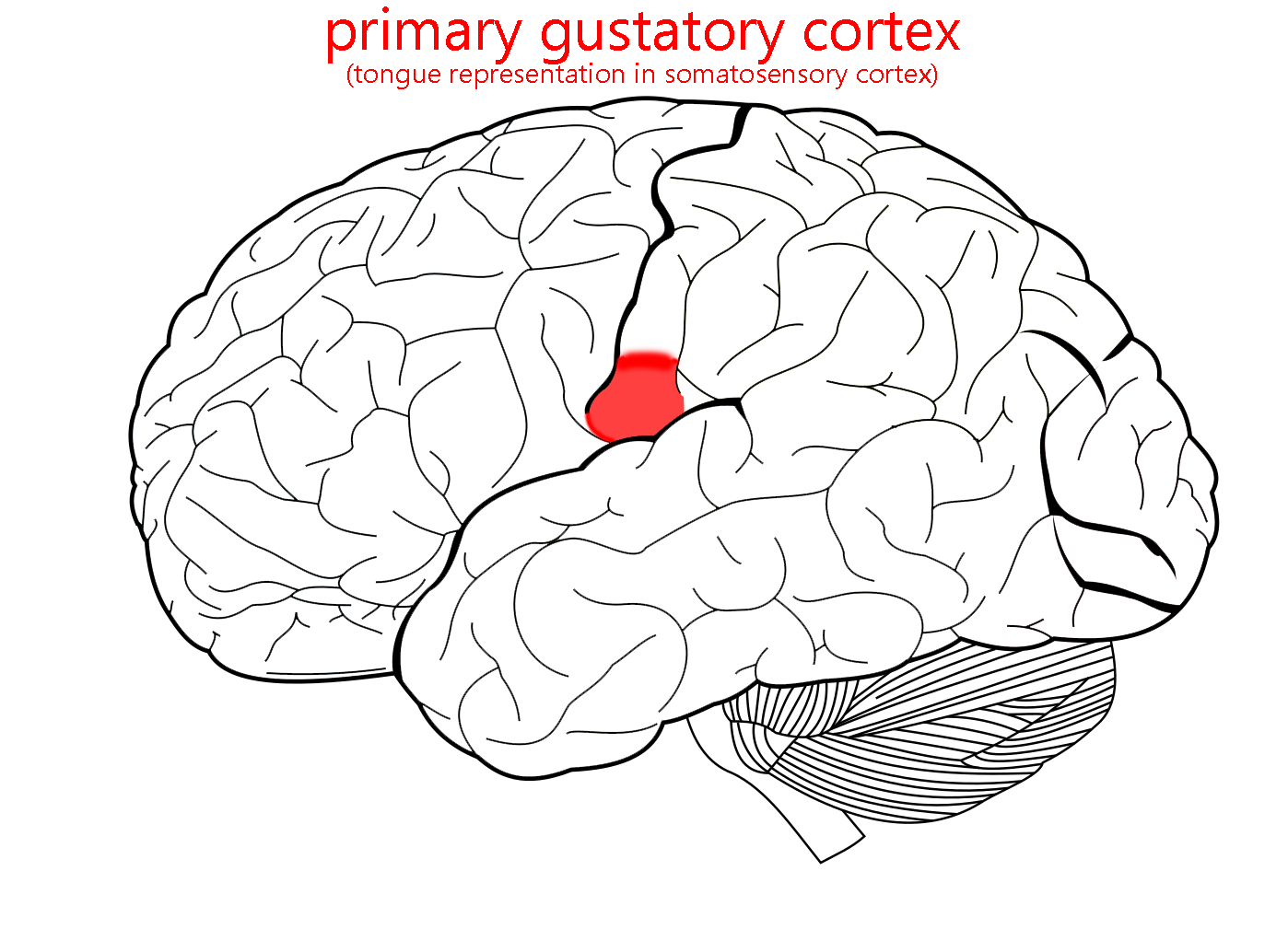
From the solitary nucleus, taste information is relayed to the thalamus (same as touch information from the oral cavity) and then to the lateral part of the postcentral gyrus. This cortical area is the same as the “mouth area” for touch in the somatosensory homunculus we saw earlier. The chef Justin Wilson used to exclaim about his Cajun cooking, “tastes so good it makes your tongue wanna slap your brains out”. In a very real sense, strong flavors make us feel as though our tongue has been physically touched, and there’s an anatomical basis for that sensation.
Media Attributions
- U12-076 Gustatory Tongue © Betts, J. Gordon; Young, Kelly A.; Wise, James A.; Johnson, Eddie; Poe, Brandon; Kruse, Dean H. Korol, Oksana; Johnson, Jody E.; Womble, Mark & DeSaix, Peter is licensed under a CC BY (Attribution) license
- U12-077 Taste Papilla Taste Bud Pink Axons © Gaillard, Dany Ph.D., and Barlow, Linda Ph.D., University of Colorado Anschutz Medical Campus is licensed under a CC BY-NC (Attribution NonCommercial) license
- U12-078 Taste Receptors © Crookston, Alexa is licensed under a CC BY-NC-ND (Attribution NonCommercial NoDerivatives) license
- U12-079 Gustatory Pathways © Wade, Alexia is licensed under a CC BY-SA (Attribution ShareAlike) license
- 12-065a Solitary Tract and Nucleus © Wade, Alexia is licensed under a CC BY-SA (Attribution ShareAlike) license
- U12-045 Sensory Homunculus © Betts, J. Gordon; Young, Kelly A.; Wise, James A.; Johnson, Eddie; Poe, Brandon; Kruse, Dean H. Korol, Oksana; Johnson, Jody E.; Womble, Mark & DeSaix, Peter is licensed under a CC BY (Attribution) license
- U12-081 Primary Gustatory Cortex © Carter, Henry Vandyke adapted by Jim Hutchins and Alexa Crookston is licensed under a CC BY-SA (Attribution ShareAlike) license
