Sensory Systems | General Features of Sensory Systems
Objective 3
Identify the general features of a sensory system. Define transduction. Define exteroceptor, interoceptor, proprioceptor. Define mechanoreceptors, thermoreceptors, nociceptors, photoreceptors, chemoreceptors, osmoreceptors.
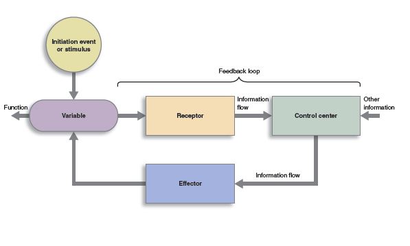
Sensory systems are the parts of the nervous system that receive information from the external or internal environment. Remember the model of homeostasis presented in Unit 1. The “receptors” in the homeostasis model (beige rectangle) are the same as “sensory systems” in the nervous system.
Let’s look at some common features of sensory systems.
Each type of receptor has a relevant stimulus. This can be photons (as in vision), or chemicals present in the environment around the receptor (as in taste and smell), or pressure waves of air (as in hearing), or temperature. Each receptor has a single relevant stimulus that it responds to. For example, photoreceptors in the eye don’t care what the temperature is.
We will see that these relevant stimuli can be quite narrowly defined. In the case of vision, only certain wavelengths of light are able to trigger a response. Only certain types of sound waves are able to activate the receptors involved in hearing. Receptors in the skin “take apart” qualities of the stimulus; different receptors are activated, and the information from these receptors takes different pathways to the brain.
 Neuroscientists call the pathways that carry these different qualities of a single stimulus sensory modalities. For example, imagine reaching out to pet your cat who is snuggled up in a blanket. Your touch (somatosensory) system is activated. You can feel the warmth of their body through the blanket. The blanket is a fleece-y texture. As they softly purr, you feel the vibrations that are created. For this single stimulus, temperature and touch and vibration are different modalities, with different pathways in the central nervous system and different destinations in the somatosensory cortex.
Similarly, different modalities in the visual system are activated. You note the difference in color and texture and distance of the cat and blanket; color and texture detection and depth perception are all different modalities within the visual system. Your visual system recognizes the shape of a cat even though most of the cat is covered by the blanket.
In many cases, relevant stimuli have no meaning for the central nervous system. For example, the CNS does not use photons to transmit or process information. For these stimuli, the relevant stimulus must be transduced to an electrical and/or chemical signal the nervous system can use, then the information must be processed by the nervous system.
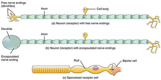
The neurons responsible for detecting these different qualities are shown here. The top neuron is anatomically a free nerve ending; this type of receptor detects the temperature of the stimulus. The middle neuron is a Pacinian corpuscle, which detects the vibrations from your cat’s soft purr as you feel them under the blanket. The bottom neuron is a photoreceptor, which detects the amount and wavelength of photons and passes that information to other cells that begin the information processing chain.
Most sensations, even those that trigger reflexes, are capable of producing a conscious perception. To do this, they must reach the region of the cerebral cortex that is responsible for that sensation, as described in Unit 11. If everything sensed in the environment reached consciousness, we would be overwhelmed with stimuli. The thalamus is responsible for deciding which stimuli reach consciousness; accordingly, all sensory systems (except olfaction, more about that later) are routed through the thalamus before they reach the cortex.
These two tables summarize the classification of different receptor types using two different schemes.
Classification by Source of Input
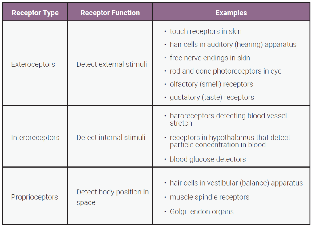
In the first table, we classify receptors based on the source of the energy that activates them: from outside the body; from inside the body; or from body movements.
Here is a little more information about each of these three types of receptor, classified based on the source of the sensory input.
Exteroceptors receive their stimuli from the external environment. Examples of these are light particle/waves that are reflected off of objects in the environment, pressure waves of air that hit the eardrum to create the perception of sound, and your friend tapping you on your shoulder to get your attention (touch). All of these stimuli come from outside your body. When we think of the five senses (sight, hearing, touch, taste, smell), we are thinking of exteroceptors.
Interoceptors receive their energy from the internal environment. Examples of these are oxygen levels, glucose levels, CO2 levels in the blood, or the state of stretch of our stomach and intestines. In general, interoceptors do not reach conscious perception. You might say, “that light is really bright”, but you would be unlikely to say, “my blood CO2 is really low”.
Proprioceptors are receptors that integrate information about the state of stretch of skin, muscles and tendons, with information about gravity, to produce a perception of where our joints are in space. For example, if you close your eyes and someone lifts your leg, you perceive that your leg is extended even though you can’t see it.
Classification by Source of Input
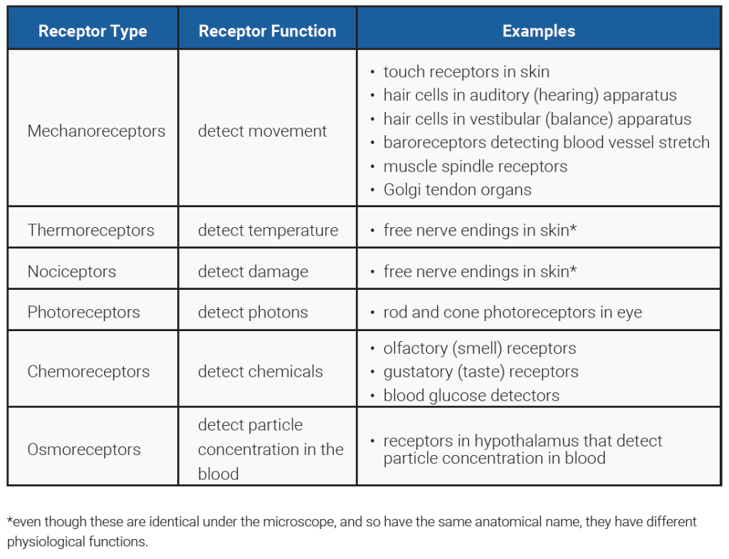
In the second table, we classify receptors based on the physics of the energy that activates them: movement; temperature; pain; photons; chemicals; or particle concentration in blood.
Notice that each of the specific receptor types in the rightmost column is represented twice, once in each classification scheme. Thus rods and cones in the eye are both exteroceptors (i.e. detect external stimuli) and photoreceptors (i.e. detect photons).
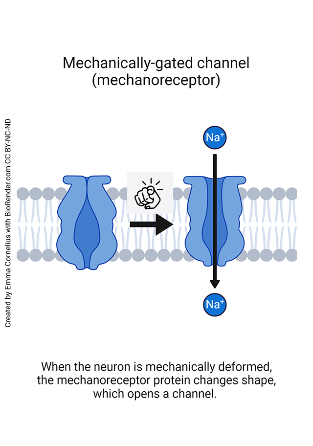
Mechanoreceptors detect movement, such as the pressure of clothing on skin, or a finger touching you, muscles being stretched, or pressure waves of air (i.e. sound). The movement of small rocks in the inner ear produce the sensation of balance.
Thermoreceptors detect temperatures between 4°C (40°F) and 50°C (122°F). Below 4°C and above 50°C, tissue is damaged and pain results. When you touch dry ice or a hot stove, you don’t measure the temperature of it. It just hurts. That’s why we describe being “burned” by a hot stove the same as by liquid nitrogen or dry ice. Scientists believe that one set of thermoreceptors operates for temperatures below body temperature (4°–37°C) and another set for temperatures above body temperature (37°–50°C).
Nociceptors derive their name from the same root as noxious and obnoxious (Latin noxa, “harm, injury, damage”). Nociceptors, then, detect harmful or damaging stimuli. When tissue is damaged, a number of chemical factors are released from the damaged cells (including K+, H+, ATP, NO, histamine, prostaglandins, bradykinin, and serotonin). These stimulate receptors on what appear histologically as free nerve endings in the skin. These are the pain receptors or nociceptors.
Photoreceptors detect photons (particle/waves of light energy). Different wavelengths of light correspond to different hues; there is one photoreceptor type for each of the primary colors (red, yellow, blue) which is what makes them “primary.”
Chemoreceptors detect chemicals in the internal or external environment. Examples of these are found in the senses of taste (sugars, acid, salt, or other substances dissolved in saliva); of smell (a wide variety of airborne molecules dissolved in mucus); and the oxygen, carbon dioxide, and pH receptors which monitor the bloodstream.
Osmoreceptors in the hypothalamus detect the solute levels in blood, and respond by secreting chemicals that regulate water retention or loss. We will look at these in detail as we study the endocrine system.
Central Pathways for Sensory Information
Once the energy from the internal or external environment is transduced into electrical and/or chemical energy that the nervous system uses, we must process that information through one or more nuclei in the central nervous system, and if the information is to reach consciousness, transfer it to the thalamus and then to the cortex.
Understanding these central pathways is therefore important for an understanding of each sensory system.
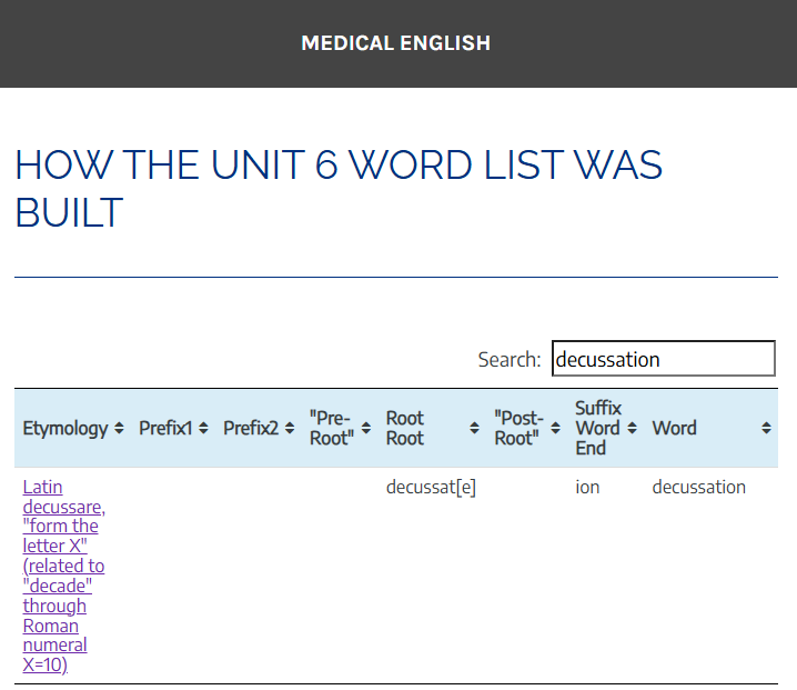 Additionally, almost all sensory information crosses the midline at some point along these central pathways. This crossing is called a decussation. For example, we’ll see that visual information crosses at the optic chiasm (because it resembles the Greek letter chi, written χ). Some of the touch information crosses in the medulla at the sensory decussation. We already saw the decussation of the pyramids in Objective 1.
Media Attributions
- U12-001 Homeostasis © Open Learning Initiative is licensed under a CC BY-NC-SA (Attribution NonCommercial ShareAlike) license
- U12-029b Sleeping Cat © Rakka is licensed under a CC BY-NC-ND (Attribution NonCommercial NoDerivatives) license
- U12-030 Sensory Receptors © Betts, J. Gordon; Young, Kelly A.; Wise, James A.; Johnson, Eddie; Poe, Brandon; Kruse, Dean H. Korol, Oksana; Johnson, Jody E.; Womble, Mark & DeSaix, Peter is licensed under a CC BY (Attribution) license
- U12-031 Type of Receptors by Location of Information table © Hutchins, Jim is licensed under a CC BY-SA (Attribution ShareAlike) license
- U12-032 Type of Receptors by Stimulus Table © Hutchins, Jim is licensed under a CC BY-SA (Attribution ShareAlike) license
- U12-033 Mechanically-gated Channel © Cornelius, Emma is licensed under a CC BY-NC-ND (Attribution NonCommercial NoDerivatives) license
- U12-033a decussation medical english screenshot © Jim Hutchins is licensed under a CC BY-SA (Attribution ShareAlike) license
