Pancreas
Objective 8
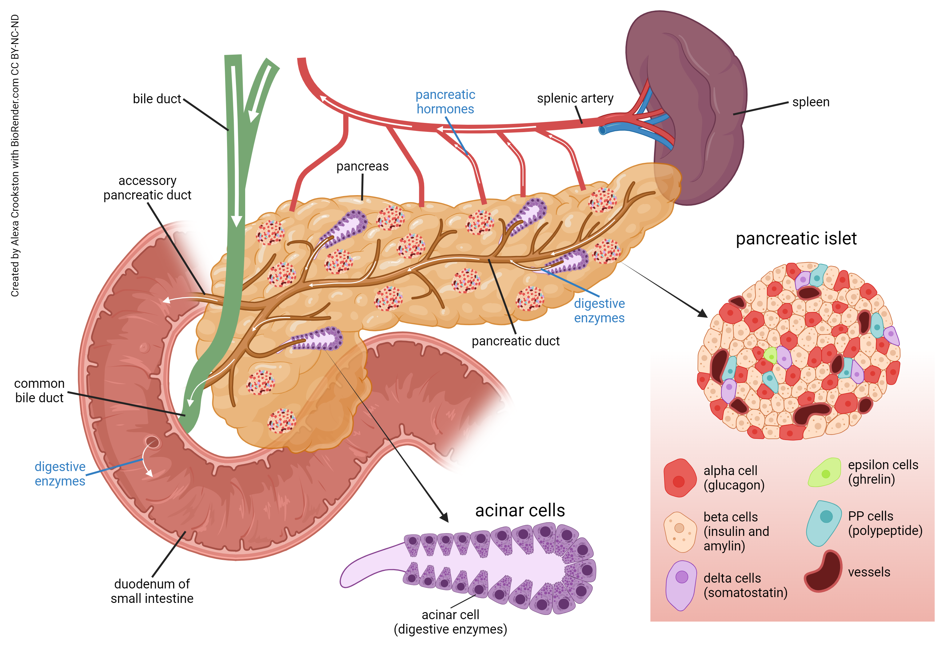
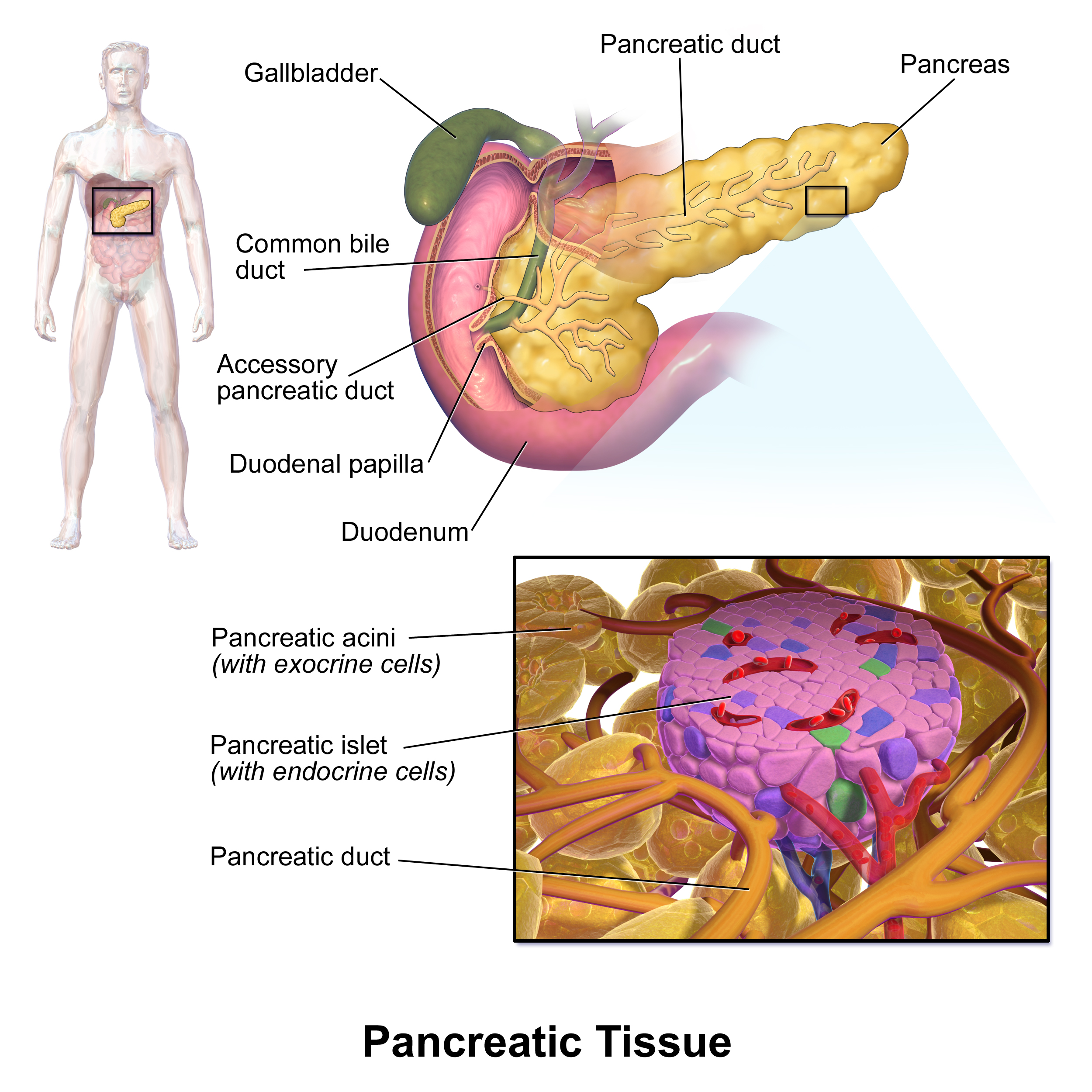
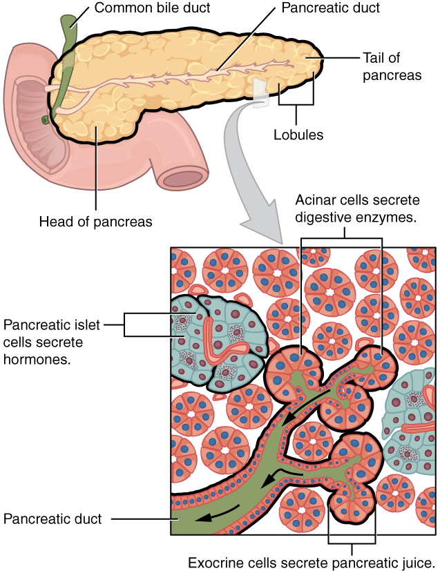
Describe or identify on a diagram the structures in a pancreatic acinus. Describe or identify on a diagram the structures in a pancreatic islet. Identify the pancreatic duct and hepatopancreatic sphincter and explain how these structures are involved in digestion.

We’ve already seen the role of the pancreas as an endocrine organ in Unit 14. Remember that the pancreatic islets are where the cells of the endocrine pancreas reside. Even though the islet cells are a small minority of the pancreatic cells, they exert a huge influence on our physiology.
The cells of the islet, and their endocrine products, are:
- alpha cells: glucagon
- beta cells: insulin
- delta cells: somatostatin
- F cell (also called PP cells): pancreatic
- epsilon cells: ghrelin (Cummings DE et al, NEJM 346:1623–1630, 2002)
We’ve covered the effects of glucagon and insulin in some detail in Unit 14, and you should take a look at them again so they’re familiar to you. Ghrelin may be new to you. It has become a popular area of study because it seems to have a profound effect on stimulating appetite. Increases in ghrelin increase appetite, and mice that are genetically altered to interfere with the ghrelin system are not only skinny, but here’s the kicker: they live longer.

The part we’ll focus on in this unit is the exocrine pancreas. Remember that “exocrine” means secreting externally (“endocrine” means secreting internally). The outside, in this case, is the lumen of the GI tract.
You may have noticed that diagrams have a lot of islets on them; everyone loves the pancreatic islet. Islets are rare, making up only about 1% of the pancreas.
Most of the pancreas is made up of the acini, the exocrine pancreas. Remember that the Latin word acinus means “berry”; the plural of acinus is acini.
Among the useful things made by the exocrine pancreas are:
- Sodium bicarbonate (NaHCO3) — good ol’ Arm and Hammer™ baking soda, the primary buffer system of the blood introduced in Unit 2.
- Amylase — a starch-digesting enzyme.
- Trypsin, chymotrypsin, carboxypeptidase, and elastase — enzymes that break proteins into amino acids.
- Pancreatic lipase — a fat-digesting enzyme.
- Ribonuclease and deoxyribonuclease — enzymes that break down nucleic acids.

That means pancreatic secretions neutralize stomach acid (sodium bicarbonate), inactivate pepsin released from the stomach, and supply enzymes needed in the intestine so that we can break down and absorb the nutrients in our food. All these join the fat-emulsifying bile in the hepatopancreatic ampulla and are released into the duodenum on demand.
Media Attributions
- U18-053 Pancreas © Crookston, Alexa is licensed under a CC BY-NC-ND (Attribution NonCommercial NoDerivatives) license
- U18-054 Islets and Acini © BruceBlaus is licensed under a CC BY (Attribution) license
- U14-047b Pancreas and Islets © Betts, J. Gordon; Young, Kelly A.; Wise, James A.; Johnson, Eddie; Poe, Brandon; Kruse, Dean H. Korol, Oksana; Johnson, Jody E.; Womble, Mark & DeSaix, Peter is licensed under a CC BY (Attribution) license

