Functions of Nervous Tissue
Objective 1
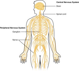
Describe the general structure and overall function of the nervous system.
There are an estimated 80 billion neurons, and the average neuron has 10,000 connections. Neuroscientists used to believe there were about 10 times as many glial cells as neurons, but now it’s believed the neurons and glia are equally abundant.
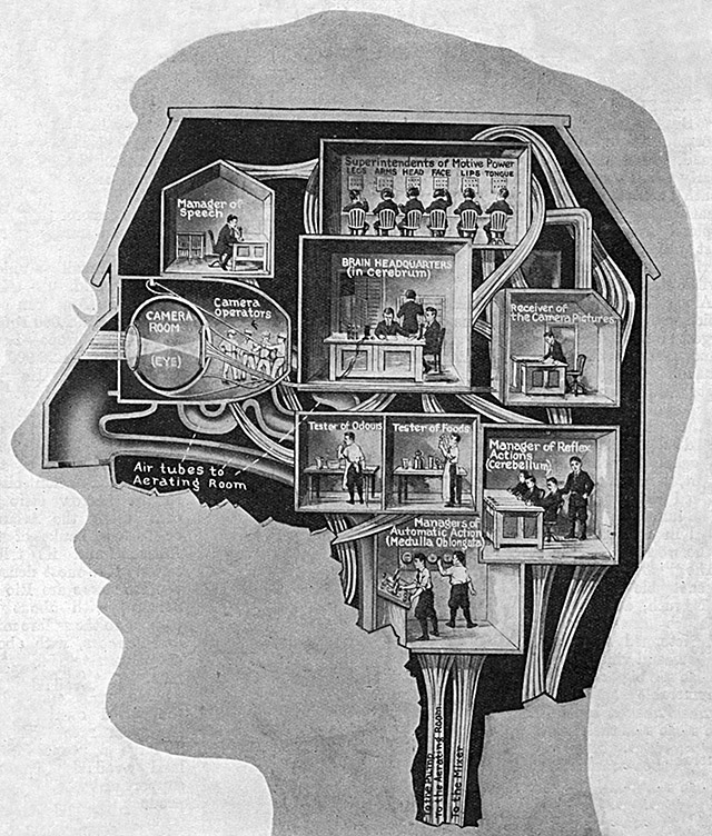
The receiving, processing, and sending of information is a neuron’s specialty. Obviously, with so many neurons engaged in this activity, the brain’s capacity for higher-order functions like learning and memory or language is really remarkable.
Component |
Part of Nervous System |
Receptor |
Sensory |
Control Center |
Central Nervous System (Brain and Spinal Cord) |
Effectors |
Muscles and Glands |
Understanding anatomy and how it relates to physiology (homeostasis) is an essential element of this course. The nervous system is a prime example of how the body maintains homeostasis; in fact, in most animals, the entire nervous system is devoted to homeostasis. Humans are unique in the complexity of their behavior. They use their brains for many things other than homeostasis, such as reading this page.
Remember that a homeostatic loop (regardless of whether it is positively or negatively controlled) requires a minimum of three elements: a receptor, a control center, and an effector.
Examples of Receptors
Information enters the nervous system on receptors. Examples of this are:
- cells in the retina of the eye
- dendrites detecting temperature changes in the skin
- receptors that detect chemical changes in the blood, such as [H+] (pH)
- muscle stretch receptors
- joint position receptorscon
- chemicals dissolved in air (smell; olfaction)
- sound waves reaching the eardrum (audition)
The receptors of the nervous system are sensory receptors (on the skin surface, in the taste buds of the mouth, in the retina of the eye, and many other places) receive many types of sensations.
The control center of the central nervous system is the brain and spinal cord.
Examples of Effectors
Information leaves the nervous system through effectors — effectors produce an effect. Examples of this are:
- muscle fibers in the biceps brachii muscle
- heart muscle
- smooth muscle in the gastrointestinal tract
- glands producing saliva
- contraction of the biceps femoris
The effectors of the nervous system are smooth muscle, cardiac muscle, skeletal (voluntary) muscle, and glands.
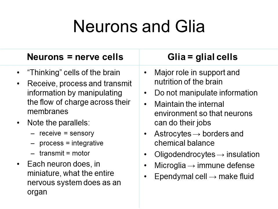
We learned in Unit 7 that there are two types of cells in the nervous system, glial cells (glia) and nerve cells (neurons).
The glial cells perform structural and support roles in the nervous system. They also remain capable of continued cell division (mitosis).
Like all cells of the body, both glial cells and neurons have a charge across their cell membrane, with a slight excess of negative charges inside. Additionally, like most cells, neurons and glial cells have receptor proteins that change the internal biochemistry of the cell in response to outside chemical signals (G protein-coupled receptors).
In Unit 13, we will study the molecular properties that distinguish neurons from all other cells. Neurons are the only cells in the body that are able to process information by changing the flow of charges across the cell membrane. This is accomplished by “gating” ion channels so they open or close in response to particular conditions.
Some sensory neurons have ion channels that open and close in response to environmental changes. Examples include:
- ion channel proteins that change shape to open when put under pressure (mechanically-gated ion channels); and
- ion channel proteins that are closed by a complicated set of chemical reactions in response to photons; and
- ion channel proteins that open in response to temperature changes.
Neurons have ion channels that open or close in response to voltage changes (voltage-gated ion channels).
All neurons have receptor ion channels that change the flow of charges across the cell membrane in response to a chemical signal (ligand-gated ion channels).
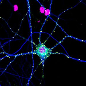
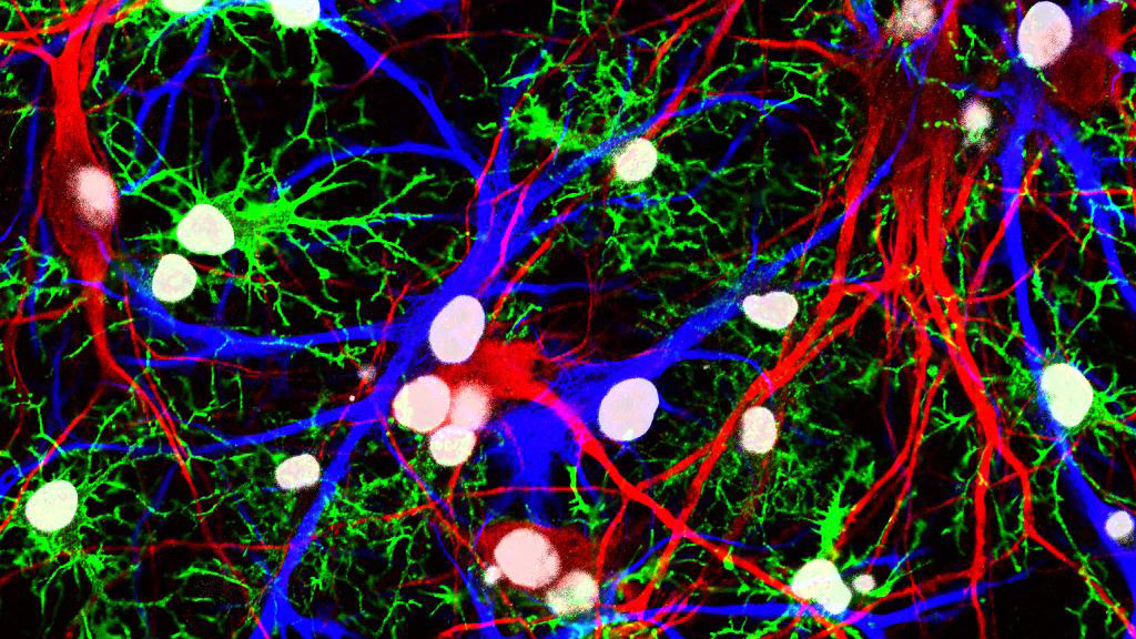 11-005-neurons-and-glia-cropped.jpg” alt=”Neurons and glia” width=”640″ height=”360″ />
