Nephron Structure and Function
Objective 3
Identify the structures that comprise the nephron. Describe the structure and function of the renal corpuscle and the juxtoglomerular apparatus. Identify and describe the three basic functions performed by the nephrons (glomerulus and the renal tubules).
The Nephron
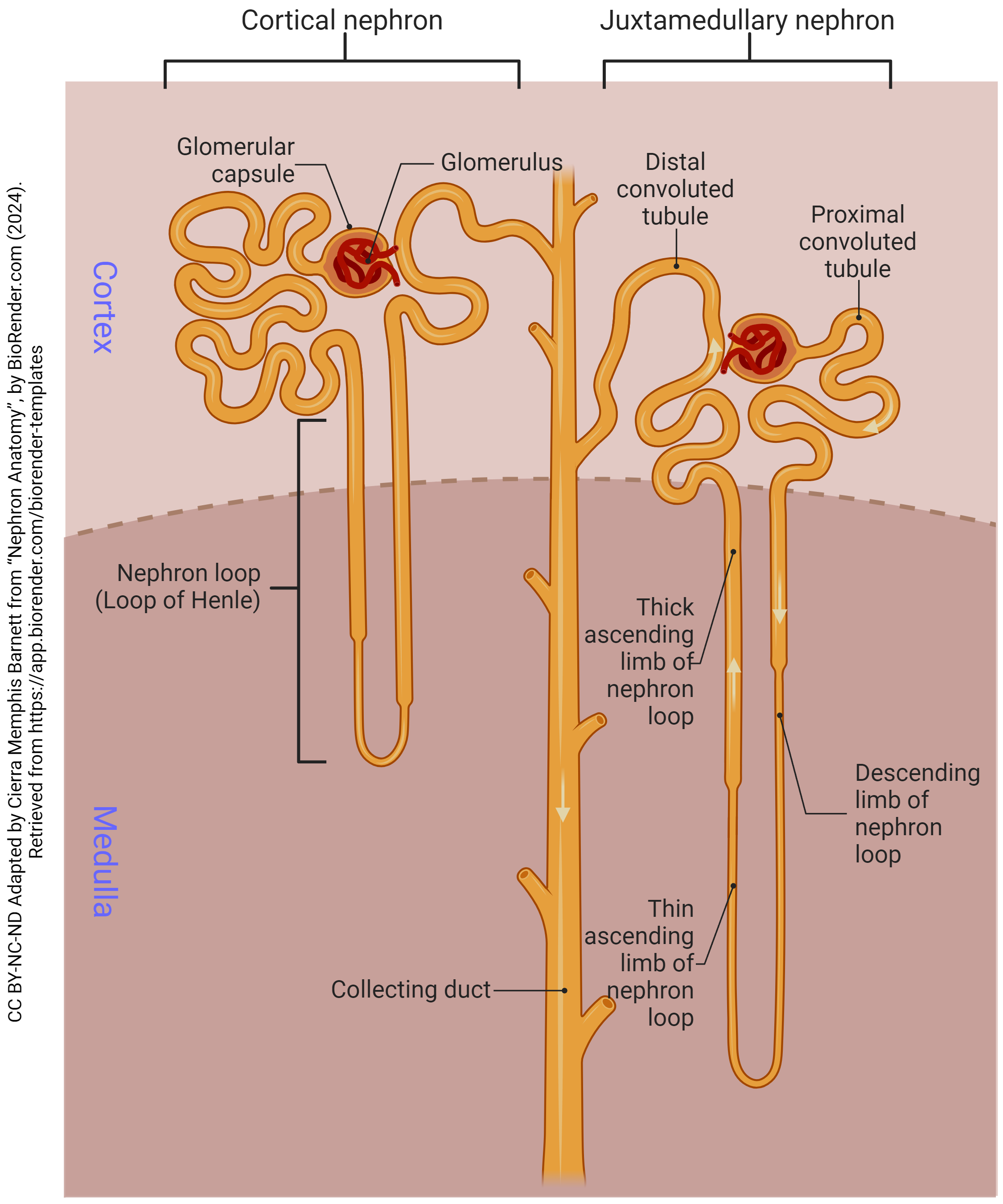
The nephron includes two groups of structures, the renal corpuscle, and the renal tubules. The renal corpuscle consists of the glomerulus (glomerular capillaries) surrounded by a glomerular capsule. The renal corpuscle is the filtering structure of the nephron.
The role of the renal tubules is to modify the filtrate (product of filtration) to facilitate normal urine production. The renal tubules are named based on their shape and/or their position related to the glomerulus. The proximal convoluted tubule is a tightly-coiled tubule attached directly to the glomerulus.
The nephron loop (loop of Henle) forms a hair-pin turn by connecting two lengths, or limbs of the tubule, the ascending and descending. The final tubule is the distal convoluted tubule. Similar to the proximal tubule it is tightly coiled, but is farther away from the glomerulus. (Note that the terms “proximal” and “distal” don’t refer to anatomy in this case, but rather, to the functional order they are found in: the proximal convoluted tubule is the first structure to receive glomerular filtrate, while the distal convoluted tubule receives glomerular filtrate much later in the process.)
Several distal convoluted tubules come together to form a single collecting duct. Many collecting ducts merge to form a papillary duct, which empties into the previously mentioned minor calyx.
There are two types of nephrons, cortical and juxtamedullary. The difference is simply the length of the nephron loops. About 80-85% of nephrons are cortical. Their nephron loops only penetrate the outer regions of the medulla. Juxtamedullary nephrons comprise the remaining 15-20%, with their nephron loops extending into the deepest regions of the renal pyramids. These long loops play a role in the ability to reabsorb water, thus concentrating the urine. In the image below, the nephron on the left is a juxtamedullary nephron. The nephron on the right is a cortical nephron. Notice the difference in the length of the nephron loops and distance extended into the medulla.
The Renal Corpuscle
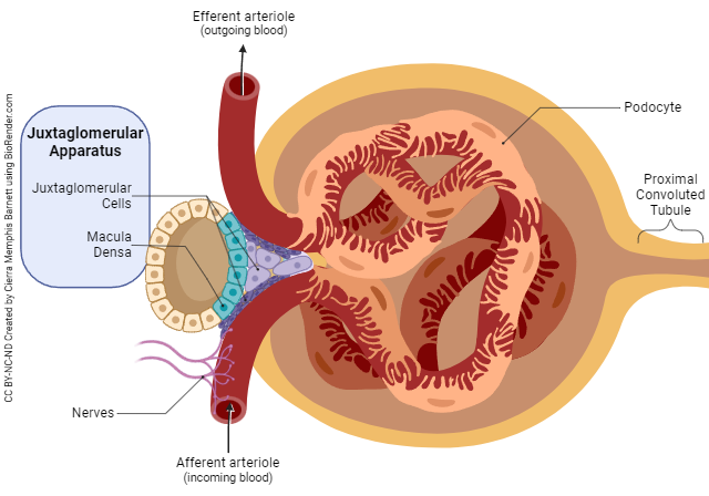
The renal corpuscle is located in the cortex and is the structure of the nephron that filters the blood. As previously mentioned, the corpuscle consists of two components, the glomerulus and the glomerular capsule. Specialized capillaries where filtration occurs make up the glomerulus.
The glomerular capsule is the receptacle for the filtered blood before it enters the tubules. This filtrate is further adjusted as it flows through the nephron before becoming urine. The fluid entering the nephron is blood, within the nephron it is glomerular filtrate.
Filtrate draining from papillary ducts into minor calyces is now urine.
The juxtaglomerular apparatus is formed by a combination of cells from the ascending limb of the nephron loop and the afferent arteriole for each nephron. At this location, the cells of the ascending limb are columnar in shape and tightly packed. This is why this group of cells is called the macula densa.
The wall of the afferent arteriole contains modified smooth muscle cells that control the arteriole’s diameter. Because of their position, these cells are called the juxtaglomerular cells, (“next to the glomerulus”), and together with the macula densa control the blood pressure and rate of filtration within the kidneys.
Glomerular Filtration, Tubular Reabsorption, and Tubular Secretion
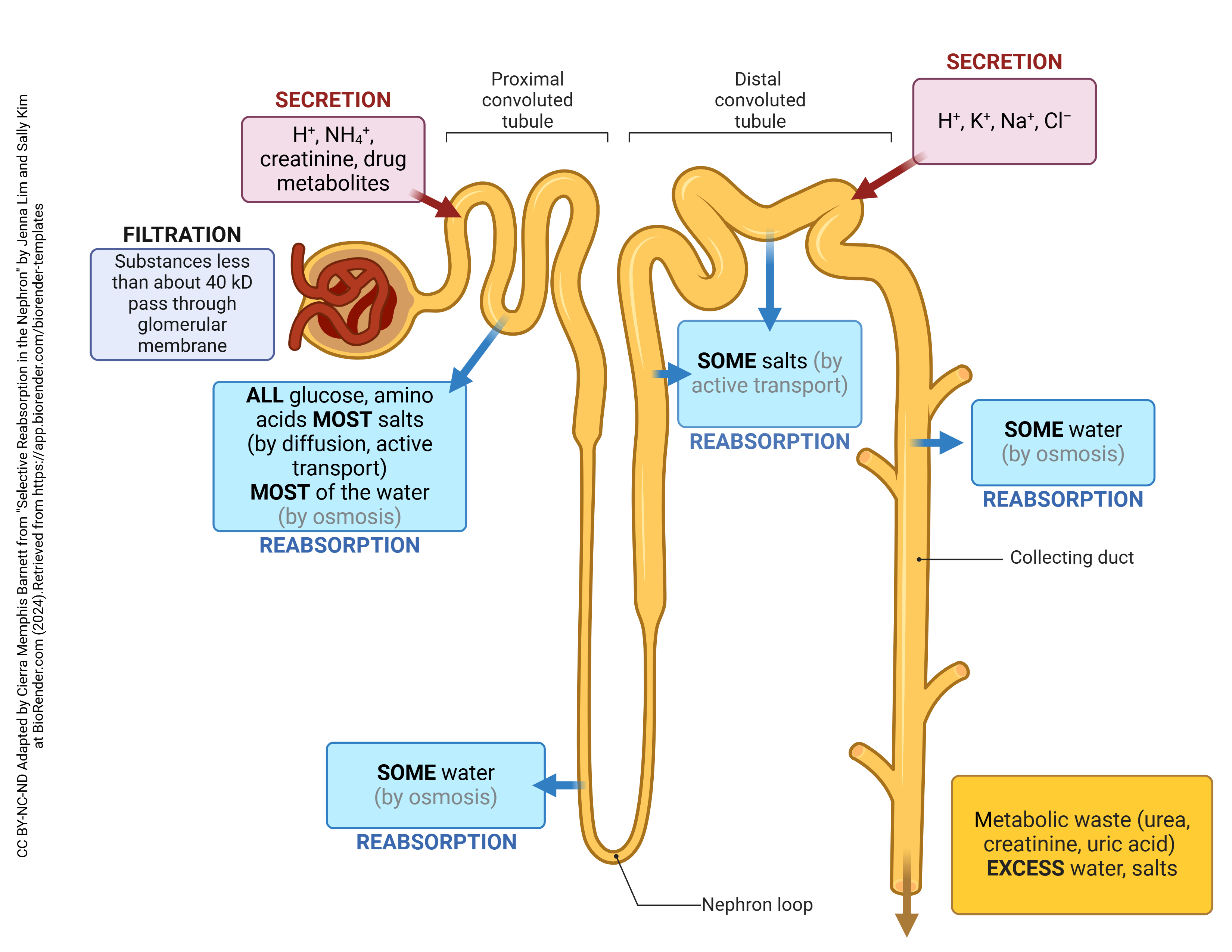
The goal of the nephron is to filter waste-laden blood and produce a final waste product (urine). The kidneys filter about 25% of the cardiac output. Wastes must be removed; pH and water balance adjusted and additional substances added to the filtrate, which will eventually become urine. Three mechanisms accomplish this: glomerular filtration, tubular reabsorption, and tubular secretion. The location of each of these processes is simple; look at the first term, glomerular (glomerulus) and tubular (tubules).
To introduce these briefly, glomerular filtration is the process of taking waste-laden blood, passing it through a membrane, and forming glomerular filtrate. The renal corpuscle facilitates this process.
Once glomerular filtrate is formed, important nutrients that would otherwise be lost in the urine are brought back into the blood stream. This process is called tubular reabsorption.
As filtrate flows through the tubules, renal tubule cells and ductal cells can secrete unwanted substances. So, tubular secretion is the process of removing a substance from the blood and transporting it into the filtrate to be excreted in the urine.
Media Attributions
- U19-009 Nephron Anatomy © Barnett, Cierra Memphis is licensed under a CC BY-NC-ND (Attribution NonCommercial NoDerivatives) license
- U19-010 Juxtaglomerular Apparatus and GFR © Barnett, Cierra Memphis is licensed under a CC BY-NC-ND (Attribution NonCommercial NoDerivatives) license
- U19-011 Reabsorption in the Nephron v3 © Cierra Memphis Barnett | BioRender is licensed under a CC BY-NC-ND (Attribution NonCommercial NoDerivatives) license
