Motor Systems | Somatic Motor
Objective 1
Name the areas of cerebral cortex responsible for production of movements. Explain how information from the motor cortex travels to the spinal cord. Trace the pathway taken by axons through the internal capsule, pyramidal tract, decussation of the pyramids, and lateral corticospinal tract. Describe the role of alpha motor neurons in the spinal cord. Compare and contrast upper and lower motor neurons. Understand how the cerebellum and basal nuclei contribute to voluntary movement.
Now that we’ve gone through all the various parts of the brain and spinal cord, we’re ready to put those parts together into circuits that carry out different functions. This area of neuroscience is called systems neuroscience.
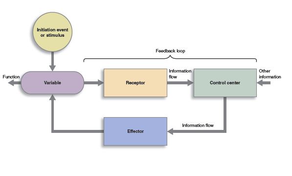
In Unit 1, we introduced the concept of a homeostatic loop. This is a perfect way to organize our thinking about systems neuroscience: a system consists of input from receptors, processing by a set of neurons who extract information from the input and organize it in various ways; and an output to effectors.
Motor Systems Are the Effectors
 The nervous system as a whole contains probably dozens of levels of homeostatic loops all nested within each other, or in computer programming terms, subroutines that carry out different tasks.
 Motor systems are all the parts of the brain and spinal cord that are devoted to the output of the nervous system. Recall that the nervous system has sensory, processing, and motor functions. The motor functions are the only outputs of the nervous system.
Divisions of the Motor Systems
We start with two kinds of motor systems:
- somatic (voluntary)
- autonomic (involuntary)
The embryonic structures that develop into voluntary (skeletal) muscle are the effectors of the somatic motor system. This objective (Objective 1) is about the neural circuitry that controls these skeletal muscle effectors.
The embryonic structures that develop into smooth muscle, cardiac muscle, and glands are collectively called the visceral motor components by neuroembryologists and the autonomic nervous system and enteric nervous system by neurophysiologists. For example, the muscles that control the diameter of blood vessels, and therefore your blood pressure, are part of the autonomic nervous system. Similarly, the system that controls the rate at which your heart contracts is also part of the autonomic nervous system. The salivary gland secretions are part of this autonomic nervous system. The movement of substances through the gut tube (stomach and intestines) is controlled by the enteric nervous system which is closely tied to the autonomic nervous system and sometimes included with it. We’ll look at these circuits in Objective 2.
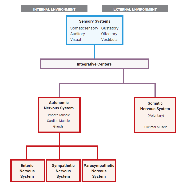
Here is the overall organization of the nervous system we introduced in Unit 11 Objective 2. Motor systems are red in this diagram.
Motor Cortex
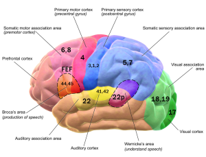 The areas of the cerebral cortex that are responsible for producing conscious, planned movement were introduced in Unit 11, as we were subdividing cortical real estate based on the Brodmann classification. Areas responsible for movement, and their Brodmann numbers, are all in the frontal lobe. They include:
- Area 4: (precentral gyrus): the primary motor cortex, sending axons to the α motor neurons of spinal cord (executing movement).
- Area 6: premotor cortex and supplementary motor area (planning or imagining movement). Among the interesting neurons found here are mirror neurons, which are only active when watching someone else perform an action that you are trying to plan.
- Area 8: Frontal eye fields. This is the area of premotor cortex that plans eye movements.
- Areas 44 and 45: Broca’s area. In most people, Broca’s area on the left side is responsible for the production of symbolic language. This can include speech, sign language, or writing. A lesion in this area produces aphasia, as we saw in Unit 11 Objective 11.
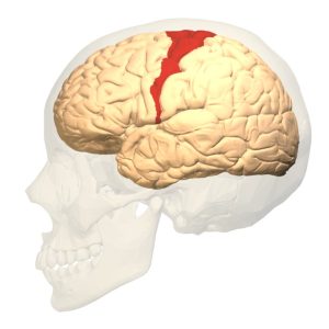 For the time being, we will focus on the primary motor cortex (Brodmann 4, or precentral gyrus).
As for other motor and sensory areas of the brain, neurons which control movement form an orderly map of the body surface within the brain. Recall from Unit 11 Objective 7 that this orderly map is called a homunculus (“little man”).
 The motor homunculus, pictured here, is very similar to the sensory homunculus. The face areas are found in the most lateral part of the precentral gyrus. As we move towards the midline, we encounter, in order: hand, arm, shoulder, trunk, hip; in the medial longitudinal fissure, the map continues with leg and toes.
The lateral surface of the brain is supplied by a different set of arteries and arterial branches than the medial surface of the brain (Unit 11 Objective 8). Recall that the right side of the brain controls the left side of the body and vice versa. Therefore, a stroke (infarction, loss of blood supply) in the right lateral part of the precentral gyrus would produce paralysis of the left side of the face while a stroke in the right medial part of the precentral gyrus would produce paralysis of the left leg.
Information about conscious, willed movement is planned in the supplemental and premotor cortices. These areas then transfer their plan to the motor cortex, which convenes a neural “committee” to “vote” on which axons are going to be activated, and how many nerve impulses will be sent through each axon. The pyramid-shaped cells in layer V of area 4 (precentral gyrus) are called Betz cells. Betz cells are the largest neurons in the human body, and at 0.1 mm across, the cell bodies are visible to the naked eye in stained tissue. Accordingly, these cells have axons that are among the largest (and fastest) in the human body.
 This image shows the motor axons, along with all other axons going to and from the cortex, in a structure that is collectively called the corona radiata (“radiating crown”). The technique shown here, diffusion tensor imaging, is useful in seeing when the Betz cell axons or any other axon bundles are damaged, as in concussion.
Corticospinal Tract
The axons of Betz cells which leave primary motor cortex (Brodmann 4, precentral gyrus) and terminate on motor neurons in the spinal cord are collectively called the corticospinal tract. The first half of the word tells us where it came from and the second half tells us where it’s going; “tract” tells us it’s a bundle of axons in the central nervous system. An older name for this system is the pyramidal tract (so named because of the medullary pyramids they travel through, see below).
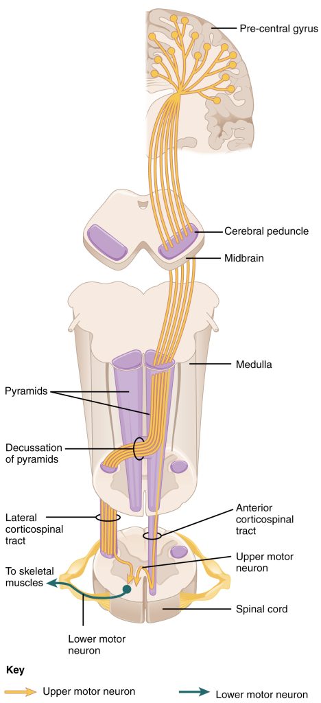 The pyramidal system is a single group of axons that, inexplicably, has seven names. Starting out as part of the
- corona radiata and part of the
- posterior limb of the internal capsule, it forms part of the
- crus cerebri which itself is part of the cerebral peduncles of the midbrain. In the pons, it reverts to the name
- corticospinal fibers before emerging on the ventral surface of the medulla as the
- pyramids, then crosses (decussates) at the
- decussation of the pyramids (at the level of the foramen magnum, the “big hole”in the bottom of the skull), then finally becomes the
- lateral corticospinal tract in the spinal cord.
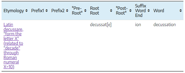
Decussation is a word that derives from the same root as “decade” in English. Because the Roman numeral ten is represented by X, an X-like crossing is called a decussation (i.e., “making an X”)
The decussation of the pyramids is where the information from the left side of the brain crosses to the right side of the body, and vice versa.
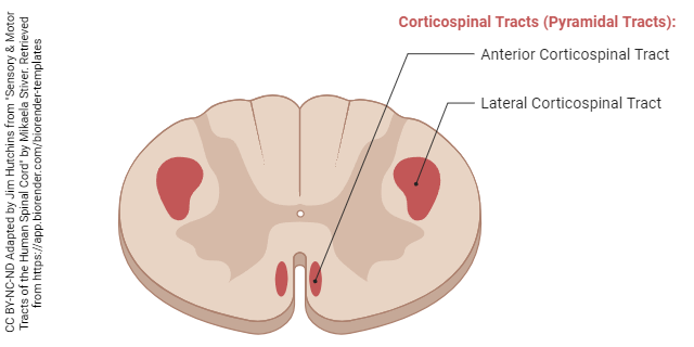
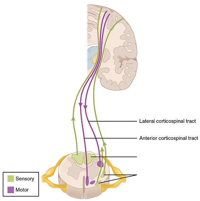
In the spinal cord, these fibers form two pathways. We’ll ignore the less important, uncrossed anterior corticospinal tract and focus on the lateral corticospinal tract. These axons, which started in the motor cortex of the brain, find the level they want (where the spinal nerve that controls the muscle is located), take a sharp right or left turn into the ventral (anterior) horn, and then make a synaptic contact onto the α motor neuron.
Upper vs. Lower Motor Neurons
 Neurologists use a slightly confusing terminology to divide the motor system into two parts.
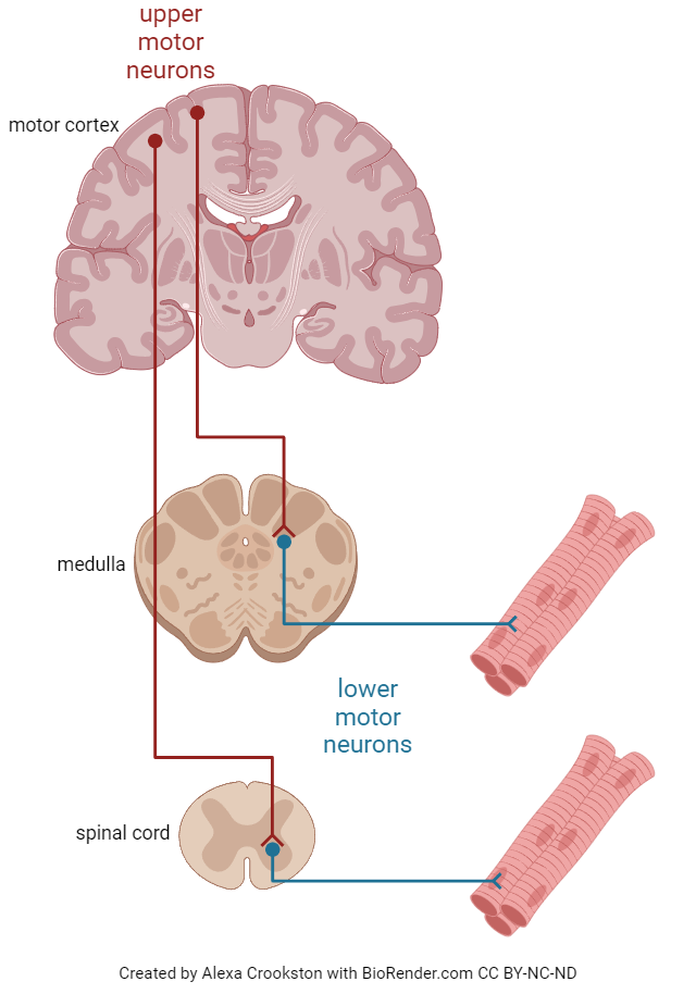
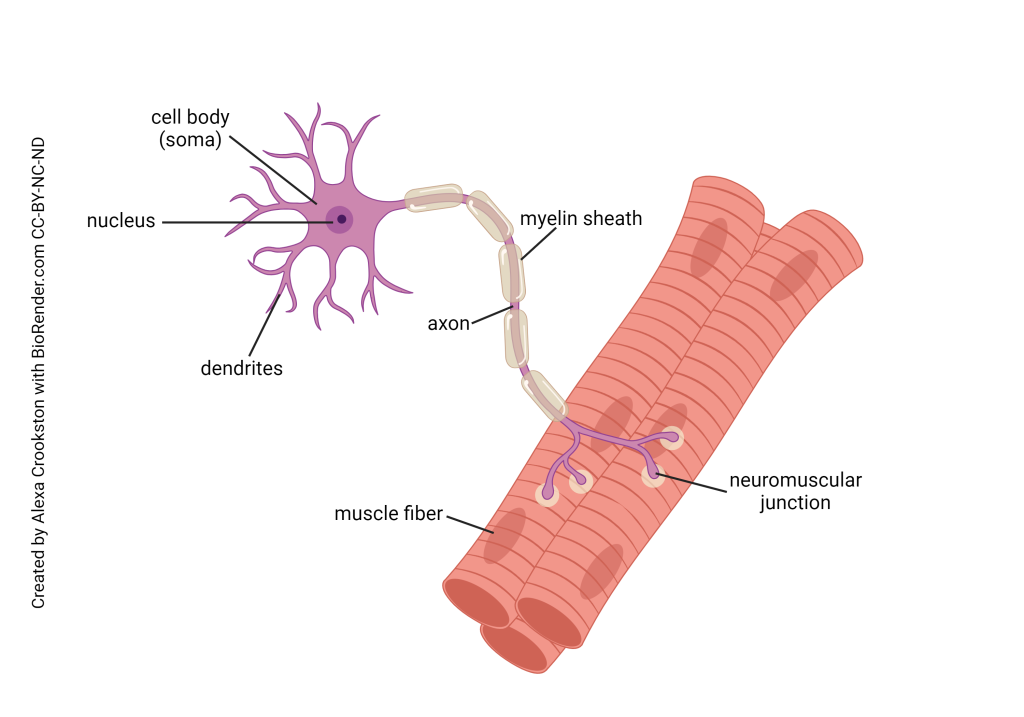
The term lower motor neuron is used to refer to the neuron that makes the final contact between the nervous system and effector organ. Lower motor neurons are the α motor neurons with cell bodies in the anterior horn of the spinal cord and an axon that travels as part of a nerve to end in a neuromuscular junction on skeletal muscle.
Lower Motor Neurons
Each effector of the motor system (glands, smooth muscle, cardiac muscle, striated muscle) is controlled by a (lower) motor neuron.
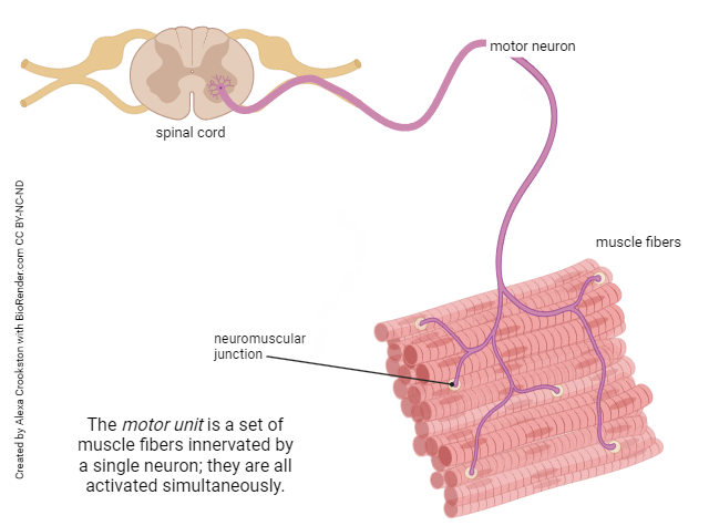 In the somatic (voluntary) motor system, each α motor neuron controls between several and thousands of individual muscle fibers. This α motor neuron and the group of muscle fibers it controls are collectively called the motor unit (Unit 10 Objective 5).
The α motor neuron is the last point of contact between the nervous system and the effector organ. You can think of it as the point where the baton is passed from one type of tissue to another.
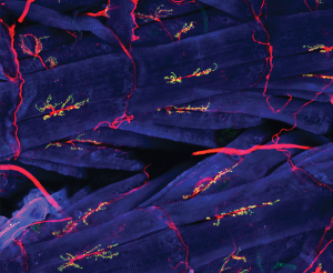
In the case of somatic motor systems, the effector organ that receives the baton is skeletal muscle. The baton is the neurotransmitter acetylcholine and it acts on nicotinic acetylcholine receptors in the muscle’s postsynaptic membrane. These nicotinic acetylcholine receptors are excitatory; they cause muscle contraction (Units 10 & 13). Remember that the entire process, from α motor neuron activation to muscle contraction, is called excitation-contraction coupling. Anatomically, the structure where nerve contacts muscle is called the neuromuscular junction or motor end-plate. It’s just a special name for the synapse between muscle and nerve.
Upper Motor Neurons
All the other neurons in the brain and spinal cord that influence movement, but do not make direct contact with a skeletal muscle fiber, are called upper motor neurons. Examples of these upper motor neurons are neurons of the precentral gyrus (primary motor cortex) of the brain; neurons in the basal ganglia (basal nuclei); and neurons of the cerebellum.
The neurons which make up the pathway from Brodmann area 4 (primary motor cortex) to the cell bodies of α motor neurons in the anterior horn of spinal cord, which we will see later in this objective and name the corticospinal (or pyramidal) tract, are examples of upper motor neurons.
Upper Motor Neurons: Cerebellar Circuits
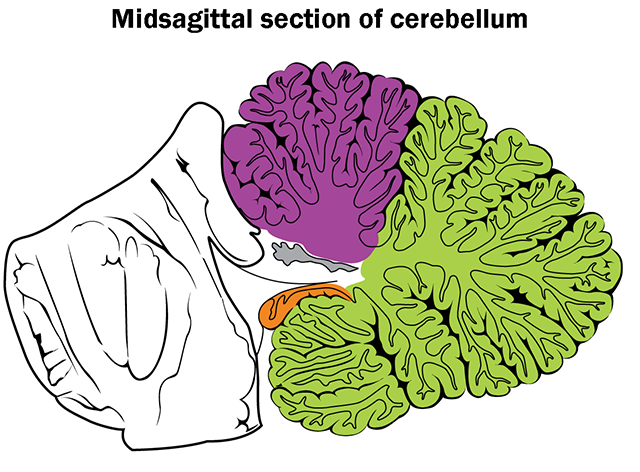
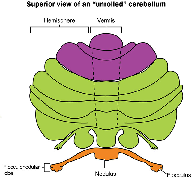 Just as the pathway from primary motor cortex to spinal cord is called the pyramidal motor system, the motor systems which don’t travel this pathway are called extrapyramidal motor systems. The two main extrapyramidal motor systems, which we’ll discuss now, are the cerebellar circuits and the basal nuclei circuits.
As with all circuits we’ll study in this unit, there are multiple interconnections between processing centers (nuclei) in the central nervous system. The cerebellum receives input (cerebellar afferents) from a number of motor nuclei, including the red nucleus in the midbrain and the motor nuclei of the thalamus (ventral anterior and ventral lateral), from the pons, and from the vestibular nuclei of cranial nerve VIII in the medulla.
In return, the cerebellum sends its output (cerebellar efferents) first to the cerebellar nuclei, and then to the inferior olive of the medulla and vestibular nuclei.
Put simply, the job of the cerebellum is to compare what is actually happening (the body position in space; the touch system) to the intended motor program developed by primary motor cortex, and make corrections as needed.
Upper Motor Neurons: Basal Nuclei Circuits
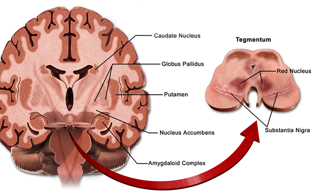
The basal nuclei (often incorrectly called the basal ganglia) are also involved in important motor circuitry. There are extensive motor loops connecting the prefrontal cortex as well as the premotor and supplementary motor cortices (area 6) to the caudate and putamen nuclei. The caudate and putamen, in turn, send their output via the globus pallidus (“pale globe”). The globus pallidus sends projections to the ventral anterior and ventral lateral thalamus, mentioned earlier in regard to cerebellar circuitry, and these thalamic nuclei project back to prefrontal cortex and area 6.
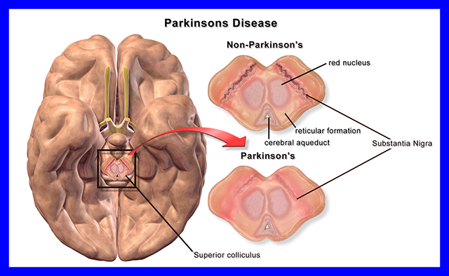
An important pathway into the caudate and putamen is from the substantia nigra (“black substance”), which provides dopamine to modify movement. These cells are lost in Parkinson disease, with tragic results.
Similarly, a structure called the subthalamic nucleus, which lies — you guessed it — underneath the thalamus at the top of the midbrain, also helps modify movement circuits in the basal nuclei. If the subthalamic nucleus is damaged, the debilitating condition hemiballismus results.
The most important function of the basal nuclei is the initiation and execution of movement. Current thinking is that the basal nuclei play a key role in action selection, choosing the “right” movement from the variety of choices the brain has, especially with regard to the emotional aspects of this choice: will the action result in reward or punishment? For example, asymmetry (left/right imbalance) in the activity of the caudate nuclei is associated with obsessive-compulsive disorder, and patients with Parkinson disease or Huntington disease have both movement disorders and emotional disturbances.
Media Attributions
- U12-001 Homeostasis © Open Learning Initiative is licensed under a CC BY-NC-SA (Attribution NonCommercial ShareAlike) license
- U12-002 Receptors © Bizzell, Lizz is licensed under a CC0 (Creative Commons Zero) license
- U12-003 Effectors © Bizzell, Lizz is licensed under a CC0 (Creative Commons Zero) license
- U12-004 Flowchart Nervous System © Hutchins, Jim is licensed under a CC0 (Creative Commons Zero) license
- U12-005 Brain Motor Sensory © BruceBlaus is licensed under a CC BY (Attribution) license
- U12-006 Brodmann Area 4 Lateral © Anatomography is licensed under a CC BY-SA (Attribution ShareAlike) license
- U12-007 Motor Homunculus © Penfield, Wilder is licensed under a Public Domain license
- U12-008 Dti Corona Radiata © NICHD/P. Basser is licensed under a CC BY (Attribution) license
- U12-009 Corticospinal Tract © Betts, J. Gordon; Young, Kelly A.; Wise, James A.; Johnson, Eddie; Poe, Brandon; Kruse, Dean H. Korol, Oksana; Johnson, Jody E.; Womble, Mark & DeSaix, Peter is licensed under a CC BY (Attribution) license
- U12-009b Decussate Medical English © Hutchins, Jim is licensed under a CC BY-SA (Attribution ShareAlike) license
- U12-009a Motor Tracts of the Human Spinal Cord v2 © Hutchins, Jim and Stiver, Mikaela is licensed under a CC BY-NC-ND (Attribution NonCommercial NoDerivatives) license
- U12-011 Upper and Lower Neurons and Pathways © Crookston, Alexa is licensed under a CC BY-NC-ND (Attribution NonCommercial NoDerivatives) license
- U12-012 Motor Neuron (anatomy) © Crookston, Alexa is licensed under a CC BY-NC-ND (Attribution NonCommercial NoDerivatives) license
- U12-013 Motor Unit © Crookston, Alexa is licensed under a CC BY-NC-ND (Attribution NonCommercial NoDerivatives) license
- U12-014 Neuromuscular Junction © NICHD/Y.J. Kim and M. Serpe is licensed under a CC BY (Attribution) license
- U12-010 Spinal Cord Pathways Motor Focus v2 © Betts, J. Gordon; Young, Kelly A.; Wise, James A.; Johnson, Eddie; Poe, Brandon; Kruse, Dean H. Korol, Oksana; Johnson, Jody E.; Womble, Mark & DeSaix, Peter adapted by Jim Hutchins is licensed under a CC BY (Attribution) license
- U12-015a Cerebellar Regions Midsagittal View © Betts, J. Gordon; Young, Kelly A.; Wise, James A.; Johnson, Eddie; Poe, Brandon; Kruse, Dean H. Korol, Oksana; Johnson, Jody E.; Womble, Mark & DeSaix, Peter adapted by Jim Hutchins is licensed under a CC BY (Attribution) license
- U12-015 Cerebellar Regions Unrolled © Betts, J. Gordon; Young, Kelly A.; Wise, James A.; Johnson, Eddie; Poe, Brandon; Kruse, Dean H. Korol, Oksana; Johnson, Jody E.; Womble, Mark & DeSaix, Peter adapted by Jim Hutchins is licensed under a CC BY (Attribution) license
- U12-016 Basal Ganglia © BruceBlaus is licensed under a CC BY (Attribution) license
- U12-017 Parkinson Disease © BruceBlaus is licensed under a CC BY (Attribution) license
