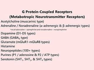
Metabotropic Receptors (G Protein-Coupled Receptors)
Objective 12
Recognize G protein-coupled receptors (metabotropic receptors).
 Most neuroscience lectures focus on the ligand-gated ion channels. They’re easier to understand, and we’ve been studying them for 100 years. They are important. But they’re not the most important or even the most common type of synaptic receptor. That distinction belongs to the G protein-coupled receptor, or metabotropic receptor.
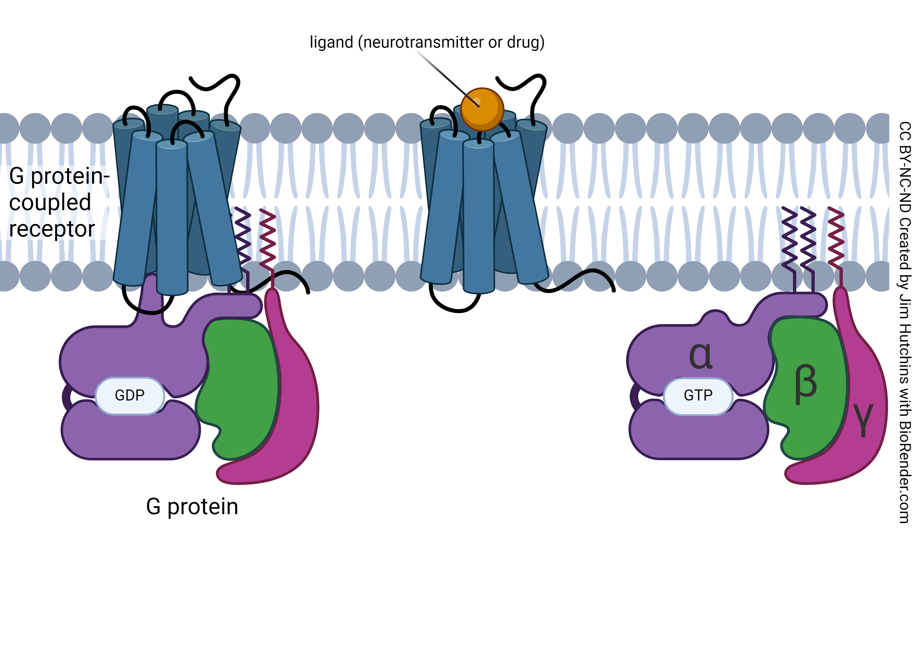
The name G protein-coupled receptor (GPCR) is given because they are receptors that are coupled to a G protein, a particular type of signaling molecule complex. There are a wide variety of G proteins, and their individual actions are why different GPCRs have different effects on the internal biochemistry of cells.
The name metabotropic describes how these receptors act. Just like ionotropic is a word smashed together from English and Greek, metabotropic is a combination of “metabolism” and the Greek τρόπος, trópos, “turn, direction, way”.
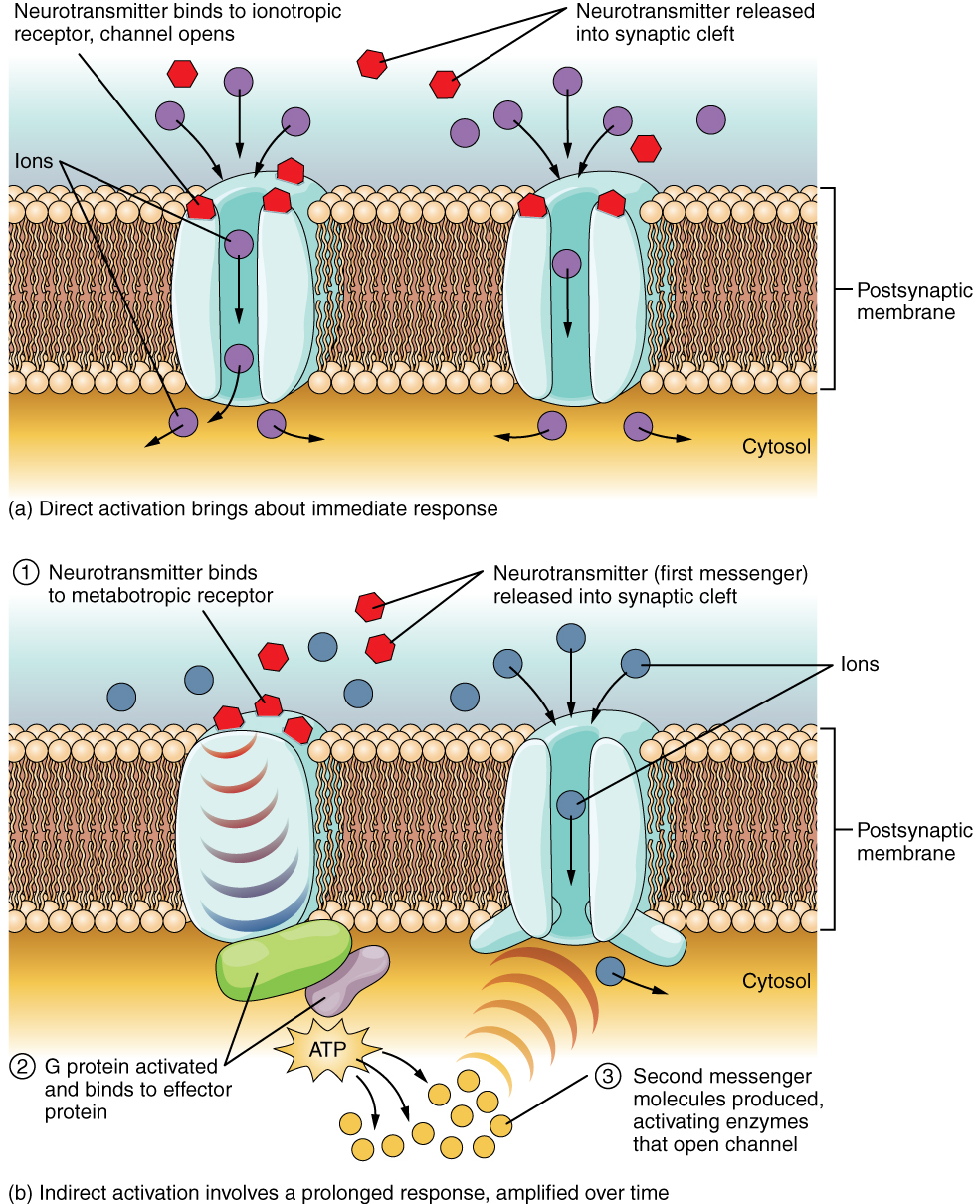 This diagram compares and contrasts the action of ionotropic receptors vs metabotropic receptors. The ionotropic receptor (top), a ligand-gated ion channel, opens and closes in response to the binding of a signaling molecule, typically a neurotransmitter.
In contrast, the metabotropic receptor has no direct effect on ionic permeability. There is no channel in the receptor protein. Rather, the binding of a signaling molecule causes a change in the shape of the receptor protein. That change then alters the intracellular association of the GPCR with a G protein, typically consisting of α, β, and γ subunits. Each of these subunits may act independently. Typically, though, the α subunit goes one way and the βγ subunits go another. These slide in the plane of the cell membrane, along the inside of the neuron, until they encounter an effector protein. The effector protein is usually an enzyme. The enzyme creates or destroys a second messenger, which in turn sets off a chain of events in a signaling pathway.
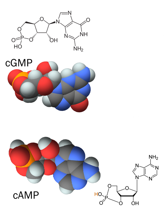
Metabotropic receptors act to alter the levels of a key intracellular signaling molecule called the second messenger. The first messenger is the neurotransmitter that binds to the receptor on the external side of the postsynaptic membrane. The second messenger is a molecule, generally either cyclic AMP (cAMP) or cyclic GMP (cGMP), which then sets into motion a biochemical cascade within the cytoplasm of the receiving cell. We were briefly introduced to these molecules in Unit 3. They consist of a nucleotide (adenine or guanine), the same as those found in RNA; a ribose sugar; and a phosphate group which makes a loop and gives the molecule the “cyclic” part of the name.
Armed with that basic information, let’s look at the steps in more detail.
- The signaling molecule binds to the receptor.
- The receptor changes shape and the associated G protein subunit adds a phosphate to guanosine diphosphate (GDP) to make guanosine triphosphate (GTP).
- The G protein subunit bound to GTP is now released and slides along the inner surface of the neuron membrane.
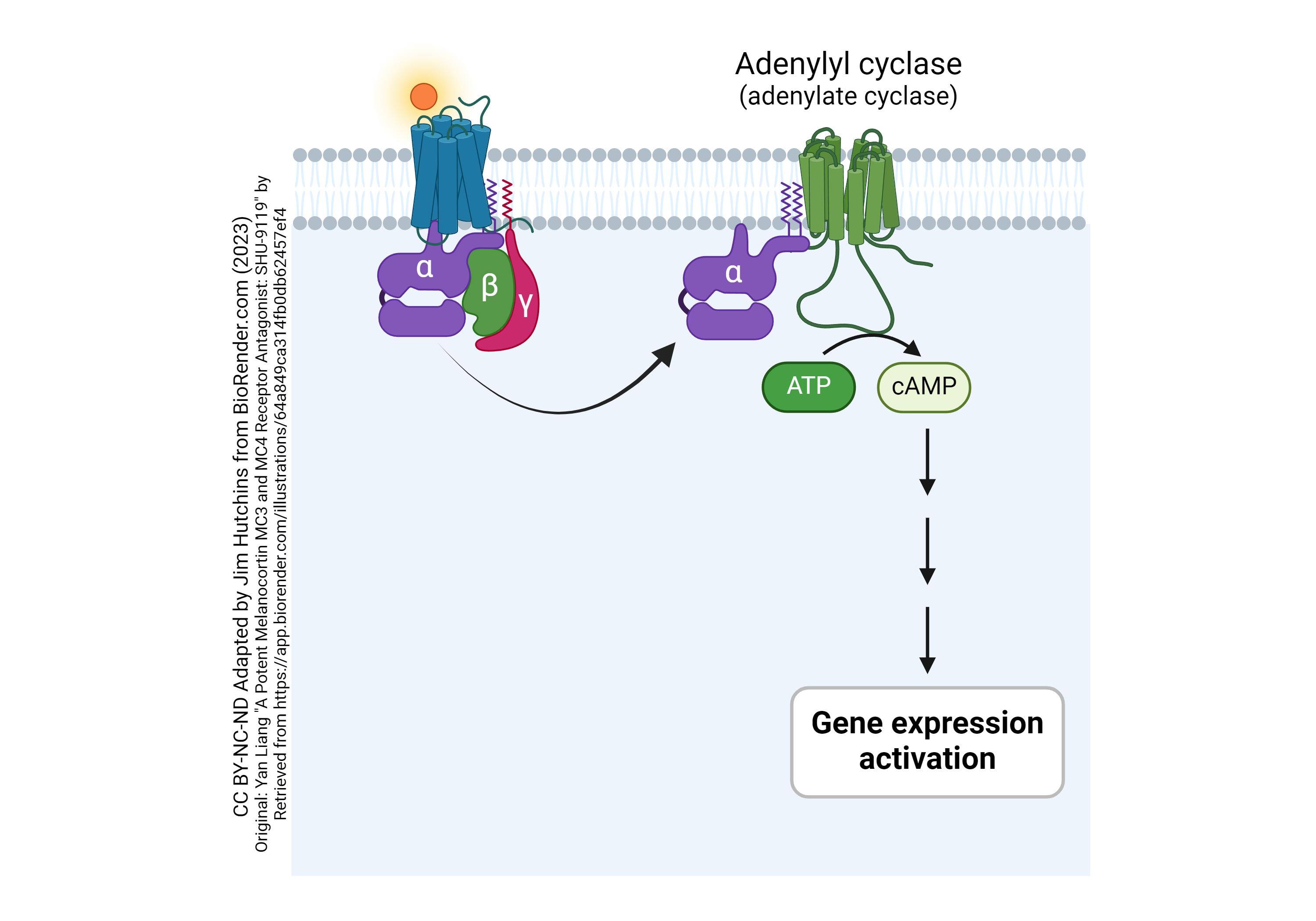
- The bang into, and activate, the enzyme adenylyl cyclase.
- Adenylyl cyclase is the effector protein. As its name indicates, it takes the substrate adenosine triphosphate (ATP), breaks off two phosphates to make adenosine monophosphate (AMP) and then forms a bond between one of the oxygens in the phosphate group and a carbon on ribose, making the molecule now cyclic AMP (cAMP).
- cAMP now acts as a second messenger to alter cellular function. In general, it acts by activating an enzyme that adds phosphate groups onto proteins. The addition of phosphate groups to proteins is called phosphorylation and the enzyme that performs this function is a kinase. Activation of a kinase often changes gene expression in the postsynaptic neuron.
The key feature of this complicated mechanism is that it is a cascade of biochemical reactions that results in amplification of the signal. One signaling molecule binds to one receptor. But that means that dozens of adenylyl cyclase effector proteins are activated, and they make thousands of cAMP molecules, which activate tens of thousands of kinases, which phosphorylate millions of protein molecules. Phosphorylated proteins behave differently than unphosphorylated proteins.
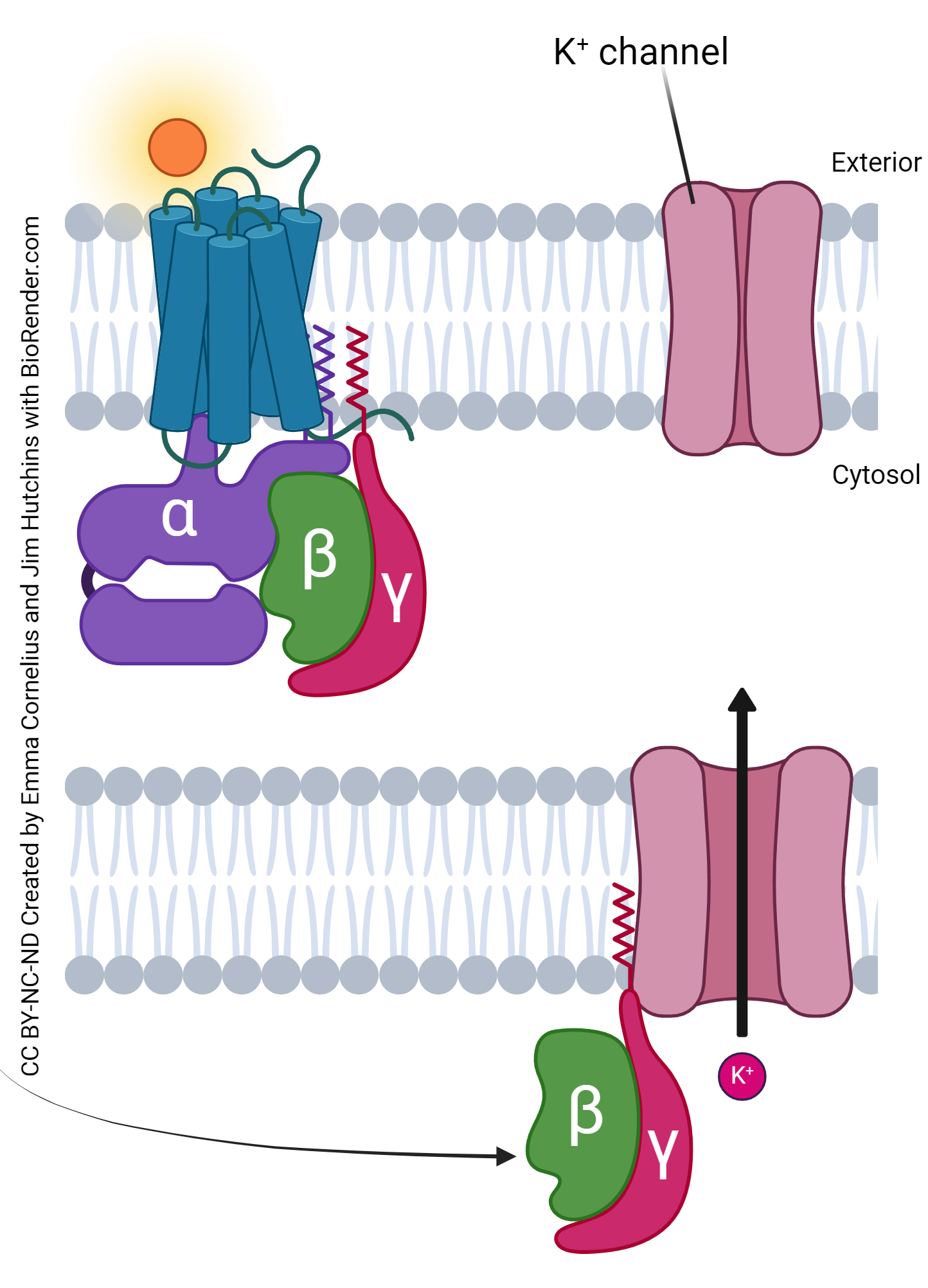
Activation of a G protein-coupled receptor does not always result in the creation or destruction of a second messenger such as cAMP or cGMP. For example, the muscarinic acetylcholine receptor (mAChR) is a G protein-coupled receptor. When ACh binds to the mAChR, it releases the β and γ subunits (GγGβ). In this case, the pathway stops at the effector protein because the effector protein is a signal-gated ion channel, as we saw in Objective 1. It is a phosphorylation-gated K+ channel, and when the GγGβ slams into it, it gets phosphorylated and sticks in the open position. Open K+ channels hyperpolarize the cell because K+ wants to leave the cell. Positive ions leaving the cell cause it to be more negative. Another way of seeing this is that when we increase the routes that K+ can take, K+ gets closer to its equilibrium potential which is almost always more negative than the resting membrane potential. This is the way that acetylcholine released from the vagus nerve slows the action of the heart.
Media Attributions
- U13-094 Gpcr Table © Hutchins, Jim is licensed under a CC BY-SA (Attribution ShareAlike) license
- U13-095 Ionotropic Receptor vs GPC Receptor © Betts, J. Gordon; Young, Kelly A.; Wise, James A.; Johnson, Eddie; Poe, Brandon; Kruse, Dean H. Korol, Oksana; Johnson, Jody E.; Womble, Mark & DeSaix, Peter is licensed under a CC BY (Attribution) license
- U13-096 cGMP and cAMP © PubChem adapted by Jim Hutchins is licensed under a CC BY-SA (Attribution ShareAlike) license
- U13-100 G Protein-coupled Receptor Basics © Hutchins, Jim is licensed under a CC BY-NC-ND (Attribution NonCommercial NoDerivatives) license
- U13-101 Activation of Adenylate Cyclase by G Protein Alpha Subunit © Hutchins, Jim and Liang, Yan is licensed under a CC BY-NC-ND (Attribution NonCommercial NoDerivatives) license
- U13-102 K+ Channel Reaction to G Protein © Cornelius, Emma and Hutchins, Jim is licensed under a CC BY-NC-ND (Attribution NonCommercial NoDerivatives) license
