Meiosis and Gametogenesis
Objective 9
Compare and contrast the homologous endocrine events between spermatogenesis and oogenesis, thoroughly review the similarities and differences between mitosis and meiosis, and relate the phases of meiosis with the processes of spermatogenesis and oogenesis.
Homologous Endocrine Events
As we’ve just covered, in females, gonadotropin releasing hormone (GnRH) from the hypothalamus directs the secretion of follicle-stimulating hormone (FSH) and luteinizing hormone (LH) from the anterior pituitary. FSH stimulates follicle development and estrogen production. LH stimulates ovulation, corpus luteum formation, and progesterone and estrogen production (by the corpus luteum).
Although named for their actions in the female reproductive system, FSH, and LH are also released in males. However, there are no follicles and no corpora lutea. Instead, FSH stimulates the spermatogenic cells and the sustentacular cells of the testes to produce sperm. A negative feedback loop controlled by inhibin ensures sperm are not over-produced. LH stimulates the interstitial cells of the testes to secrete testosterone.
Mitosis
Mitosis and meiosis – you’ve heard these terms before, back in Unit 6, The Molecular Biology of the Cell. We need to review mitosis and meiosis, including the differences between the two processes, so we can understand how these concepts apply directly to male and female gametogenesis.
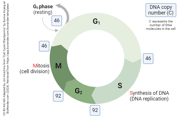 The vast majority of our body cells are somatic cells. When somatic cells divide they undergo mitosis, a process where the DNA content is doubled (DNA replication during S phase), and then divided equally (M phase), resulting in two daughter cells that are genetically identical to each other and to the parent cell. In doing so, we preserve a principle called ploidy, which refers to the number of complete sets of chromosomes in each cell of the organism.
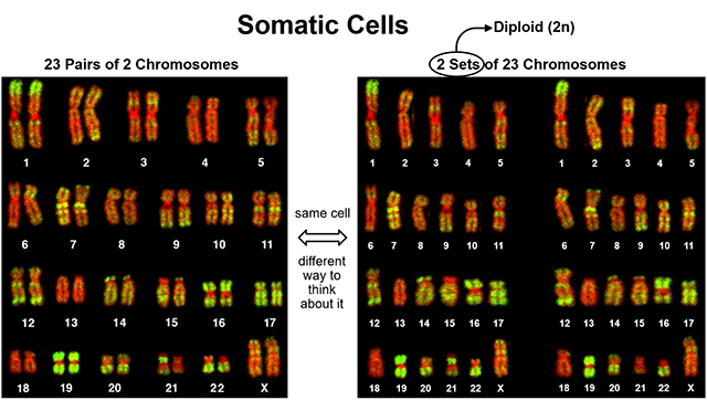 You’re already aware that human somatic cells have 23 pairs of chromosomes (two chromosome 1s, two chromosome 2s, …two chromosome 22s, and two sex chromosomes; 23 chromosome pairs, 46 individual molecules of DNA). To better understand ploidy, let’s take that same somatic cell and describe its chromosomes a little bit differently. Rather than saying it has 23 pairs (of two) chromosomes, lets describe it as having two sets of 23 chromosomes (one set came from Mom and has a copy of chromosome 1, a copy of chromosome 2, …a copy of chromosome 22, and a sex chromosome; the other set came from Dad and contains the same list of chromosomes). Because there are two complete sets, we say the cell is diploid or 2N. All somatic cells are diploid, 2N.
N counts the number of chromosomes; C counts the number of DNA molecules. A diploid somatic cell has 46 DNA molecules (two copies of each numbered chromosome 1-22, plus two sex chromosome DNA molecules). During the cell cycle, those 46 DNA molecules briefly become 92 DNA molecules before the cell divides into two daughter cells and restores the diploid number (C=46) again.
Meiosis
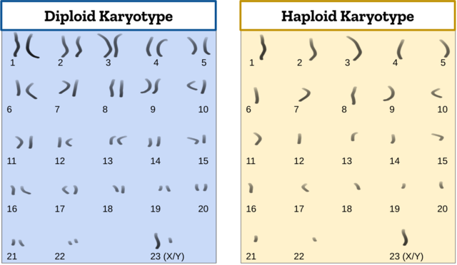
The only cells in the body that are not somatic cells are gametes (sperm, ova). Each gamete has just one set of 23 chromosomes, which makes them haploid, or N (C=23).
Think about the two gametes (haploid) – one sperm and one ovum – that united to form the zygote (diploid) that became a fetus that became you. Where did they come from? We’ve already covered male and female gametogenesis in this unit (Objectives 7 and 8), so you should be thinking “sperm, from the seminiferous tubules of the testes” and “oocyte, from a mature ovarian follicle.” But, in those gender-specific gamete production processes, how do we start with diploid (2n) somatic cells and end up with haploid (n) gametes? Hopefully, you’re thinking, “Oh, yeah, that other kind of cell division — meiosis!”
In the ovaries and testes, the process begins with somatic gametogenic stem cells (oogonia and spermatogonia). These diploid (46C) cells first duplicate their DNA in S phase (92C) and then divide via mitosis; one 46C daughter cell remains attached to the basement membrane (to maintain the stem cell population) and the other 46C daughter cell will undergo meiosis to produce two haploid (23C) gametes.
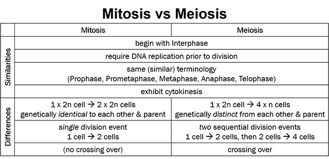
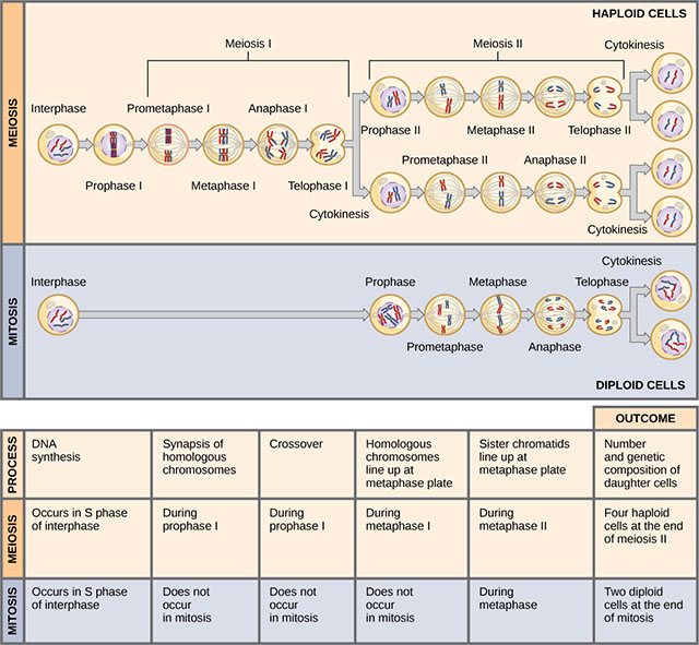
Notice the similarities between mitosis and meiosis:
- both begin with Interphase
- both require DNA replication, during S Phase of Interphase, prior to cell division
- both use the same basic terminology – Prophase, Prometaphase, Metaphase, Anaphase, Telophase – to outline and describe the steps in the process
- both exhibit cytokinesis
Now, note the differences between mitosis and meiosis:
- mitosis begins with one diploid cell and ends with two diploid cells, genetically identical to each other and to the parent cell; meiosis begins with one diploid cell and ends with four haploid cells, each genetically distinct from the other and from the parent
- mitosis includes a single cell division (one cell to two cells) which can occur over and over; meiosis is actually two sequential cell divisions (one cell to two, then each of those two cells to two cells, for a total of four cells) and is a dead end for those cells
- perhaps the biggest difference is the crossing over that occurs during Prophase 1 of Meiosis (but does not occur anywhere in mitosis) is the event called crossing over.
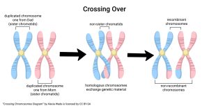 Crossing over occurs only in meiosis and leads to the first difference noted above, ie that meiosis results in cells that are genetically distinct from each other and from the parent cell. It takes place in Prophase I, after DNA replication and before the first cell division. Instead of immediately lining up on the metaphase plate to be pulled apart, replicated chromosomes first form tetrads and crossing over occurs between adjacent non-sister chromatids. Remember (from Unit 6) that crossing over involves the exchange of genetic information between two homologous chromosomes. Mendel was basically correct that each offspring receives one copy of chromosome 1 from the father, and one copy of chromosome 1 from the mother, etc. What Mendel could not have envisioned is the 50:50 chance that the chromosome 1 received from his father did not exactly match either copy of his father’s own chromosome 1; rather, it had pieces of both his father’s chromosomes 1. Likewise, the chromosome 1 he could have received from his mother did not exactly match either of her chromosomes 1.
Crossing over introduces genetic variability into the reproduction process. It ensures that each offspring is at least a little (or a lot, depending on how much crossing over occurs) genetically distinct from all others offspring from the same parents. Without crossing over, all siblings would regularly inherit exactly the same set of chromosomes from their parents, resulting in identical twins (triplets, quads…) born years apart!
To summarize, here are the differences between mitosis and meiosis:
- Mitosis is a cell division where the DNA of the daughter cells is exactly the same sequence in the same order as that of the parent cell (the daughter cells are clones of the parent). Meiosis involves tetrad formation and crossing over, which leads to genetically distinct daughter cells.
- Mitosis preserves the ploidy (n number) of the cell; multiplication of the n number occurs during S phase; division of the n number occurs during M phase. Meiosis is a reduction division; the precursors of gametes (spermatogonia, primordial oocytes) are diploid (2n), the resulting cells are haploid (n); when two haploid gametes meet and the nuclei combine, the resulting cell is now diploid (2n); the fertilized ovum is now a zygote, which will divide mitotically to become an embryo, then a fetus.
- Mitosis is a cyclical process. Meiosis is a “one-way street;” once cells become gametes, they cannot ever produce new cells except by joining another gamete in fertilization.
Gametogenesis Stages
Now, let’s combine what we’ve learned about meiosis and gametogenesis to see how the two line up.
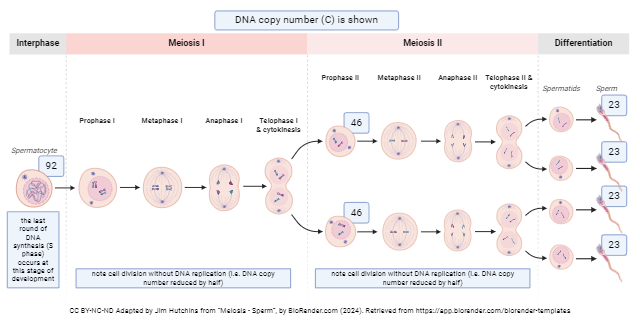
In males, gametogenesis begins with a stem cell, a spermatogonium (diploid). Spermatogonia undergo mitosis to produce a clonal population (diploid). The daughter cells that stay near the basement membrane remain stem cells and continue to divide by mitosis (diploid). Daughter cells that move away from the basement membrane toward the lumen of the seminiferous tubule begin a process of differentiation to become primary spermatocytes (diploid). Primary spermatocytes undergo crossing over and then the reduction division of meiosis I to become secondary spermatocytes (haploid). These undergo meiosis II to produce spermatids (haploid). Spermatids further differentiate (with no additional changes in DNA content or ploidy number) and mature become spermatozoa (sperm, haploid).
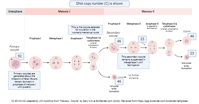
Female gametes are generated through a different and somewhat more complex process. Stem cells, oogonia (diploid), undergo their last mitotic division in the female fetus. Meiosis I also begins before birth, as primary oocytes complete crossing over but then stop before completing prophase I. At birth, female ovaries are full of primary oocytes suspended in prophase I. Each month after menarche, 5-12 primary oocytes respond to the hormonal signals from the pituitary and begin to develop. In general, only one of these will complete meiosis I at ovulation to become a secondary oocyte. Remember that, in males, meiosis I yields two haploid secondary spermatocytes. In females, because the end goal of the process is a single viable ovum, meiosis I yields only one haploid secondary oocyte; the remaining genetic material, called the first polar body, is disposed of.
The ovulated secondary oocyte then starts, but does not complete, meiosis II. Meiosis II is only completed if a spermatozoon penetrates the secondary oocyte. If this occurs, the secondary oocyte completes meiosis II and becomes an ovum. Again, the excess genetic material, the second polar body, is disposed of. For a time, the haploid female DNA and haploid male DNA remain separate, within the ovum, as female and male pronuclei. These eventually fuse to form the diploid nucleus of the zygote; this fusion marks time zero of embryonic development.
In males, mitotic spermatogonia (stem cell) production starts in embryonic life and continues for a lifetime. Differentiation, meiosis I, and meiosis II begin at puberty and occur continuously and as rapidly as possible thereafter. In this way, a male produces 500 billion sperm over an average lifetime.
In females, mitotic oogonia (stem cell) production begins and ends before birth, with a peak of several million primary oocytes in fetal ovaries. At birth, the numbers have declined to hundreds of thousands. At menarche (first menstruation), there remain tens of thousands. An average woman has between 400 and 500 menstrual cycles in her lifetime. At each cycle, 5 to 12 (about 10) eggs begin follicular development – about 5000 follicles in a lifetime; less than 500 of these will ovulate as secondary oocytes and travel in the uterine tubes. Perhaps 1% of these will be successfully fertilized and an estimated ⅓ of zygotes are spontaneously aborted (miscarried). By menopause, zero primary oocytes remain.
As you can see, gamete “strategy” is completely different between male and female.
Media Attributions
- U20-037c Cell Cycle Mitosis © Hutchins, Jim and Huang, Eunice is licensed under a CC BY-NC-ND (Attribution NonCommercial NoDerivatives) license
- U20-038 Karyotype Diploid © Bolzer, Andreas; Kreth, Gregor; Solovei, Irina; Koehler, Daniela; Saracoglu, Kaan; Fauth, Christine; Müller, Stefan; Eils, Roland; Cremer, Christoph; Speicher, Michael R.; and Cremer, Thomas adapted by Brad Winterton is licensed under a CC BY (Attribution) license
- U20-039 Diploid vs Haploid Karyotype © SadiesBurrow is licensed under a CC BY-SA (Attribution ShareAlike) license
- U20-040 Mitosis vs Meiosis © Clark, Mary Ann; Douglas, Matthew and Choi, Jung is licensed under a CC BY (Attribution) license
- U20-041 Mitosis vs Meiosis Table © Winterton, Brad is licensed under a CC BY-SA (Attribution ShareAlike) license
- U20-042 Crossing Over © Wade, Alexia is licensed under a CC BY-SA (Attribution ShareAlike) license
- U20-045 Meiosis – Sperm v2 © Hutchins, Jim is licensed under a CC BY-NC-ND (Attribution NonCommercial NoDerivatives) license
- U20-044 Meiosis – Oocyte © Hutchins, Jim and Kim, Sally is licensed under a CC BY-NC-ND (Attribution NonCommercial NoDerivatives) license
