Lymphatic Organs
Objective 7
Differentiate primary from secondary lymphatic organs/structures. Give examples of each and explain their functions. Describe the locations, structure, and function of lymph nodes.
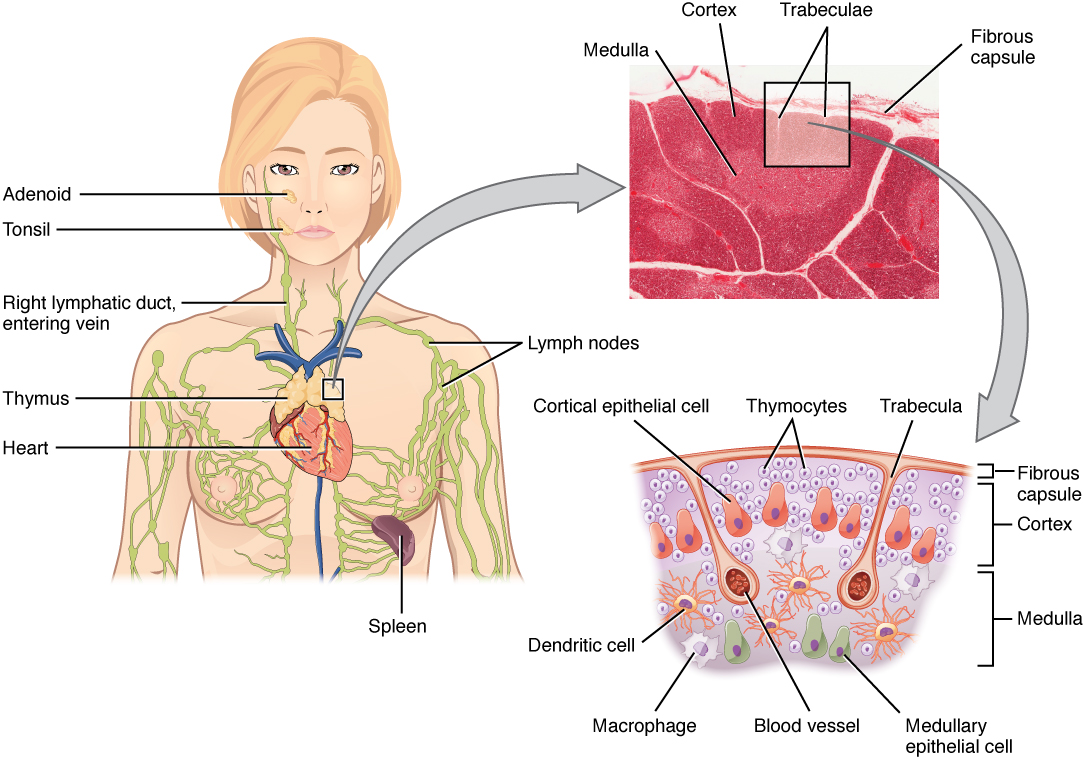
The primary lymphatic organs are the locations where stem cells divide to produce immune system cells or where these cells develop and mature. Once mature, cells can migrate out of the primary lymphoid organs into the secondary lymphoid organs and the blood. The primary lymphatic organs are the bone marrow and the thymus gland. Hematopoietic stem cells in the bone marrow divide to produce both B lymphocytes and T lymphocytes. B lymphocytes will remain in the bone marrow to mature. T lymphocytes will leave as T lymphocyte progenitor cells and migrate to the thymus gland to become mature T cells.
Weird fact: B cells were originally discovered by Bruce Glick of Mississippi State University and were named for the bursa of Fabricius, a lymphoid structure found only in birds. It is located near the chicken’s cloaca, the common port for egg laying, defecation, and urination. Imagine scientists’ relief when they discovered that in the human, B cells mature in the bone marrow, which also starts with the letter B.
Both B and T lymphocytes are born from stem cells in the bone marrow.
B lymphocytes mature in bone marrow.
T lymphocytes mature in the thymus.

The thymus gland is located in the mediastinum between the sternum and the aorta. In infants, the thymus gland is approximately 70 g. It will remain approximately this size until after puberty, when connective and adipose tissue will replace the thymic cells. By the time an individual reaches old age, the thymus will only weigh about 3 g, but it will continue to release some mature T cells.
Although mature, cells leaving the primary lymphoid organs are said to be naïve or inactivated. The secondary lymphatic organs and tissues provide a place for these cells to encounter foreign antigens and become activated. This activation determines how that cell will perform and what its target will be for the rest of its life. Lymph nodes and the spleen are secondary lymphatic organs; lymphatic nodules are secondary lymphatic tissues as well. The difference is the presence or absence of a capsule. Lymph nodes have a connective tissue capsule, nodules do not. Examples of lymphatic nodules include: portions of the appendix, the tonsils (pharyngeal, adenoid and palatine), and the Peyer’s patches in the intestinal tract.
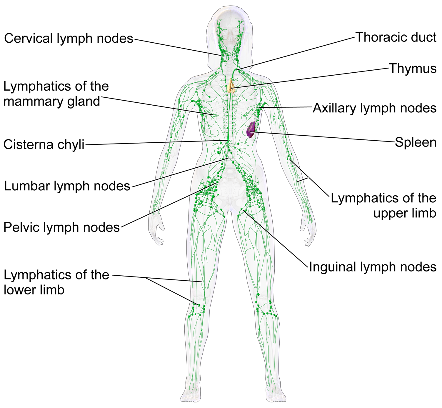
Lymph nodes are bean-shaped lymphatic organs and are anywhere from 1 to 25 mm in length. There are approximately 600 lymph nodes in the body, and they occur at intervals along the lymphatic vessels, serving as check points where immune cells can encounter foreign antigens. There are regions in the body where the lymph nodes are grouped together more prominently. These groups of lymph nodes are named for the location they are found and include cervical, submandibular, axillary, and inguinal regions. Swelling in the different groups of lymph nodes often indicates nearby infection.

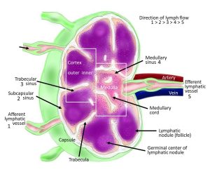
A lymph node is surrounded by a dense, irregular connective tissue capsule. Extensions of the capsule (trabeculae) divide the node into compartments. Reticular connective tissue inside the lymph node forms a mesh wherein immune cells lie in wait to encounter antigens. There are two regions of a lymph node, the cortex and the medulla. Both regions contain large numbers of white blood cells. The types of cells present in each region differ slightly but typically comprise a combination of T cells, B cells, dendritic cells, and macrophages.

Lymphatic fluid passes through the lymph nodes, allowing the cells to check the fluid and respond to invading pathogens. Lymph enters a node through afferent lymphatic vessels. The lymph flows through the cortex, where it comes into contact with large populations of B lymphocytes, dendritic cells, and macrophages. The lymph continues to flow through the node into the medulla. There it is exposed to more B lymphocytes, plasma cells (activated B cells), and more macrophages. The lymph will then exit the lymph node through efferent vessels and returns to the lymphatic vessel system.
Media Attributions
- U15-036 Thymus and Spleen © Betts, J. Gordon; Young, Kelly A.; Wise, James A.; Johnson, Eddie; Poe, Brandon; Kruse, Dean H. Korol, Oksana; Johnson, Jody E.; Womble, Mark & DeSaix, Peter is licensed under a CC BY (Attribution) license
- U15-037 Lymphatic System © BruceBlaus is licensed under a CC BY (Attribution) license
- U15-038 Lymphatic Immune System Lymph Node © Sullivan, Chris is licensed under a CC BY-SA (Attribution ShareAlike) license

