Kidney Anatomy
Objective 2
Describe the external and internal anatomy of the kidney. Trace the path of blood flow through the kidneys and explain what makes the vascular system of the kidneys unique compared to other organs.
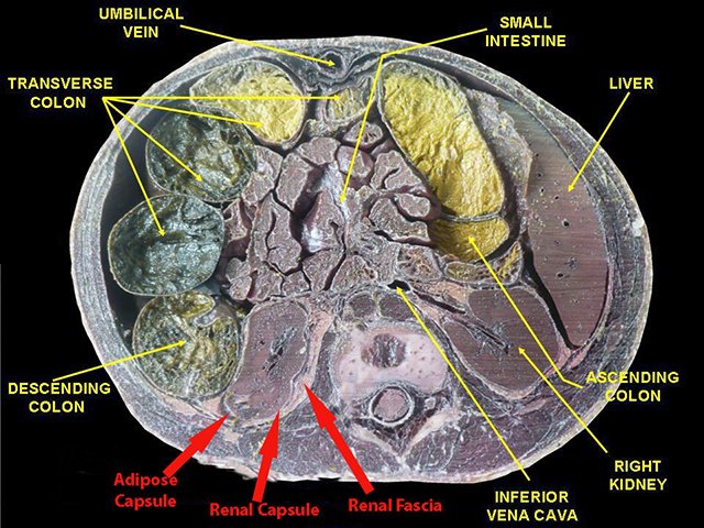
Externally, three layers of tissue (from deep to superficial) cover the kidney: renal capsule, adipose capsule, and renal fascia. The renal capsule helps protect and maintain the shape of the kidney. The adipose capsule also protects, as well as maintains the position of the kidney in the abdominal cavity. Lastly, the renal fascia anchors the kidney to the abdominal wall and neighboring structures.
Internally, the kidney has two distinct regions, the renal cortex and medulla. The cortex is more superficial. It extends between the renal pyramids in the medullary region to form the renal columns. The renal pyramids are triangular structures within the medulla that appear striated (striped) due to the presence of the renal tubules and ducts.
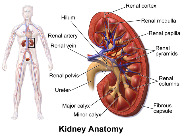
Each kidney contains approximately 1 million nephrons. The nephron is the functional unit of the kidney. Urine produced by the nephrons drains from the apices (renal papillae) of the pyramids. The urine from each pyramid will enter a cup-like structure called a minor calyx. Two or three minor calyces drain into a major calyx. Each kidney has two or three major calyces, which will drain into one large cavity, called the renal pelvis.
Path of Renal Blood Flow
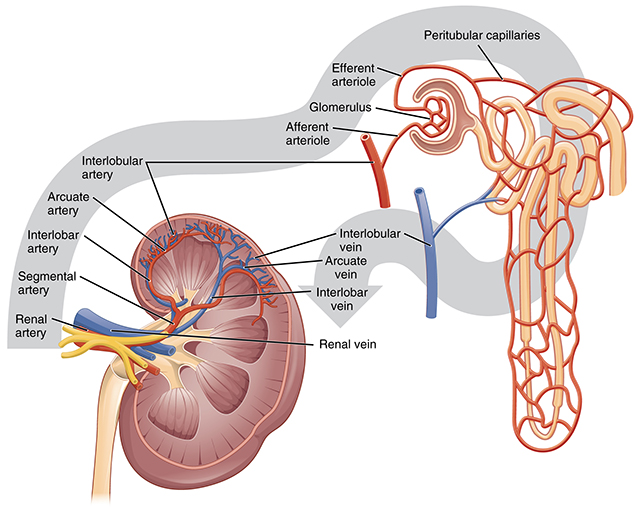
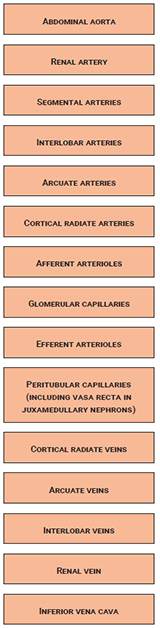 There are two unique vascular features of the kidneys:
First, the glomerular capillaries are positioned between two groups of arterioles.
Second, unlike any other organ of the body, there are two sets of capillaries, the glomerular capillaries and the peritubular capillaries.
Media Attributions
- U19-005 Kidney Capsule Labeled © Anatomist90 adapted by Travis Price is licensed under a CC BY-SA (Attribution ShareAlike) license
- U19-006 Kidney Anatomy © BruceBlaus is licensed under a CC BY-SA (Attribution ShareAlike) license
- U19-008 Blood Flow in the Kidney © Betts, J. Gordon; Young, Kelly A.; Wise, James A.; Johnson, Eddie; Poe, Brandon; Kruse, Dean H. Korol, Oksana; Johnson, Jody E.; Womble, Mark & DeSaix, Peter is licensed under a CC BY (Attribution) license
- U19-007 Flowchart of Blood Flow in Kidneys © Price, Travis is licensed under a CC BY-SA (Attribution ShareAlike) license
