Arteries
Objective 6
Name and label the major arteries of the body.
You will use the diagrams given in this section to name all of the arteries listed on a diagram with the labels removed.
The Aorta and Its Branches

Note that the origin of the right subclavian artery and right common carotid artery is different than for the left subclavian artery and left common carotid. The pattern shown here is typical; it is rare but possible to have a brachiocephalic trunk on the left side as well. The old name for the brachiocephalic artery is the innominate artery, which is weird, because “innominate” means “no name” and it clearly has one. It is also sometimes called the brachiocephalic trunk.
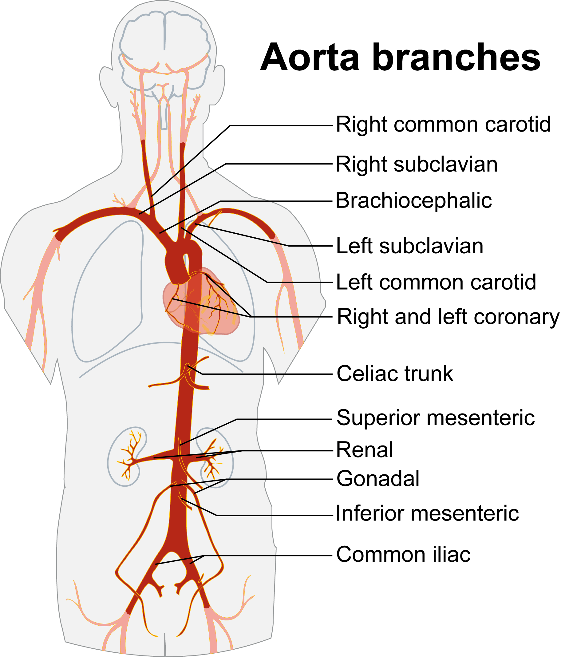
Once the descending aorta crosses the diaphragm muscle, it changes name from the thoracic aorta to the abdominal aorta. The first major branch of the abdominal aorta is the celiac trunk. The celiac trunk branches into arteries that supply the distal esophagus, stomach, second part of the duodenum, liver, pancreas, gallbladder, and spleen. The superior mesenteric arteries supply part of the intestines (the name “mesenteric” means “pertaining to the middle of the intestines”). The inferior mesenteric arteries also supply the intestines. The renal arteries supply the kidneys, and the gonadal arteries supply the gonads.
The abdominal aorta ends by branching into two common iliac arteries, which will eventually become the right and left femoral arteries as they pass into the lower extremity.
The Aorta and Its Branches
Divisions of the Aorta
- Ascending aorta
- Aortic arch
- Descending aorta
- Thoracic aorta
- Abdominal aorta
Arteries that Branch Off the Thoracic Aorta (in order)
- Coronary artery (right and left)
- Brachiocephalic artery (typically only right)
- Right subclavian artery
- Right common carotid artery
- Left common carotid artery
- Left subclavian artery
Arteries that Branch Off the Abdominal Aorta (in order)
- Celiac trunk
- Superior mesenteric artery
- Renal artery
- Gonadal artery
- Inferior mesenteric artery
Terminal Branches of the Abdominal Aorta
- Common iliac artery
Arteries of the Head and Neck
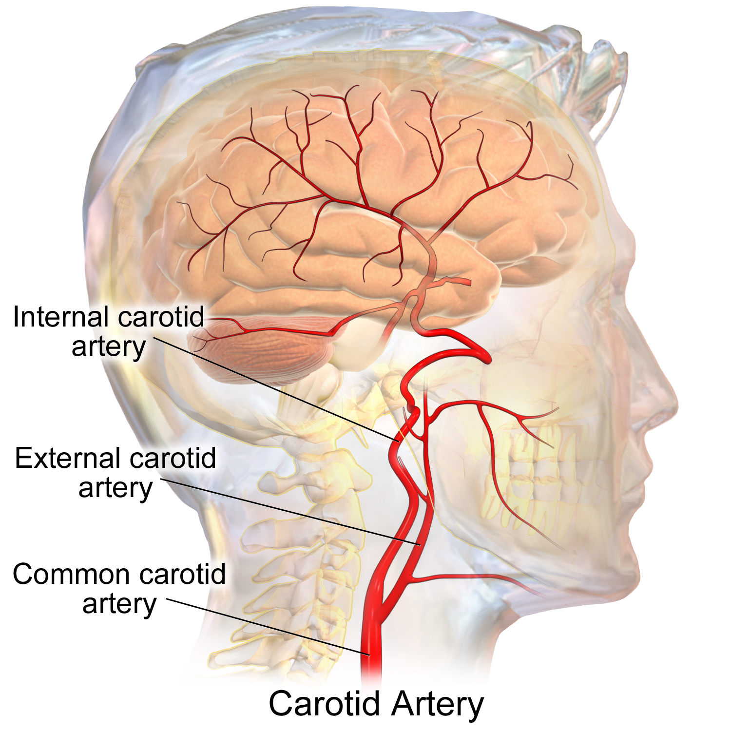 The common carotid artery divides into the external carotid artery, which supplies the face; and the internal carotid artery, which supplies about 3/4 of the blood used by the brain.
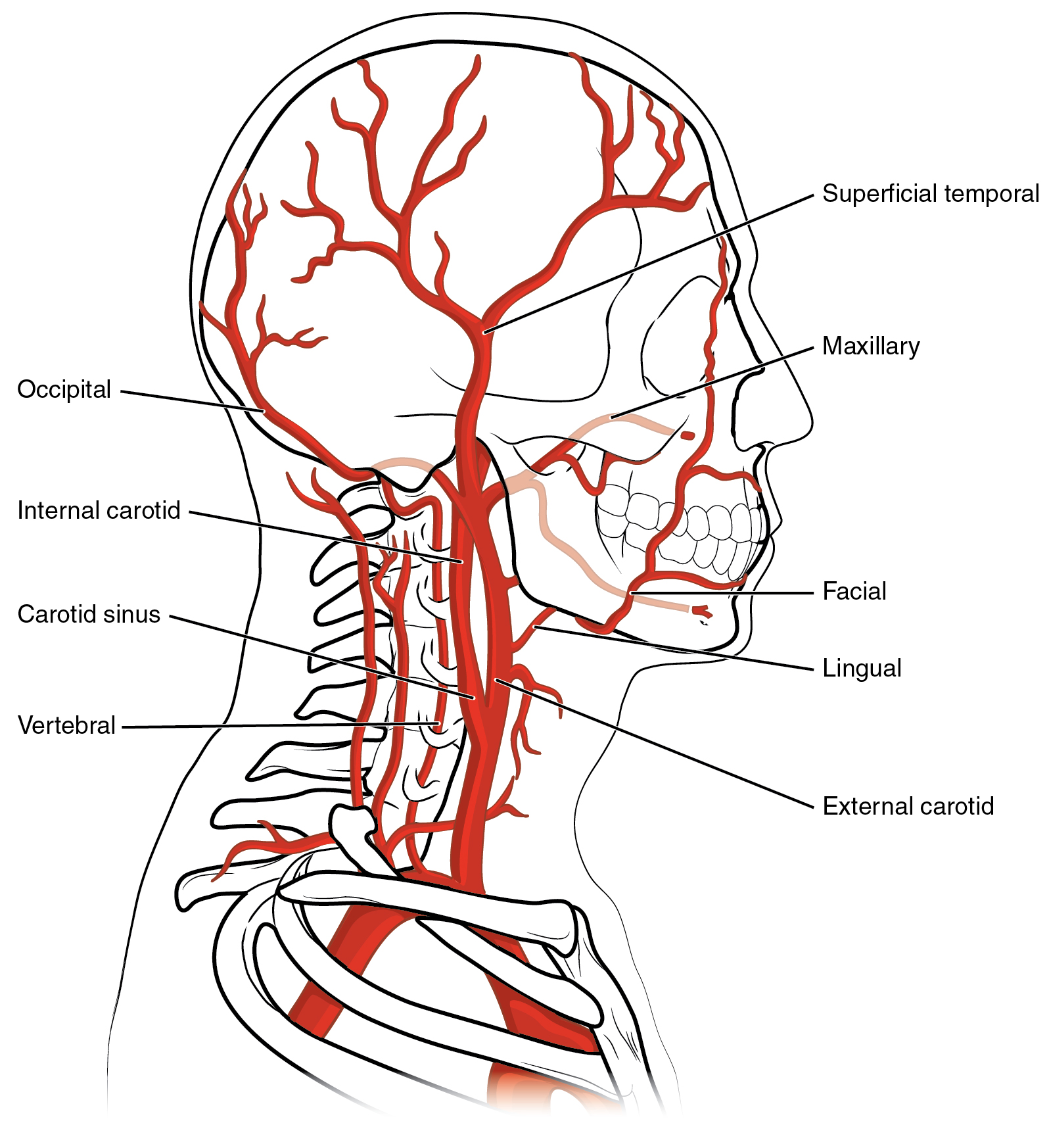 The external carotid artery and its branches supply the face.
The internal carotid artery and its branches supply the brain. There is an organ called the carotid body at the root of the internal carotid artery. The carotid body detects the level of oxygen, carbon dioxide, and hydrogen ion (pH) in the blood, and supplies that information to the respiratory and cardiovascular centers so they can respond appropriately. (For example, if blood oxygen levels drop — hypoxemia — then it would be a good idea to increase the respiratory rate to get more oxygen into the blood.)
The internal carotid artery is the source of the anterior circulation of the brain.
The vertebral artery arises as a branch of the subclavian artery. It threads its way through the foramina formed by the lateral part of the vertebrae, and gives off two branches near its superior ending: the anterior spinal artery, which supplies much of the blood to the spinal cord; and the posterior inferior cerebellar artery (PICA), which supplies blood to the dorsal medulla and the inferior surface of the cerebellum.
Then the vertebral arteries do a weird thing: they fuse together (anastomose) to form the midline basilar artery, which as the name implies runs along the base of the brainstem. The basilar artery gives rise to branches as it rises superiorly: the anterior inferior cerebellar artery (AICA); the pontine arteries; and the superior cerebellar artery. At about the level of the midbrain, the basilar divides into two (bifurcates) to form the posterior cerebral arteries. This system comprises the posterior circulation of the brain.
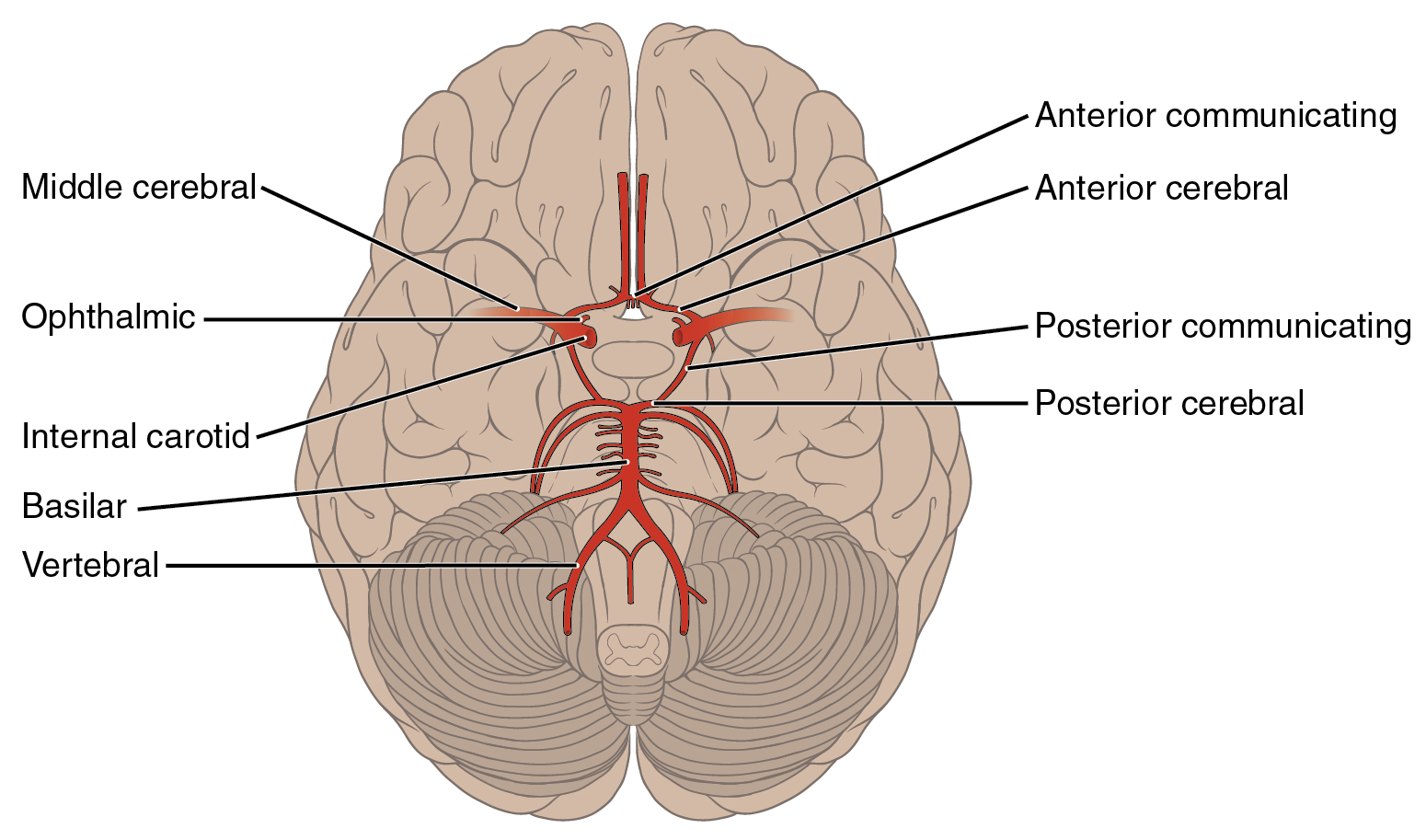 The anterior and posterior circulation of the brain are joined at an complex anastomosis called the circle of Willis (cerebral arterial circle).
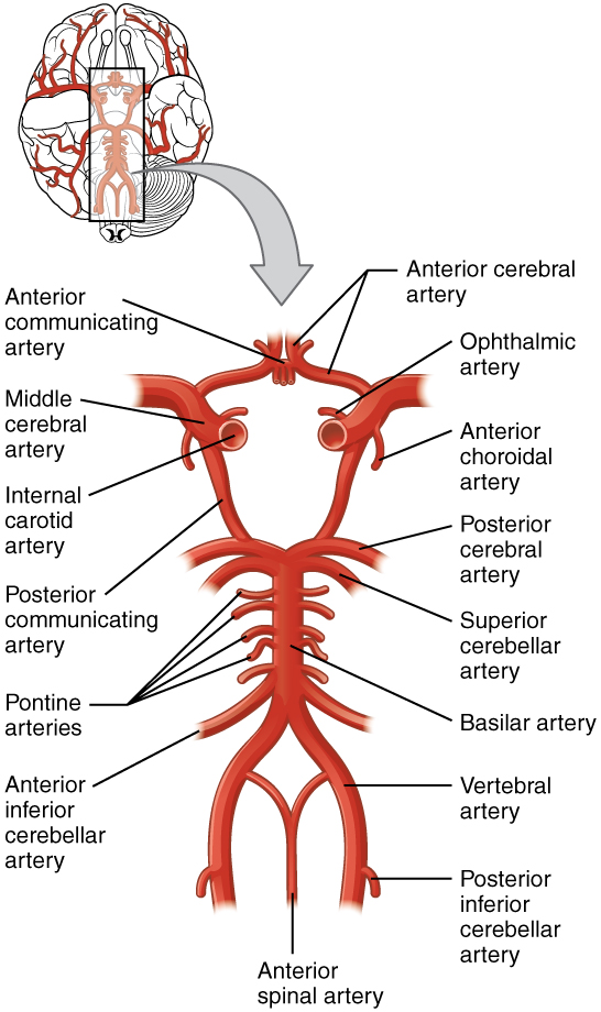
As the basilar bifurcates into the left and right posterior cerebral artery (PCA), those large arteries each give off a branch of much smaller caliber called the posterior communicating arteries. Each posterior communicating artery joins with the internal carotid arteries at the point where they bend sharply to form the left and right middle cerebral arteries. Each anterior cerebral artery (ACA) also branches off the internal carotid at this point so in a sense the internal carotid divides itself to form the middle and anterior cerebral arteries. The two anterior cerebral arteries are joined by a tiny vessel called the anterior communicating artery. This completes the circle of Willis.
The internal carotid also gives off two branches, the anterior choroidal artery and the ophthalmic artery, which supply parts of the thalamus and the eye, respectively.
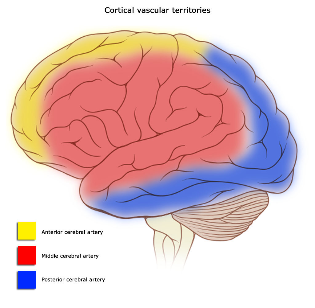
You may recall from Unit 11 that we discussed the territories of the anterior cerebral artery (yellow shading), middle cerebral artery (red shading), and posterior cerebral artery (blue shading). The anterior cerebral artery also supplies most of the midline structures of the brain (not shown) and the posterior cerebral artery also supplies most of the inferior surface of the brain (also not shown).
Arteries of the Head and Neck
Divisions of the Common Carotid Artery
- Common carotid artery
- External carotid artery
- Internal carotid artery
Branches of the External Carotid Artery
- Lingual
- Facial
- Maxillary
- Superficial temporal
- Occipital
The Posterior Circulation
- Vertebral artery
- Posterior inferior cerebellar artery (PICA)
- Anterior spinal artery
- Basilar artery
- Anterior inferior cerebellar artery (AICA)
- Pontine arteries
- Superior cerebellar artery
The Anterior Circulation
- Internal carotid artery
- the internal carotid bends and branches to form the anterior cerebral artery (ACA) and middle cerebral artery (MCA)
- Ophthalmic artery
- Anterior choroidal artery
The Circle of Willis
- Posterior cerebral artery (PCA)
- Posterior communicating artery
- Anterior cerebral artery
- Anterior communicating artery
Arteries of the Upper Extremity
The entire blood supply of the upper extremity is supplied by the subclavian artery. Remember there is a difference between right and left: the right subclavian artery arises from the brachiocephalic trunk, while the left subclavian artery branches directly off the aorta.
Now anatomists do something annoying. They change the name of the artery two more times. As the subclavian artery crosses the first rib, it becomes the axillary artery. As the axillary artery reaches the inferior border of the teres major muscle, it becomes the brachial artery.
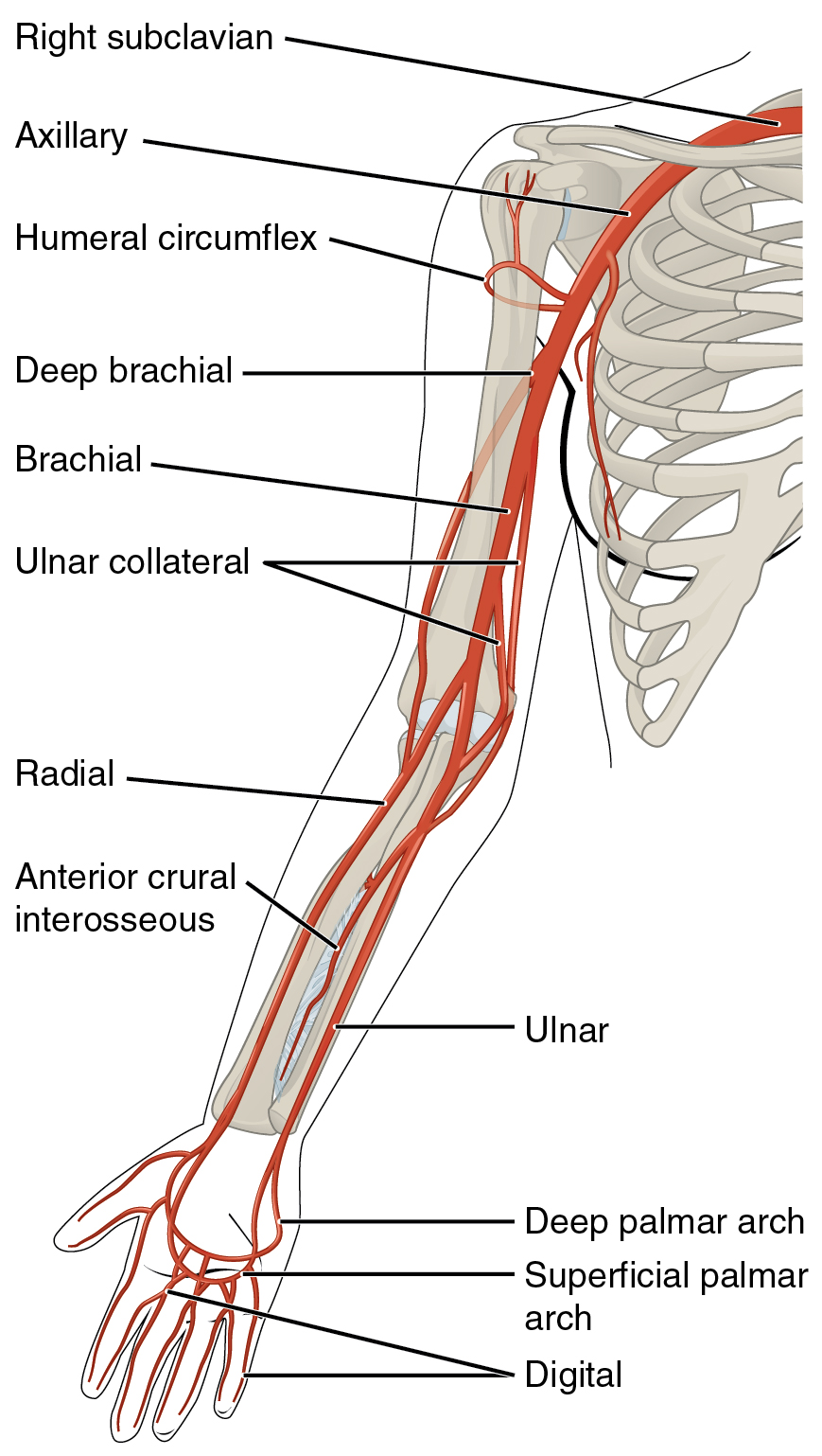 The humeral circumflex artery, as the name implies, wraps around the humerus. The deep brachial artery supplies deep parts of the arm. (In anatomical terminology, the “arm” is between the shoulder and elbow, and the “forearm” is between the elbow and wrist.) The ulnar collateral artery sends branches that supply the elbow region.
In the cubital (elbow) region, the brachial artery ends by bifurcating into the radial artery and ulnar artery.
A short branch of the ulnar, not seen in this diagram, is the common interosseous artery. It branches off the ulnar artery in the cubital region. It gives rise to the anterior interosseous artery (AIA).
The palm of the hand is supplied by looping arteries that join the ends of the radial and ulnar arteries. These are the deep palmar arch and superficial palmar arch.
Each of these loops gives rise to a number of digital arteries which supply the fingers (digits).
Arteries of the Upper Extremity
- Humeral circumflex artery
- Deep brachial artery
- Ulnar collateral artery
- Radial artery
- Ulnar artery
- Common interosseous artery (short; not seen)
- Anterior interosseous artery
- Deep palmar arch
- Superficial palmar arch
- Digital arteries
Arteries of the Lower Extremity
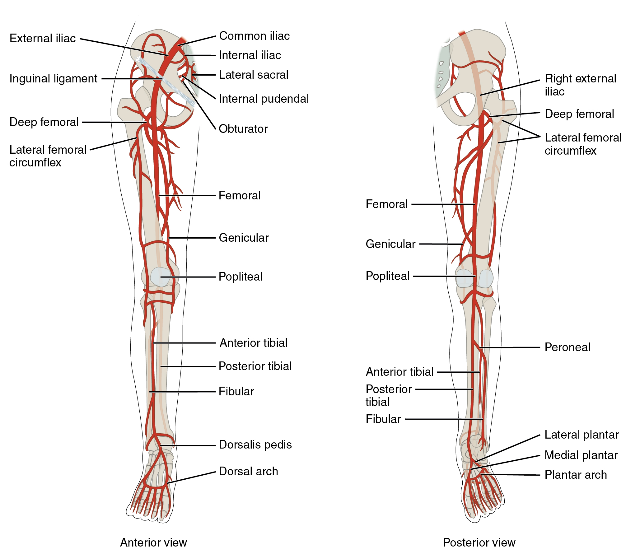
The right and left common iliac arteries were last seen as the terminal branches of the abdominal aorta.
The common iliac artery sends off a branch, the internal iliac artery, which in turn branches to the lateral sacral artery, internal pudendal artery, and obturator artery. As the common iliac artery branches to give rise to the internal iliac artery, it changes name to the external iliac artery.
As the external iliac artery passes beneath the inguinal ligament, it changes name to the femoral artery.
Branches of the femoral artery include the deep femoral artery and lateral femoral circumflex artery.
The knee region is supplied by the genicular artery. (“Genu” is Latin for “knee”; “geniculate” means “pertaining to the knee”.) Other arteries that supply the knee region are the popliteal artery and peroneal artery.
Anatomists use “leg” to refer to the region of the lower extremity between the knee and ankle. In this region, we find the tibia bone and fibula bone. Accordingly, we also find the anterior tibial artery; posterior tibial artery; and fibular artery.
The dorsal (upper) surface of the foot is supplied by the dorsalis pedis artery and the dorsal arch. The plantar (under) surface of the foot is supplied by the lateral plantar artery; the medial plantar artery; and the plantar arch.
Arteries of the Lower Extremity
- External iliac artery = Femoral artery
- Internal iliac artery
-
Lateral sacral artery
-
Obturator artery
-
- Deep femoral artery
- Lateral femoral circumflex artery
- Genicular artery
- Popliteal artery
- Peroneal artery
Arteries of the leg
- Anterior tibial artery
- Posterior tibial artery
- Fibular artery
Arteries of the foot
- Dorsalis pedis artery
- Dorsal arch
- Lateral plantar artery
- Medial plantar artery
- Plantar arch
Media Attributions
- U16-041 Aorta © Betts, J. Gordon; Young, Kelly A.; Wise, James A.; Johnson, Eddie; Poe, Brandon; Kruse, Dean H. Korol, Oksana; Johnson, Jody E.; Womble, Mark & DeSaix, Peter is licensed under a CC BY (Attribution) license
- U16-042 Aorta Branches © Häggström, Mikael is licensed under a CC BY-SA (Attribution ShareAlike) license
- U16-043 Carotid Arteries © BruceBlaus is licensed under a CC BY (Attribution) license
- U16-044 Arteries Supplying the Head and Neck © Betts, J. Gordon; Young, Kelly A.; Wise, James A.; Johnson, Eddie; Poe, Brandon; Kruse, Dean H. Korol, Oksana; Johnson, Jody E.; Womble, Mark & DeSaix, Peter is licensed under a CC BY (Attribution) license
- U16-045 Arteries Serving the Brain © Betts, J. Gordon; Young, Kelly A.; Wise, James A.; Johnson, Eddie; Poe, Brandon; Kruse, Dean H. Korol, Oksana; Johnson, Jody E.; Womble, Mark & DeSaix, Peter is licensed under a CC BY (Attribution) license
- U11-095 Circle of Willis © Betts, J. Gordon; Young, Kelly A.; Wise, James A.; Johnson, Eddie; Poe, Brandon; Kruse, Dean H. Korol, Oksana; Johnson, Jody E.; Womble, Mark & DeSaix, Peter is licensed under a CC BY (Attribution) license
- U11-094 Cerebral Vascular Territories © Gaillard, Frank Dr. is licensed under a CC BY-SA (Attribution ShareAlike) license
- U16-046 Major Arteries Serving the Thorax and Upper Limb © Betts, J. Gordon; Young, Kelly A.; Wise, James A.; Johnson, Eddie; Poe, Brandon; Kruse, Dean H. Korol, Oksana; Johnson, Jody E.; Womble, Mark & DeSaix, Peter is licensed under a CC BY (Attribution) license
- U16-047 Major Arteries Serving the Lower Limb © Betts, J. Gordon; Young, Kelly A.; Wise, James A.; Johnson, Eddie; Poe, Brandon; Kruse, Dean H. Korol, Oksana; Johnson, Jody E.; Womble, Mark & DeSaix, Peter is licensed under a CC BY (Attribution) license
