Fetal Circulation
Objective 9
Compare and contrast fetal circulation to adult circulation.
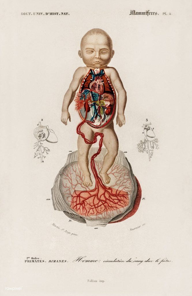
The fetal circulation allows the fetus to benefit from the mother’s ability to oxygenate and nutrify the blood. The fetal lungs and digestive organs will not swing into action until shortly after birth.
Fetal circulation begins at the placenta, a joint project between the mother and child.
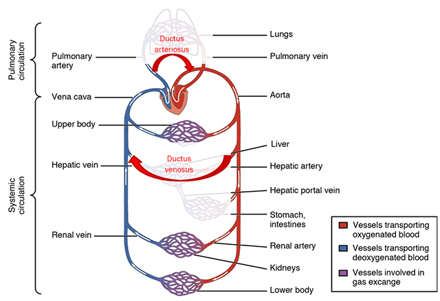
The umbilical vein carries oxygen-rich, nutrient-rich blood from the placenta to the fetal inferior vena cava, joining the vena cava near the liver. This connection is called the ductus venosus.
The oxygen-rich, nutrient-rich blood then enters the right atrium of the fetal heart. There, it finds another bypass called the foramen ovale, which joins the right and left atria of the fetal heart. Just enough blood enters the right ventricle and pulmonary trunk to help in development of the lungs, but fetal lungs do not oxygenate blood.
Most of the blood entering the fetal pulmonary trunk is returned to the fetal heart without going through the lungs. The channel that performs this function is the ductus arteriosus.
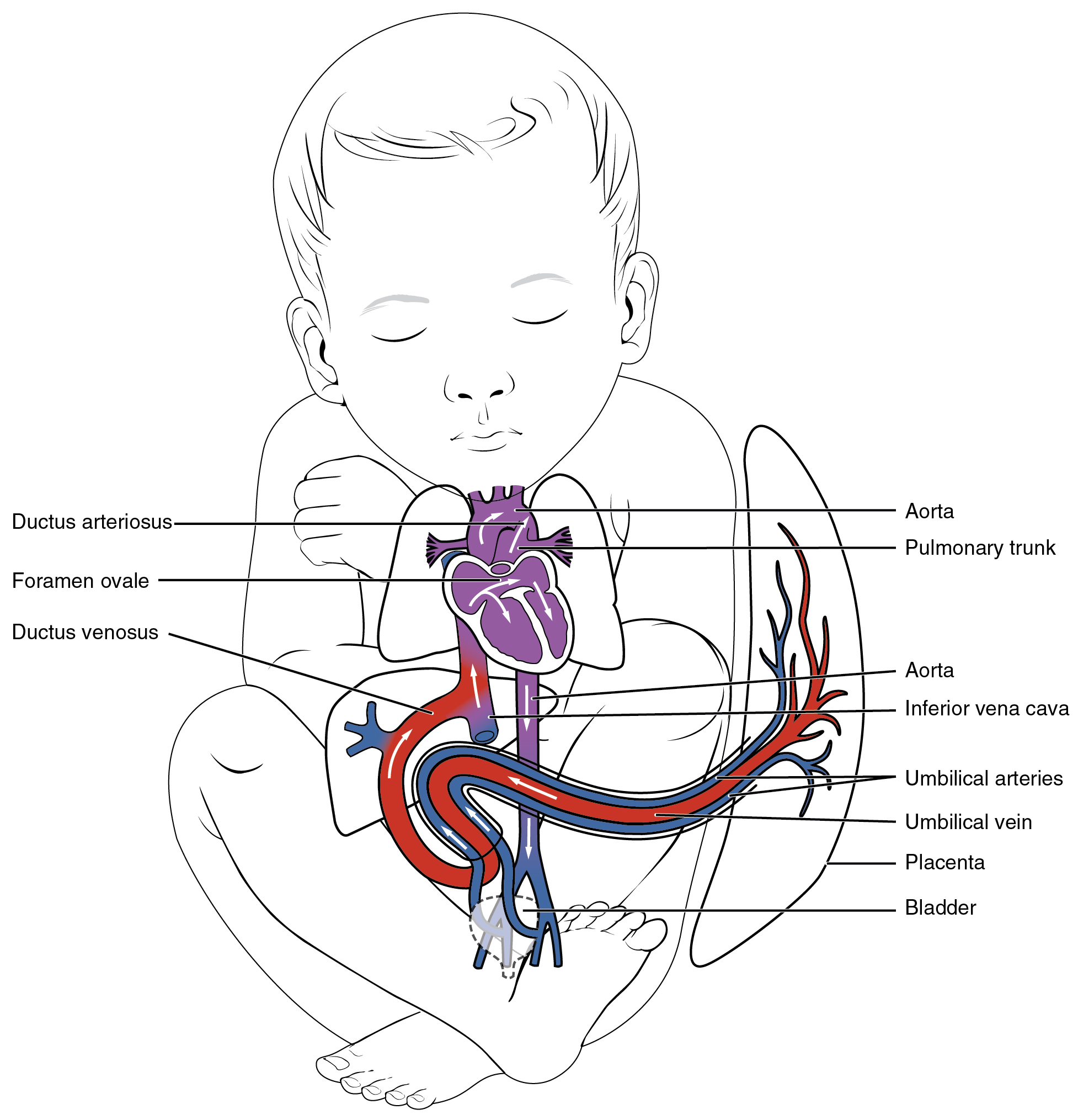
The oxygen-rich, nutrient-rich blood then shifts from the left atrium to the left ventricle. The left ventricle pumps this blood into the systemic circulation of the fetus via the aorta. It returns to the mother via the umbilical arteries.
Thus, the ductus arteriosus allows fetal blood to bypass the lungs; the ductus venosus allows fetal blood to bypass the liver and digestive organs.
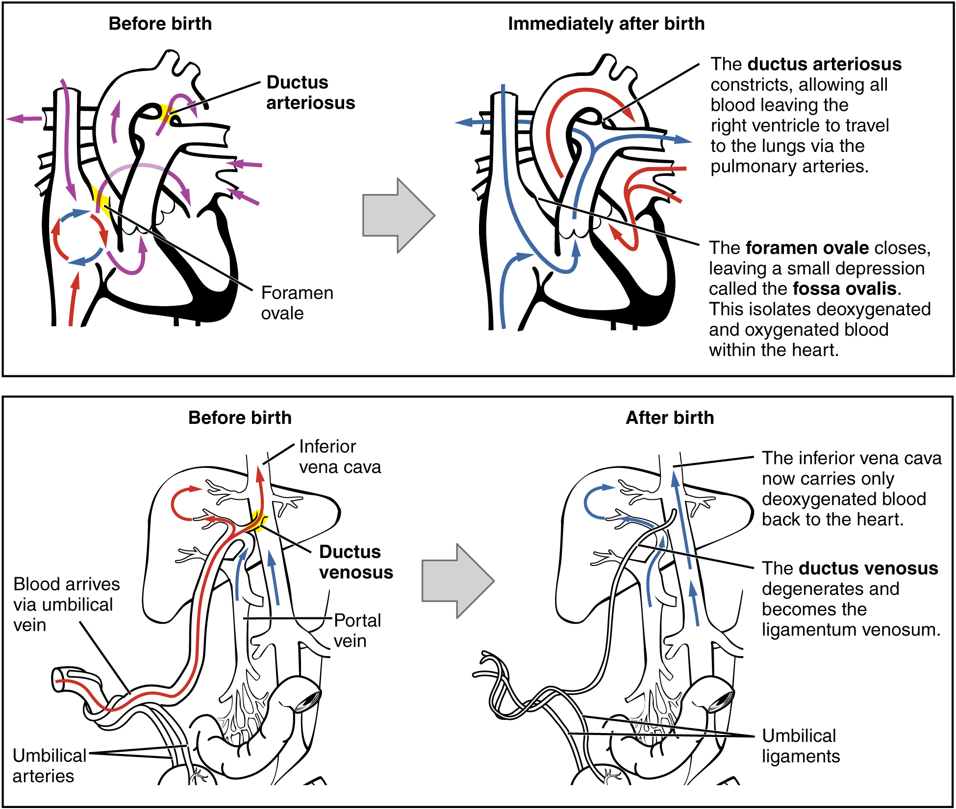
When oxygen levels in the blood increase and acetylcholine and other chemical signals are released from the lungs, muscular tissue which surround the ductus arteriosus is activated and constricts the bypass. This is helped by a decrease in pressure in the shunt. The ductus arteriosus is mostly closed within 12 to 24 hours of birth, and completely closed at 2 to 3 days of age.
The foramen ovale becomes the fossa ovalis in the heart wall between the right and left atrium. About 1/4 of people have a patent foramen ovale.
Similarly, the ductus venosus closes because of a decrease in blood pressure in the shunt when the umbilical vein is detached from the placenta. The vessel takes about 1 to 3 months to completely close and become a remnant called the ligamentum venosum.
Media Attributions
- U16-056 Circulation of the Blood in a Fetus. © Dictionnaire universel d'histoire naturelle is licensed under a Public Domain license
- U16-058 Fetal Shunts © Betts, J. Gordon; Young, Kelly A.; Wise, James A.; Johnson, Eddie; Poe, Brandon; Kruse, Dean H. Korol, Oksana; Johnson, Jody E.; Womble, Mark & DeSaix, Peter is licensed under a CC BY (Attribution) license
- U16-057 Circulation Modified to Show Fetal © Betts, J. Gordon; Young, Kelly A.; Wise, James A.; Johnson, Eddie; Poe, Brandon; Kruse, Dean H. Korol, Oksana; Johnson, Jody E.; Womble, Mark & DeSaix, Peter adapted by Jim Hutchins is licensed under a CC BY (Attribution) license
- U16-059 Neonatal Circulatory System © Betts, J. Gordon; Young, Kelly A.; Wise, James A.; Johnson, Eddie; Poe, Brandon; Kruse, Dean H. Korol, Oksana; Johnson, Jody E.; Womble, Mark & DeSaix, Peter is licensed under a CC BY (Attribution) license
