Digestive System Histology
Objective 2
Describe and be able to recognize the histology of the alimentary canal (gastrointestinal tract). Identify, and describe the function of, the following layers: mucosa, submucosa, muscularis, serosa.
One continuing theme of this course is how function follows form (and form follows function). Because the entire GI tract, from mouth to anus, carries out some combination of the six functions listed previously, the histology (microscopic structure) of the entire GI tract follows the same basic plan.
This simplifies our study of digestive system histology: once we learn the basic plan, then everything that follows is just “variations on a theme.”

The GI tract is an elongated tube. The inside of the tube is called the lumen. The outside of the tube is covered with a serous membrane.
The four layers seen throughout the GI tract, from lumen to serous membrane, are:
- Mucosa
- Submucosa — areolar connective tissue; see Unit 7 for a definition of “areolar connective tissue”)
- Muscularis — also called muscularis externa to distinguish it from the thinner, weaker muscle layer of the mucosa above
- Serosa — in the abdomen, the serosa forms part of the peritoneal cavity.
Let’s take a look at each of these layers individually.
Mucosa
The mucosa is the layer that lines the lumen. It has three sublayers, in order from deep to superficial (lumen out):
- Epithelium – stratified squamous in the mouth and simple columnar in the stomach and intestines.
- Lamina propria – a layer of areolar connective tissue with blood and lymphatic vessels to pick up material absorbed by the epithelium.
- Muscularis mucosae – a thin muscle layer that makes the inside of the GI tract all crinkly and folded.
Submucosa
The submucosa comprises areolar connective tissue. In it, we find blood and lymphatic vessels and the submucosal plexus of the enteric nervous system.
Muscularis
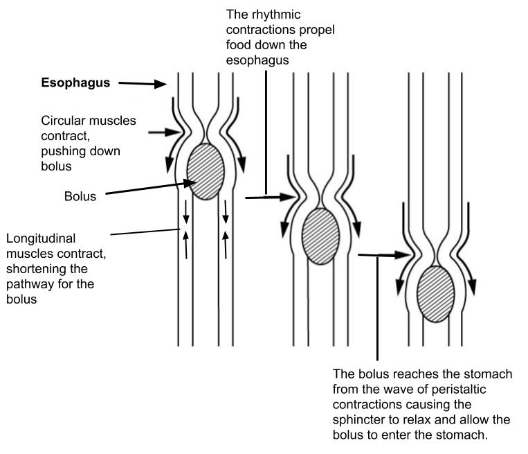
Striated (voluntary) muscle in the mouth and pharynx controls swallowing. Smooth (involuntary) muscle, usually in two layers, lines the rest of the GI tract and keeps materials moving through peristalsis.
Serosa
The serosa is made up of areolar connective tissue covered by a simple squamous epithelium (mesothelium). In the abdominal cavity, the serosa is called the visceral peritoneum (Latin viscera, “bowels”) because it forms the “guts” (visceral) side of the peritoneal cavity.
Media Attributions
- U18-003 Histology © Betts, J. Gordon; Young, Kelly A.; Wise, James A.; Johnson, Eddie; Poe, Brandon; Kruse, Dean H. Korol, Oksana; Johnson, Jody E.; Womble, Mark & DeSaix, Peter is licensed under a CC BY (Attribution) license
- U18-004 Peristalsis © Betts, J. Gordon; Young, Kelly A.; Wise, James A.; Johnson, Eddie; Poe, Brandon; Kruse, Dean H. Korol, Oksana; Johnson, Jody E.; Womble, Mark & DeSaix, Peter adapted by Allison Calabrese is licensed under a CC BY (Attribution) license
