Cells of the Nervous System | Glia
Objective 4
State the different types of glial cell and their functions.
There are two main cell types in the brain. Neurons are what we generally think of when we think of “nerve cells”. Remember that these are the cells that receive, process and transmit information in a point-to-point fashion in the nervous system. There are about 80 billion neurons in the human nervous system, but no one can possibly count them, so that’s just an estimate.
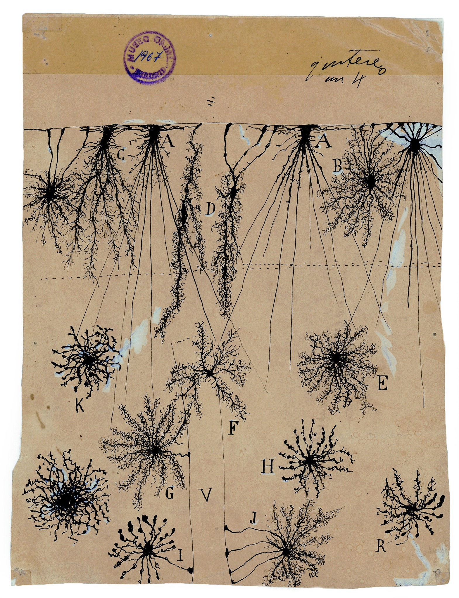
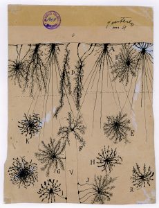
Another roughly 80 billion cells in the brain are glial cells or glia (Greek λοια, gloia, “glue”). For a long time, no one wanted to study cells whose name made people think of “Elmer’s” or the kid in first grade who ate paste. Recently, however, glial cells have come into their own as a field of study. Glial cells maintain the structural and chemical environment of the brain, and function in ways we probably don’t fully appreciate. For example, Einstein’s brain had the same number of neurons as you and I, but more glial cells than normal1. The image here, from Ramón y Cajal’s drawings, shows the glial cells in the cerebral cortex of a human child.
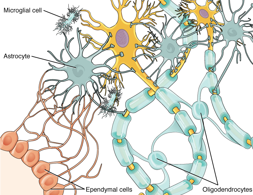
There are four types of glial cells:
- astrocytes (astroglia),
- oligodendrocytes (oligodendroglia),
- microglia, and
- ependymal cells
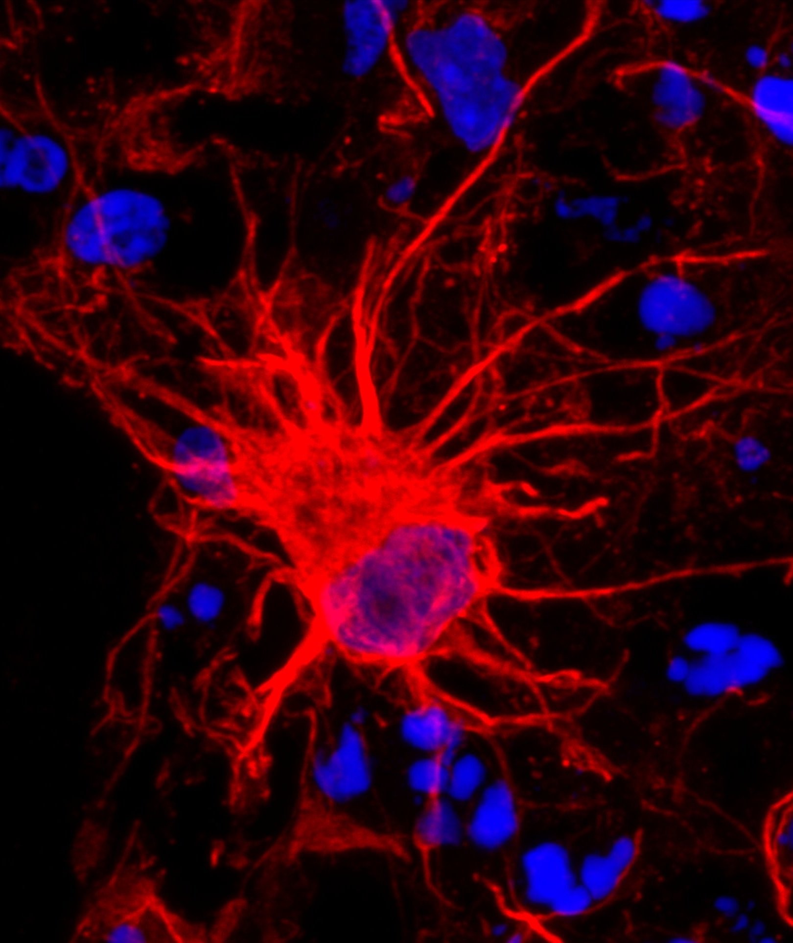
Astrocytes (also called astroglia) form the borders of the CNS. They cooperate with blood vessels to form the blood-brain barrier (BBB), which permits only lipid-soluble substances, certain amino acids, and glucose to pass (we will examine the BBB in detail in Objective 8). This helps protect the brain from chemical or microbiological damage, but also makes it hard for the body to mount a defense against something that circumvents the BBB. The end-feet of astrocytes make up the pia mater, one of the coverings on the outside of the brain we will also study in Objective 8. Astrocytes also wall off groups of neurons and act as sponges to soak up potentially harmful ions and waste products.
Oligodendrocytes (also called oligodendroglia) have only one job, but they do it well. They form myelin sheaths which insulate nerve axons that must send information over long distances. For technical reasons we’ll discuss in Unit 13, the myelin sheath must have gaps every few hundred micrometers. These gaps are called the nodes of Ranvier.
In Unit 15, we will learn about immune cells of the tissues called macrophages. Macrophages swallow and digest invaders and waste products.
Microglia are the brain’s equivalent of macrophages. The brain is termed an “immunologically privileged site”, so no immune cells can pass the BBB. Only microglia are normally allowed to carry out immune functions in the brain.
Ependymal cells, along with the border-forming astrocytes, make up a single layer of border cells lining the ventricles. These open spaces in the brain are filled with cerebrospinal fluid (CSF), which bathes, floats, and cushions the brain. CSF circulates continuously, being made by a specific tissue in special locations, traveling through the chambers of the brain and spinal cord, and finally being absorbed into veins at another location. Accordingly, ependymal cells have cilia that continuously “row” CSF through the ventricular system. Again, the function of ependymal cells and the fluid they produce will be discussed in Objective 8.
The CNS has immune privilege and ventricles. The PNS lacks both of these features, so the glial cell types carrying these two functions (microglia and ependymal cells, respectively) are absent in the PNS.
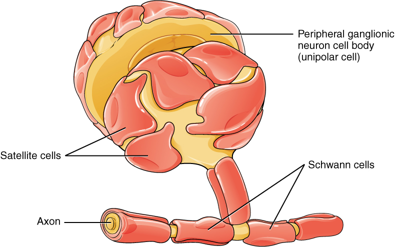
Satellite cells in the PNS perform the same basic functions as astrocytes in the CNS: maintenance of a favorable chemical environment and mechanical/structural support. Satellite cells are found mostly in ganglia, collections of nerve cell bodies in the PNS.
Schwann cells are found in the PNS in place of the oligodendrocytes of the CNS. The distinction is not only in its name. There is a functional difference as well: axons ensheathed by oligodendrocytes in the CNS cannot regrow if cut or damaged, while those surrounded by Schwann cells in the PNS can regenerate. For example, if you cut nerves in your finger with a deep knife cut, the finger may be numb for several weeks but eventually, the nerves will regrow.
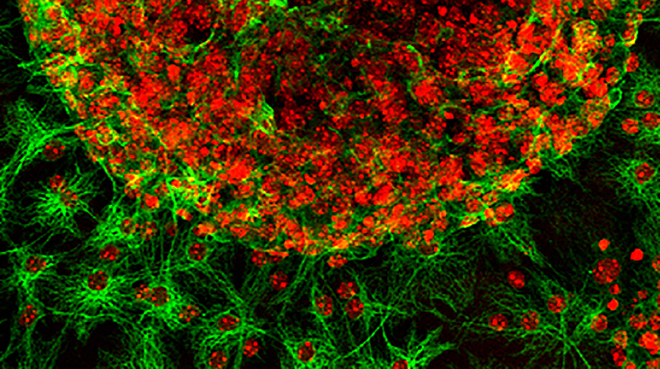
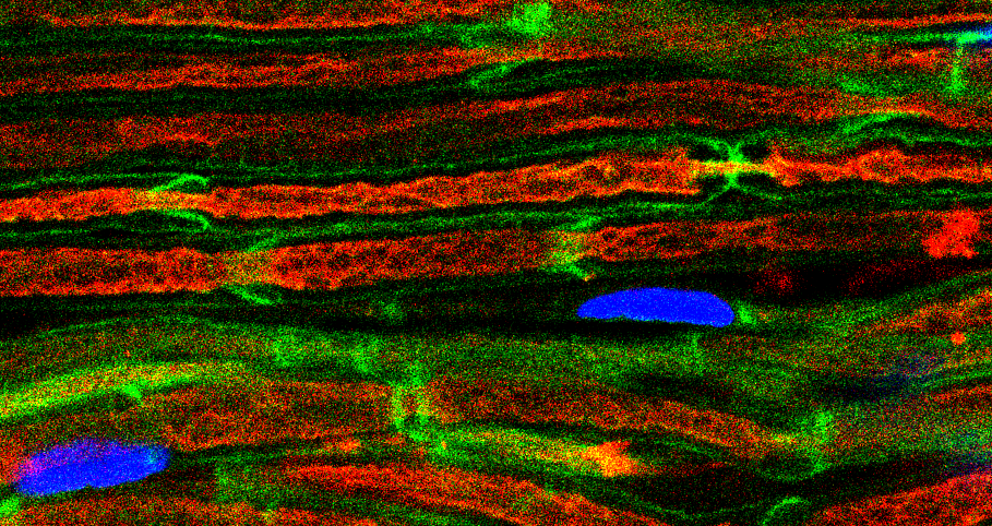 Similarly, PNS nerves damaged by surgery can regrow, albeit slowly (about 1 mm per day). In this image, Schwann cells are stained green; axons are red; and nuclei are blue.
1 See, for a thorough analysis of the data on Einstein’s brain: Colombo JA, Reisin HD, Miguel-Hidalgo JJ and Rajkowska G. (2006) Cerebral cortex astroglia and the brain of a genius: A propos A. Einstein’s. Brain Res Rev 52(2): 257-263.
Media Attributions
- U11-039 Astrocytes Green © NIH/NICHD is licensed under a CC BY (Attribution) license
- U11-040 Glial Cells in a Child © Santiago Ramón y Cajal is licensed under a Public Domain license
- U1-041 Glial Cells © Betts, J. Gordon; Young, Kelly A.; Wise, James A.; Johnson, Eddie; Poe, Brandon; Kruse, Dean H. Korol, Oksana; Johnson, Jody E.; Womble, Mark & DeSaix, Peter is licensed under a CC BY (Attribution) license
- U11-042 Astrocytes © Pasca Lab, Stanford University is licensed under a CC BY-NC (Attribution NonCommercial) license
- U11-043 Glial Cell © Betts, J. Gordon; Young, Kelly A.; Wise, James A.; Johnson, Eddie; Poe, Brandon; Kruse, Dean H. Korol, Oksana; Johnson, Jody E.; Womble, Mark & DeSaix, Peter is licensed under a CC BY (Attribution) license
- U11-044 Schwann Cells Green, Axons Red, Nuclei Blue © Alvarez-Prats, A. and Balla, T., NICHD/NIH is licensed under a CC BY-NC-ND (Attribution NonCommercial NoDerivatives) license
